Abstract
Anderson, D. L. (University of Minnesota, Minneapolis), M. E. Pollock, and L. F. Brower. Morphology of Mycoplasma laidlawii type A. I. Comparison of electron microscopic counts with colony-forming units. J. Bacteriol. 90:1764–1767. 1965.—Cells of Mycoplasma laidlawii A grown in dialyzing flask cultures were counted with the electron microscope by use of a spray technique which deposited mixtures of polystyrene latex of known concentration and M. laidlawii in discrete droplet patterns on specimen films. Glutaraldehyde and formaldehyde were effective in preserving gross morphology of cells in spray preparations. The standard deviation of the mean ratio of latex-M. laidlawii was 5.5% when 2,000 total particles were counted in 18 droplet patterns. Microscopic counts resembled counts of colony-forming units (CFU) at various culture ages when cells larger than 0.25 μ were enumerated. If small bodies ranging from 0.1 to 0.25 μ in diameter were included in microscopic counts of cultures older than 70 hr, these counts exceeded the numbers of CFU by 4 to 10 times.
Full text
PDF
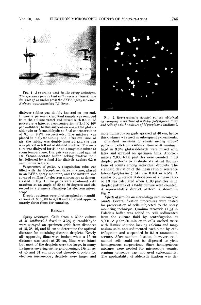
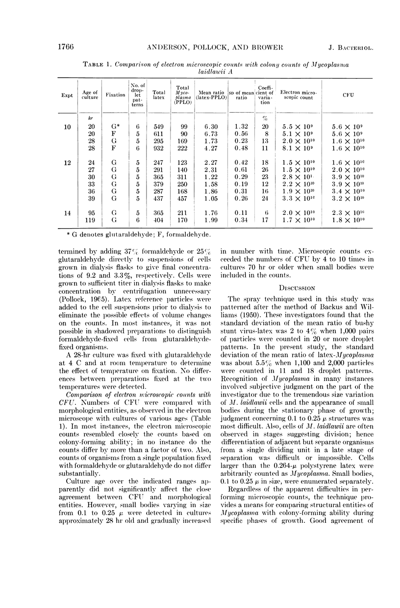
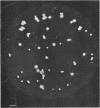
Images in this article
Selected References
These references are in PubMed. This may not be the complete list of references from this article.
- MOROWITZ H. J., TOURTELLOTTE M. E. The smallest living cells. Sci Am. 1962 Mar;206:117–126. doi: 10.1038/scientificamerican0362-117. [DOI] [PubMed] [Google Scholar]
- Pollock M. E. Use of dialyzing culture technique for high yield of Mycoplasma. J Bacteriol. 1965 Dec;90(6):1682–1685. doi: 10.1128/jb.90.6.1682-1685.1965. [DOI] [PMC free article] [PubMed] [Google Scholar]


