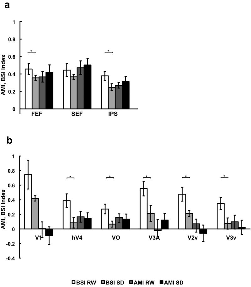
Fig. 3. Mean Baseline Shift Index (BSI) and Attentional Modulation Index across different cortical regions.
fMRI signal change normalized for activation magnitude was assessed using BSI (Baseline shift/Attend) and AMI (Attend – Non-attend)/Attend), as calculated separately for each state and for each individual before being averaged across individuals for display purposes. Across the volunteers, SD reduced BSI in right FEF and IPS, as well as all visual cortical regions except V1 indicating a deficit in endogenous attention. AMIs were not significantly changed due to state in any region indicating regions that show an effect of attention on activation - higher cognitive areas and the extrastriate cortex show comparable reduction in activation across state whether the peripheral stimulus was attended or not. These findings also highlight that SD has a relatively more pronounced effect on the preparatory / prestimulus signal than on stimulus-related activity. Error bars indicate SEM.

