Abstract
To protect natural coral reefs, it is of utmost importance to understand how the growth of the main reef-building organisms—the zooxanthellate scleractinian corals—is controlled. Understanding coral growth is also relevant for coral aquaculture, which is a rapidly developing business. This review paper provides a comprehensive overview of factors that can influence the growth of zooxanthellate scleractinian corals, with particular emphasis on interactions between these factors. Furthermore, the kinetic principles underlying coral growth are discussed. The reviewed information is put into an economic perspective by making an estimation of the costs of coral aquaculture.
Keywords: Corals, Growth, Aquaculture, Zooxanthellate Scleractinia
Introduction
Being the main builders of coral reefs, zooxanthellate scleractinian corals (i.e., calcifying corals that live in symbiosis with microalgae—the zooxanthellae) are of crucial importance for marine ecology. In addition, coral reefs represent a high economic value as a source of food (Bryant et al. 1998) and natural products (Fusetani 2000), as an attractive resource for tourism (Bryant et al. 1998) and by forming a natural protection of coastlines. It has been estimated that approximately 10% of the world’s population is directly or indirectly depending on coral reefs. However, reefs are currently under high pressure, mainly caused by anthropogenic disturbances such as overfishing, pollution, eutrophication, and human-induced climate change (Hughes et al. 2003). Also the trade in aquarium ornamentals has increased in the last decades and is now also becoming a threat for natural populations of reef organisms including scleractinian corals (Wabnitz et al. 2003; Knittweis et al. 2009). This has resulted in an increased effort to develop cost-effective in situ (sea-based) and ex situ (aquarium) coral aquaculture methods. An example of this is the CORALZOO project, in which scientists and public aquaria collaborated to improve techniques for breeding and husbandry of scleractinian corals (Osinga 2008).
To understand reef development in a changing environment, it is crucial to identify the factors that determine the growth rates of corals and to understand how these factors interact (Langdon and Atkinson 2005). The same knowledge is needed for efficient breeding of corals ex situ. Furthermore, in this respect, it is important to understand the kinetics of coral growth, which determines how proliferation of biomass develops in time.
This mini-review presents an overview of studies describing effects of environmental factors on coral growth rates. Based on this overview, we will try to explain how coral growth is controlled. Our views will be further supported by new experimental data obtained during the CORALZOO project. Secondly, we will discuss the kinetic principles underlying coral growth. Finally, the information will be put into an economic perspective: the costs of coral culture will be analyzed in the view of the biological information provided.
The Coral Growth Process
Zooxanthellate Scleractinia corals represent a true symbiosis. The coral provides shelter and nutrients to the algae, while the algae translocate a substantial proportion of their photosynthetically acquired organic carbon to the coral host. The translocated photosynthetates are used by the host for respiration and biomass buildup (Muscatine and Cernichiari 1969; Muscatine 1990). The coral also acquires organic carbon through feeding on a wide range of particulate and dissolved organic materials (reviewed by Houlbrèque and Ferrier-Pagès 2009). A third important characteristic of scleractinian corals is that they form massive calcium carbonate skeletons through a process called “calcification” (see review by Gattuso et al. 1999). To enable calcification, scleractinian corals synthesize an organic matrix around which calcium carbonate is deposited (Allemand et al. 1998). For a more detailed description of the physiology of zooxanthellate corals, we refer to reviews by Muscatine (1990), Dubinsky and Jokiel (1994), Titlyanov and Titlyanova (2002), and Furla et al. (2005).
Factors Influencing Coral Growth
Taking into account the three major physiological processes described above (photosynthesis, heterotrophic feeding, and calcification), the following basic requirements (building blocks) for coral growth can be identified: light, carbon dioxide (CO2), and inorganic nutrients (needed for photosynthesis); organic food (needed for organic tissue synthesis and organic matrix synthesis); and calcium and carbonate ions (Ca2+ and CO2−3, needed for skeleton formation). In addition to these basic requirements, water movement (flow) is an important factor facilitating coral metabolism. Flow enhances the exchange of gasses (O2, CO2) and dissolved compounds (nutrients, metabolic waste products) between the coral and its environment. Hence, insufficient flow may lead to depletion of resources (thus inducing resource limitation) and/or accumulation of inhibiting substances.
Several other factors have been reported to influence coral growth, either positively or negatively. These factors include temperature and pH (Reynaud et al. 2003; Langdon and Atkinson 2005; Anthony et al. 2008), iron (Ferrier-Pagès et al. 2001), zinc (Ferrier-Pagès et al. 2005), competition and predation (Fabricius 2005 and references therein), polluting substances such as herbicides (Jones 2005), oil (Haapkylä et al. 2007) and sunscreens (Danovaro et al. 2008), sedimentation (Van Katwijk et al. 1993; Torres 2001; Fabricius 2005), UV radiation (Jokiel and York 1982; Kuffner 2001; Torres et al. 2007), and dissolved oxygen (DO). Despite its key role in metabolism, very few scientists have investigated the potential role of DO as a growth-controlling agent for corals, probably due to the technical complexity of working under low DO concentrations. Rinkevich and Loya (1984) found that aeration of the water significantly enhanced dark calcification in Stylophora pistillata. They suggested that under non-aerated conditions, dark calcification in this species was limited by low DO due to the absence of photosynthesis. Hence, flow-dependent mass transfer of oxygen may control dark calcification. Fossil records suggest that reductions in DO concentrations were one of the causes of prehistoric mass extinction events of Scleractinia (Van de Schootbrugge et al. 2007). In addition to being a potentially limiting factor, high DO concentrations inside coral tissue are assumed to have a negative effect on coral metabolism (Lesser 1997; Finelli et al. 2006; Mass et al. 2010).
In the following subsections, we will more extensively review studies on the primary requirements for coral growth: light, inorganic nutrients, food, dissolved inorganic carbon (DIC, which includes carbon dioxide, bicarbonate and carbonate), calcium, and water flow. We will also discuss the role of genetic variability.
Light
There is no doubt that light plays an important role in the growth of zooxanthellate corals. The coral host is very well adapted to facilitate light capture by its symbiotic algae due to the optimal light reflecting properties of the calcium carbonate skeleton: multiple scattering on coral skeletons enhances light absorption by symbiotic algae (Enriquez et al. 2005). Photon flux density (PFD also known as irradiance) and growth/calcification are often positively correlated (Goreau 1959; Chalker 1981; Marubini et al. 2001; Reynaud et al. 2004; Schlacher et al. 2007; Schutter et al. 2008). Although a direct stimulation of calcification by light was suggested by Al-Horani et al. (2003), it is important to realize that the corals themselves are mainly indirectly influenced by light, whereas the zooxanthellae can be directly light limited. Light-related growth limitation in corals may have three causes: (1) insufficient production of photosynthates (Titlyanov et al. 2001); (2) insufficient translocation of photosynthates, for example after enrichment of seawater with inorganic nutrients (Marubini and Davies 1996, see also section on nutrients below); (3) a decrease of the internal pH due to lower photosynthesis, leading to less favorable conditions for calcification (Schneider and Erez 2006). In addition, light may become inhibiting at high photon flux densities. After long exposure to high PFD, the increase in maintenance energy required to repair the light-induced damage to the photosystem will exceed the gain in photosynthetic energy, leading to a retarded growth (photoinhibition, Iglesias-Prieto et al. 1992).
The coral-zooxanthellae holobiont adjusts its photosynthetic potential to the prevailing environmental conditions. Such photoacclimation is achieved either by increasing/decreasing the number of zooxanthellae per square centimeter of coral surface (probably a host-controlled mechanism: adaptive bleaching—Kinzie et al. 2001; Fautin and Buddemeier 2004) or by adjusting the pigment density (a zooxanthellae-controlled mechanism). Both processes occurred simultaneously within a period of 30 days after transplanting fragments of S. pistillata from high to intermediate PFD and from intermediate to low PFD (Titlyanov et al. 2001). In addition, also the pigment composition of the zooxanthellae is variable and adjusted to the available spectrum of light (Dustan 1982).
The in hospite photosynthetic potential of zooxanthellae in corals is usually determined by measuring a photosynthesis/irradiance (PI) curve (Fig. 1). PI curves can either be obtained using direct assessment of electron transport rates as a measure for photosynthesis using pulse-amplitude modulated fluorometry (e.g., Ulstrup et al. 2006) or indirectly from oxygen evolution measurements. Oxygen-based PI curves provide characteristic numbers such as the compensation point (i.e., the irradiance at which photosynthetic oxygen production equals respiratory oxygen consumption) and the onset of saturation point I k (the point on the x-axis of the curve where the initial, linear slope of the curve intersects with the horizontal asymptote resembling maximal photosynthesis. This point is also referred to as Talling index—Barnes and Chalker 1990). Due to photoacclimation, specimens of the same species growing under different light regimes may show different PI curves. For example, Fig. 1 shows two PI curves of Galaxea fascicularis. The two curves represent averages of two groups of four genetically identical colonies that had been raised under a PFD of 300 and 600 μE m−2 s−1, respectively, in a single 600 l aquarium system under controlled, stable conditions (for details, see Schutter et al. 2011, the colonies used for the experiment described here experienced a high water flow of 15–25 cm s-1). Photosynthesis and respiration rates were assessed from oxygen evolution measurements under a range of photon flux densities (methodology according to Schutter et al. 2008; corals were incubated in 1,500 cm3 incubation chambers equipped with a magnetic stirrer, corals were incubated for 30 min under each PFD level). The curves show that corals raised under high light had a lower maximal photosynthesis, a higher Talling Index and a higher compensation point. The example shows that corals raised under low light may exhibit the same rate of net photosynthesis as corals growing under high light. Hence, to assess the PFD at which light becomes limiting, it is better to use a normalized PI curve, based upon measurements done on corals only at their ambient PFD. The saturation point of such a normalized curve represents a species specific saturation point (hereinafter referred to as I k s), below which photoacclimation cannot longer compensate for the reduced influx of photons. I k s may vary as a result of variability in other environmental conditions such as the flow regime around the corals and the availability of inorganic nutrients and food (see next subsections).
Fig. 1.
Photosynthesis–irradiance curves based on oxygen evolution measurements on two groups of four colonies of G. fascicularis that had been raised under 300 (solid line) and 600 (dotted line), respectively. The Talling index I k is indicated for both curves (the intersect of the vertical dotted line with the x-axis)
Inorganic Nutrients
Both partners of the coral-zooxanthellae holobiont need nitrogen (N) and phosphorous (P) as building blocks for synthesis of proteins and other biomass components. Whereas the zooxanthellae can directly take up N and P in their inorganic forms (dissolved inorganic nitrogen (DIN) and dissolved inorganic phosphorous (DIP)), the coral host acquires its N and P through heterotrophic feeding (see “Food” section) and via translocated organic substances produced by the zooxanthellae. According to Falkowski et al. (1984), translocated substances can become very low in nitrogen when the zooxanthellae are DIN limited. They introduced the term “junk food” to describe the low-N organic excretion products of N-limited zooxanthellae; these substances only provide the coral host with metabolic energy, and not with nitrogen-rich building blocks needed for biosynthesis. It was suggested that the coral host expels the majority of this “junk food” as mucus.
Following this “junk food hypothesis”, it seems logic to assume that addition of DIN can promote coral growth. Many authors have reported that addition of DIN promotes zooxanthellae growth and augments the pigment production of the zooxanthellae, thus stimulating the overall net photosynthesis rates of the holobiont (Hoegh-Gulberg and Smith 1989; Dubinsky et al. 1990; Stambler et al. 1991, 1994; Marubini and Davies 1996; Marubini and Thake 1999; Ferrier-Pagès et al. 2000, 2001; Grover et al. 2002; Langdon and Atkinson 2005; Tanaka et al. 2007), although the photosynthesis rate per algal cell can decrease due to self-shading effects (Dubinsky et al. 1990). Most of these authors (Stambler et al. 1991; Marubini and Davies 1996; Marubini and Thake 1999; Ferrier-Pagès et al. 2000, 2001; Langdon and Atkinson 2005; Tanaka et al. 2007) also tested the effects of DIN addition on skeletal growth of the corals, which was inhibited by DIN or (in the case of moderate nitrate enrichment—Tanaka et al. 2007) only slightly elevated. Both forms of DIN applied (nitrate and ammonium) imposed a similar effect on corals (Marubini and Davies 1996). In general, it can be concluded that raising the external DIN concentration above ambient natural concentrations does not promote coral growth. Apparently, coral growth is not limited by DIN under ambient natural DIN concentrations, i.e., less than 2 μM ammonium or nitrate (Grover et al. 2002). Grover et al. (2002) suggested an external concentration of ammonium as low as 0.6 μM to be sufficient for sustaining zooxanthellae growth.
To explain the observed inhibition of skeletal growth by elevated (DIN), it has been suggested that DIN enrichment disrupts the delicate balance between host metabolism and zooxanthellae metabolism that is needed for optimal functioning of the symbiosis (e.g., Marubini and Davies 1996). It is important to note here that the studies describing effects of DIN enrichment have all been done under relatively high irradiance levels (200 μE m−2 s−1 and higher), i.e., under conditions where light is not likely to be limiting. DIN addition under low light (i.e., below I k s) is not expected to have any direct effect on either the zooxanthellae or the coral.
Some studies on nutrient enrichment (eutrophication) on natural coral reefs (see reviews by Dubinsky and Stambler 1996 and Fabricius 2005) confirm the experimental observations described above (e.g., Kinsey and Davies 1979; Tomascik and Sander 1985; Tomascik 1990; Koop et al. 2001). However, other studies showed a positive correlation between eutrophication and coral growth (Meyer and Schultz 1985; Grigg 1995; Bongiorni et al. 2003a, b). The conflicting results can be ascribed to indirect effects of DIN/DIP enrichment. Enrichment will lead to higher concentrations of particulate and dissolved organic matter in the water column, which may enhance coral growth (by providing additional food) in free-floating nurseries (Bongiorni et al. 2003a, b) and in coral reefs subjected to high water movement (Fabricius 2005). On the other hand, increased turbidity will reduce the penetration of light, which may negatively affect corals growing at greater depths where light limits growth (Fabricius 2005 and references therein). In stagnant waters, high particle loads may inhibit coral growth due to increased sedimentation (Genin et al. 1995). Eutrophication also indirectly affects coral growth by stimulating the growth of turf algae that compete for space with corals (Genin et al. 1995; Fabricius 2005).
Most studies describing effects DIP on corals show that DIP negatively affects coral growth, in particular when supplied without a corresponding increase in DIN (Snidvongs and Kinzie 1994; Ferrier-Pagès et al. 2000). The negative effect of elevated DIP may be caused by the formation of poisonous polyphosphate crystals (Simkiss 1964). There is also a record of DIP limitation in zooxanthellate corals. Steven and Broadbent (1997) found increased growth of Acropora palifera after pulsed additions of phosphate, with or without concurrent enrichment in nitrate. A good overview of studies relating to effects of both DIN and DIP is presented by Fabricius (2005).
In addition to DIN and DIP, iron and zinc have also been reported as agents that influence coral growth. Iron enrichment can have effects on the coral-zooxanthellae symbiosis that are comparable to DIN enrichment (Ferrier-Pagès et al. 2001). The role of zinc in coral growth and metabolism has been clearly outlined in another paper by Ferrier-Pagès et al. (2005). Zinc is an essential structural component of many enzymes, among which carbonic anhydrase (CA). CA is a ubiquitous enzyme in corals; it is involved in the uptake of dissolved inorganic carbon. As such, CA plays a key role in both photosynthesis and calcification and therefore, zinc limitation may limit overall coral growth. Conversely, high zinc concentrations may inhibit coral growth due to the formation of toxic free radicals, which have been reported to inhibit microalgae growth (Sunda 1991).
Bicarbonate, Carbonate, Calcium, and pH: The Aragonite Saturation State
In order to calcify, corals need Ca2+ and CO2−3. Ca2+ and CO2−3 are commonly referred to as Ω, the aragonite saturation state, which is the temperature-dependent solubility product of aragonite (Mucci 1983). Aragonite is the chrystalline form of calcium carbonate produced by corals. Both Ca2+ ions and CO2−3 ions are actively concentrated in the calicoblastic fluid (this is a thin liquid layer between the skeleton and the calicoblastic cells, the cellular layer that secretes the organic matrix of the skeleton) to facilitate precipitation of calcium carbonate. Ca2+ is actively transported across the calicoblastic membrane into the calicoblastic fluid by a Ca2+-dependent adenosine triphosphate (ATP)-ase, which exchanges Ca2+ for H+ ions (Al-Horani et al. 2003). This is a process that consumes metabolic energy (ATP). The mechanisms by which HCO−3 and/or CO2−3 are transported across the calicoblastic membrane are hitherto unknown. However, by removing protons from the calicoblastic fluid, the pH of the calicoblastic fluid is increased, which shifts the equilibrium between HCO−3 and CO2−3 in favor of the latter: a pH of 9.28 and an Ω of 25 were measured inside the calicoblastic fluid of G. fascicularis (Al-Horani et al. 2003), which is well above reference seawater levels (8.2 and 4, respectively). These measurements were done under simulated daylight conditions. The calicoblastic pH and Ω were not elevated when the corals were incubated in the dark, indicating that light stimulates calcification. Indeed, calcification is on average three times higher during the day than at night (light-enhanced calcification—Gattuso et al. 1999). The mechanism by which light promotes calcification is most likely a combination of a higher availability of ATP and a higher internal pH inside the coral, which both result from photosynthetic activity of the zooxanthellae.
It is generally agreed that Ω is positively correlated with coral growth (Schneider and Erez 2006; Marubini et al. 2008) and reef growth (Anthony et al. 2008; Jokiel et al. 2008; De’ath et al. 2009). The concentrations of both ionic components of Ω have been reported to influence coral growth in a similar way (for effects of [Ca2+], see Chalker 1981; Gattuso et al. 1998; Marshall and Clode 2002; for effects of [HCO−3] and [CO2−3], see Marubini and Thake 1999; Marubini et al. 2001, 2008; Schneider and Erez 2006; Herfort et al. 2008). Hence, [Ca2+] and [CO2−3], are of equal importance in controlling coral growth. Whereas [CO2−3] may vary due to short- and long-term changes in ocean pH (Gattuso et al. 1999; Kleypas et al. 1999), [Ca2+] is rather stable in oceanic waters. Therefore, [Ca2+] is not considered as a very relevant factor with respect to the effect of climate change on calcifying organisms. However, in an aquarium situation, where the ratio between water volume and coral volume is orders of magnitude lower than in nature, the concentration of Ca2+ can diminish rapidly and should be adequately monitored and controlled.
Food
The heterotrophic feeding biology of zooxantellate corals has recently been reviewed by Houlbrèque and Ferrier-Pagès (2009). Here, we will briefly summarize some important observations on the effects of feeding on coral metabolism and growth.
One of the proposed benefits of feeding is that it supplies the coral holobiont with nitrogen (Dubinsky and Jokiel 1994). In contrast to the effect of DIN addition, which stimulates zooxanthellae, but inhibits growth (see “Inorganic Nutrients” section), it was shown by Ferrier-Pagès et al. (2003) and Houlbrèque et al. (2003, 2004) that feeding stimulated both zooxanthellae (numbers, pigmentation, and photosynthetic activity) and growth of S. pistillata. Organic food provides the coral holobiont with nitrogen, carbon, and phosphorous in an appropriate biological ratio. Hence, in contrast to enrichment with DIN and/or DIP, providing organic food is not expected to disturb the nutrient balance inside the coral.
Further evidence supporting this view was obtained for another branching coral species (Seriatopora caliendrum) during the CORALZOO project. Three genetically identical colonies of this species were grown for a period 7 weeks in a 500 dm3 aquarium system under ambient feeding conditions (see “Genotype” section for a description of the aquarium; conditions were the same as described in “Genotype” section, except that the alkalinity and the calcium concentration were maintained at 2.0–2.5 mEq dm−3 and 440 mg Ca2+ dm−3, respectively). Specific growth rate was determined weekly (see “Growth Kinetics” section for calculation details) based upon the increase in buoyant mass (determined according to Schutter et al. 2008). After the initial period of 7 weeks, the colonies were subjected to a high feeding regime. Five days per week, each colony was taken out of the main tank and was incubated for a period of 2 h in a 1,500 cm3 aerated incubation chamber with an additional supplement of 20,000 Artemia nauplii. Conditions in the incubation chamber (light regime, mixing, temperature, and water quality) were the same as in the main tank. Specific growth rate was determined weekly until no further change in growth was observed (week 4). PI curves were determined for the colonies at the end of the initial period of 7 weeks and after the second period of 4 weeks. The methodology for measuring photosynthetic capacity was the same as described in “Light” section for G. fascicularis. We found that high feeding increased both the specific growth rate (Fig. 2a) and the photosynthetic capacity (Fig. 2b) in colonies of S. caliendrum. The beneficial effects of feeding on growth and photosynthesis appear not to be directly coupled. Food-stimulated photosynthesis is likely to occur only under high light. Both in our study on S. caliendrum and in the study by Houlbrèque et al. (2004) on S. pistillata, the stimulating effect of feeding on photosynthesis became apparent only above 200 μE m−2 s−1.
Fig. 2.
a Specific growth rates of colonies of S. caliendrum cultured at ambient aquarium feeding and high feeding (ambient aquarium feeding + 20.000 Artemia nauplii per colony per day); n = 3 for both treatments. b Photosynthesis–irradiance curves for ambient fed colonies (solid line) and highly fed colonies (dotted line). The differences between ambient and high feeding are significant at 100, 200, and 400 μE m−2 s−1 (paired t test, n = 3, p < 0.05)
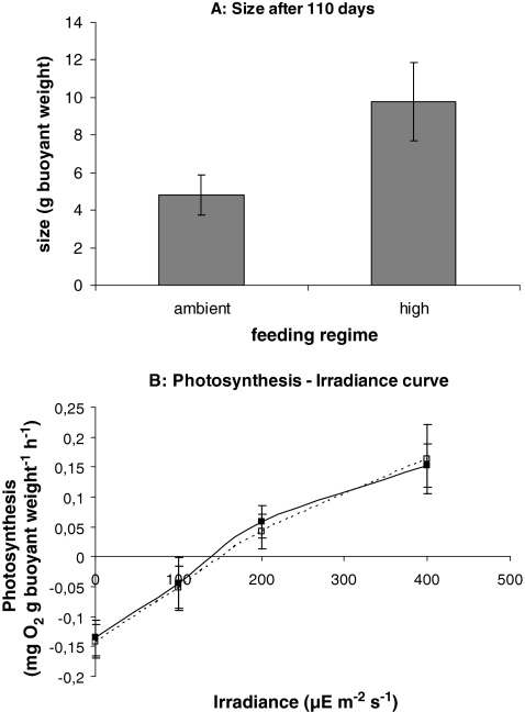
In another study that was performed during the CORALZOO project, we found increased growth of Pocillopora damicornis as a result of additional feeding, without a concurrent increase in photosynthetic activity (Fig. 3). For this experiment, series of nubbins of P. damicornis were prepared and cultured as described in Lavorano et al. (2008). Two feeding regimes were tested: low, ambient feeding (through the regular supply of fresh natural seawater to the system) versus high feeding, where ambient food was supplemented with a daily batch of freshly hatched nauplii of Artemia (starting concentration: 2,000 nauplii dm−3) and Tetraselmis suecica cells (starting concentration 30,000 cells dm−3). A photon flux density of 200 μE m−2 s−1 was applied. To substantiate differences in growth, the buoyant mass of the colonies was measured (cf. Schutter et al. 2008) after an incubation period of 110 days. The methodology for measuring photosynthetic capacity was the same as described in the “Light” section for G. fascicularis. In this experiment, the observed differences in growth could not be attributed to food-induced differences in photosynthetic activity. Either, feeding stimulated the utilization of photosynthetic products by the corals (a food-light interaction leading to a more efficient use of the photosynthetically produced resources), or the effect of feeding was just additive to growth on photosynthetically acquired resources. The possible interrelationships between feeding and photosynthesis will be further discussed in the section on interactions.
Fig. 3.
a Buoyant mass of colonies of P. damicornis grown under ambient feeding and high feeding after 110 days of culture; n = 3 for both treatments. b Photosynthesis–irradiance curves for colonies of P. damicornis grown under ambient feeding (dotted line) and high feeding (solid line)
An aspect of heterotrophic feeding that has often been overlooked is the direct uptake of nonliving dissolved organic carbon (DOC) by corals. Sorokin (1973) measured uptake rates of DOC by six common reef-building corals by adding radiolabelled DOC to coral colonies in closed incubation chambers. He found that daily DOC uptake among the six species studied ranged from 13.3% to 29% of the total amount of carbon present in the coral tissue. Hence, DOC uptake may represent a significant proportion of total food uptake and should not be neglected when estimating a coral’s carbon budget. In addition, supply of DOC to corals in culture may be a useful alternative to the commonly used live planktonic or particulate feeds.
Water Movement (Flow)
Water movement (flow) can affect coral growth in different ways. Since corals cannot actively generate water movement, they are dependent on ambient flow for the supply of basic requirements such as oxygen and inorganic carbon (Dennison and Barnes 1988; Lesser et al. 1994), inorganic nutrients (Stambler et al. 1991; Atkinson and Bilger 1992; Thomas and Atkinson 1997) and food (Sebens and Johnson 1991; Sebens et al. 1998). Flow-dependent mass transfer of oxygen may explain why Rinkevich and Loya (1984) found aeration-enhanced dark calcification. Second, flow controls the efflux rate of potentially toxic metabolic products such as oxygen and oxygen radicals (Nakamura et al. 2005; Finelli et al. 2006). Third, flow may indirectly promote coral growth by removing sediment and by preventing settlement of fouling organisms such as algae (Fabricius 2005; Box and Mumby 2007). High flow rates may inhibit coral growth. Sebens et al. (1997) observed that polyps of Madracis mirabilis located at the upstream side of the colony started to deform (flattening) at flow rates above 20 cm s−1, which reduced their prey capture efficiency. Flow-induced deformation of polyps may also explain why Atkinson et al. (1994) did not find profound effects of water flow velocity on nutrient uptake and respiration in flume experiments with Porites compressa: they compared relatively low flow rates (∼5 cm s−1) with rates exceeding 25 cm s−1, at which polyp deformation may have reduced the mass transfer of dissolved gasses and inorganic nutrients.
Experimental data demonstrate that different corals show various responses to changes in flow. Both increased growth (Jokiel 1978; Montebon and Yap 1997; Nakamura and Yamasaki 2005; Schutter et al. 2010; 2011) and decreased growth (Kuffner 2001) have been reported in relation to increases in flow.
Genotype
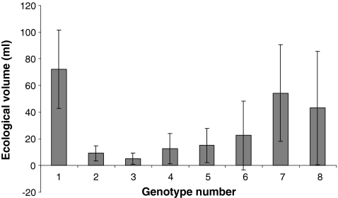
Apart from external factors influencing coral growth, there are also genetic factors that strongly affect the specific growth rate of a genetic individual. Each genet of a particular species has its own specific set of genes and will thus respond differently to different combinations of environmental conditions. Whereas some genotypes will invest more in growth, others may be better in resisting overgrowth and diseases. Here, we present an experimental example obtained during the CORALZOO project, which shows how genotypic variability affects coral growth. Groups of 10 clones originating from 10 genetically different individuals of S. pistillata were grown for 1.5 years under nearly identical conditions in a single 500 dm3 aquarium system equipped with a protein skimmer. Parent colonies were obtained from the Gulf of Eilat (Israel), their genetic independence was confirmed by AFLP analysis following Amar et al. (2008). Ten series of ten nubbins were prepared from these parent colonies according to Shafir et al. (2006), which were shipped to The Netherlands after a short period of recovery. The conditions in the aquarium were as follows: a temperature of 26 ± 1°C, a salinity of 35‰, daily feeding with 50–100 Artemia nauplii per liter; a photon flux density of 200 μE m−2 s−1 (supplied under a 12:12 h light/dark cycle), a moderate flow velocity of 5–10 cm s−1, a calcium concentration of 375–450 mg Ca2+ dm−3 and an alkalinity ranging from 2.0 to 3.5 mEq dm−3. After being in culture for 1.5 years, the ecological volume of each colony was determined according to Rinkevich and Loya (1983). Two out of ten genotypes did not survive in the aquarium. The remaining eight genotypes showed remarkable differences in growth (Fig. 4).
Fig. 4.
Biological volumes of eight genotypes of S. pistillata after being in culture for 1.5 years. Error bars represent standard deviations (n = variable, depending on the number of clones that survived). These results are part of a larger study on genotypic variability, which will be described elsewhere
This example clearly demonstrates that studies aiming to provide general information on the growth of a species should take into account genetic heterogeneity and present averages obtained from different genotypes. Working with clones obtained from a single genotype only provides information on that particular genotype and cannot be extrapolated to the species level. On the other hand, general physiological mechanisms are best studied using coral fragments that are genetically identical. It remains to be determined to what extent genetic differences in zooxanthellae populations can account for the observed genetic variability.
Interactions
Interactions between factors influencing coral growth can be defined as the extent to which one factor increases or decreases the effect of another factor. Many factors described in “Factors Influencing Coral Growth” section interact; only the most important interactions will be highlighted here.
Light will interact with water flow because water flow determines both the rate of supply of DIC and inorganic nutrients needed for photosynthesis and the efflux rate of oxygen and oxygen radicals that may inhibit photosynthesis (Finelli et al. 2006; Mass et al. 2010). Indeed, a significant interaction between light and flow was found to affect the growth of G. fascicularis. A combination of high light and flow had a stronger positive effect on growth than the individual factors. Hence, a positive correlation between irradiance and growth will be stronger under high-flow conditions (Schutter et al. 2011).
Light also interacts with the concentration of bicarbonate (HCO−3) in seawater. The positive effect of irradiance on growth is enhanced by adding HCO−3 (Marubini et al. 2001), which suggests that DIC is limiting coral growth. This is explained by the fact that two of the major physiological processes in corals, calcification and photosynthesis, compete for the same substrate (DIC). Such internal competition for DIC was also suggested by Marubini and Davies (1996) to explain the negative effects of DIN addition on calcification: enhanced photosynthesis resulting from DIN enrichment increases the photosynthetic demand for DIC at the expense of calcification. Marubini and Thake (1999) demonstrated that the inhibiting effects of DIN could indeed be stopped by doubling the concentration of HCO−3 in the seawater.
Conversely, it has been suggested also that calcification supports photosynthesis by converting HCO−3 into membrane-permeable CO2 (the substrate for the photosynthetic key enzyme RuBisco), thus reducing the need for carbonic anhydrase-based carbon concentrating mechanisms (Furla et al. 2005) to supply DIC to the zooxanthellae. This so-called “trans-calcification model” (McConnaughey and Whelan 1997) provides an elegant evolutionary explanation for the massive formation of external skeletons by scleractinian corals. Marshall and Clode (2002) presented data that support this view. These authors used enrichment with Ca2+ (the other component of Ω), which stimulated both calcification and the incorporation of photosynthetically acquired carbon into coral tissue. Increasing [Ca2+] may lower the amount of metabolic energy required for transport of [Ca2+] into the calicoblastic layer and may in this way compensate for the increased metabolic effort to acquire CO2−3 for calcification. However, the trans-calcification model appears to be in contradiction with the suggested internal competition for DIC. Moreover, it is based upon the questionable assumption that HCO−3 freely diffuses into the central cavity of the coral (see review by Allemand et al. 2004).
An interesting addition to this discussion is the potential role of heterotrophic feeding in the internal dynamics of DIC, N, P, and pH. The reason that feeding gives a similar response of zooxanthellae to enrichment with inorganic nutrients, but without a concurrent decrease in coral growth may be found in the fact that respiration of organic food will simultaneously yield DIN, DIP, and DIC in a biologically appropriate ratio. However, since the DIC provided by food digestion is in the form of CO2, excessive feeding may decrease the pH inside a coral, thus slowing down calcification. Since this effect will be stronger in the absence of light (in the light, the CO2 produced by respiration will be quickly assimilated by the zooxanthellae), it appears logical to feed aquarium corals during daytime. Indeed, Lavorano et al. (2008) found that daytime feeding of P. damicornis had a stronger positive effect on growth than nocturnal feeding.
Autotrophy versus Heterotrophy: Interactions between Light and Feeding
As reviewed by Houlbrèque and Ferrier-Pagès (2009), there is an ongoing debate on the roles of autotrophy and heterotrophy in zooxanthellate corals. The prevailing views, however, are that heterotrophy provides an alternative for autotrophy (additive effect) and that the balance between autotrophy and heterotrophy is dependent on light conditions. The shifting roles of autotrophy and heterotrophy as described by Anthony and Fabricius (2000) for corals facing increased turbidity and by Grottoli et al. (2006) for bleached corals support this view.
It is very likely that apart from providing an additional source of carbon, nitrogen, and phosphorous for both host and symbionts, heterotrophy also supplies several essential components (building blocks) for the biosynthesis of the coral host that it can hardly or not obtain from translocated photosynthetic products. Such a dependency on heterotrophy for specific components implies that the rate of heterotrophic feeding can actually directly limit coral growth. There are three facts that support this view:
There are, to the best of our knowledge, no records of 100% autotrophy in scleractinian corals.
Supplementing the water with nutrients stimulates photosynthesis and symbiont density, but does not augment coral growth (Marubini and Davies 1996), even when bicarbonate is added to prevent internal DIC-limitation (Marubini and Thake 1999).
The organic component of reef coral biomass is to a large extent of heterotrophic origin (Muscatine and Kaplan 1994; Grottoli 2000). Some components, in particular in the organic matrix of the coral skeleton, appear to be almost exclusively of nonphototrophic origin (e.g., Allemand et al. 1998).
These observations indicate that the observed beneficial effects of feeding on coral growth (Anthony and Fabricius 2000; Bongiorni et al. 2003a, b; Ferrier-Pagès et al. 2003; Houlbrèque et al. 2003, 2004; Lavorano et al. 2008) may include both additive and interactive effects. Feeding does not only provide alternative sources of carbon, nitrogen, and phosphorous, it may also provide some essential organic components that cannot sufficiently be obtained through photosynthesis. As such, feeding may augment the efficiency by which phototrophically acquired resources are used, leading to a reduced loss by excretion and mucus production (a lower release of “junk food”).
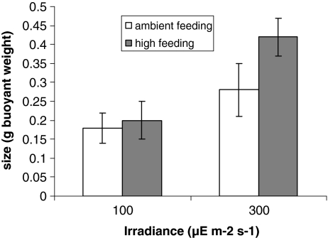
There are two studies that support this view. Houlbrèque et al. (2004) found an increased calcification coinciding with an increased deposition of aspartate when comparing fed colonies of S. pistillata to starved colonies and proposed that feeding-induced organic matrix synthesis is determining calcification rates. A second study was done recently by members of our team. The interactive effects of irradiance and food availability on the growth of the branching coral P. damicornis were tested. Nubbins of this species were prepared and cultured as described in Lavorano et al. (2008). Two photon flux densities (100 and 300 μE m−2 s−1) were tested against two feeding regimes: low, ambient feeding (through the regular supply of fresh natural seawater to the system) versus high feeding, where ambient food was supplemented with a daily batch of freshly hatched nauplii of Artemia (starting concentration: 2,000 nauplii dm3). The buoyant mass of the nubbins was determined after a period of 145 days. High feeding stimulated growth at the highest PFD, while no effect of adding food was observed at 100 μE m−2 s−1 (Fig. 5), indicating an interaction between light and feeding.
Fig. 5.
Interacting effect of irradiance and feeding on the growth of P. damicornis
Contrasting with these results, however, is the study by Ferrier-Pagès et al. (2003), who found no interaction between light and feeding on the growth of S. pistillata. Their data suggested that the effect of feeding was additive to the effect of light. Ferrier-Pagès et al. (2003) gave no details about the ambient flow velocity in their experimental aquaria, except for the general remark that water motion was generated by an air stone. Hence, it cannot be excluded that flow-related limitations have occurred under the high light conditions applied in this study, which can mask potential interactive effects between light and feeding.
In general, the uptake of organic food appears to be the most balanced way for a coral to supply itself with a number of resources, including DIN, DIP, and DIC for photosynthesis and calcification and essential organic building blocks that cannot be provided by photosynthesis. If food supply is in good balance with light supply and flow velocity, the beneficial effect of feeding on coral growth is likely to be more than just additional to the effect of light.
Synopsis: What Determines Coral Growth?
Obviously, there is not one single factor that limits the growth of zooxanthellate Scleractinia. Multiple, interacting factors influence coral growth, which may all become limiting/inhibiting within their naturally occurring ranges. Furthermore, there is variation among species and within species with respect to growth limitation: genotypic variability is large, different genotypes may have developed different strategies to survive in a fluctuating environment. For example, under a given combination of environmental conditions, light availability may limit the growth of a specific coral individual, but under the same set of environmental conditions, another factor may be limiting the growth of another individual, even when these individuals are conspecifics. This explains why many aquarists report conflicting results when growing individuals of the same species. It also shows that optimization of coral culture is a tedious process, in which many factors should be taken into account. Therefore, large, multi-factorial growth experiments are desired, not only to maximize the productivity for coral aquaculture, but also to further unravel the interactions between potentially limiting and inhibiting factors. Information on interactions may shed new light on the mechanisms that determine coral growth rates.
Despite this complexity, we have attempted to deduce some general mechanisms that may help to explain the phenomena that we observe both in nature and in culture and that may provide guidance for future research. The following working mechanism is proposed for corals growing under natural oceanic conditions in nonstagnant water:
Under a broad range of photon flux densities, the coral-zooxanthellae holobiont is capable to adjust its photosynthetic apparatus in such a way that photosynthesis is always optimal for coral growth (photoacclimation). Below the specific saturation irradiance (I k s), photoacclimation can no longer compensate for the lower photon flux. Under these circumstances, light availability can limit coral growth due to a reduced input of translocated photosynthetates. Increased heterotrophy can partially compensate for this reduced photosynthetic input. Above I k s, light is not limiting: the zooxanthellae may become nutrient limited and the coral host is limited by another factor (e.g., essential food components or DIC). Taking away these limitations will result in a higher growth rate, partially caused by a more efficient use of translocated photosynthetates. Crucial in this respect is the role of water movement: when water flow is low or absent, mass transfer of DIC or food may become limiting for coral growth, even below I k s. In addition, I k s itself may decrease due to DIC limitation of the zooxanthellae. Furthermore, insufficient water movement will cause inhibition of coral growth under higher irradiance levels, because an increasing proportion of the photosynthetically acquired carbon will then be used for stress responses (i.e., mechanisms to cope with negative effects of accumulated oxygen, oxygen derivatives, and metabolic wastes) instead of growth.
Growth Kinetics
Most studies concerning growth of corals deal with growth rates (see review by Dullo 2005), factors influencing growth (see references in earlier sections) and morphogenesis (Kaandorp and Kuebler 2001; Kruszynski et al. 2007; Shaish et al. 2006). Only few researchers analyzed the kinetics of coral growth. An elegant and extensive study on the growth of five Caribbean coral species was done by Bak (1976), who reported that the growth of all species studied developed in an exponential way. However, the specific exponential growth rate (i.e., the percentage increase in body mass per unit of time) decreased with increasing colony size. It is important to realize that a growing proportion of the scleractinian coral body mass—the skeleton—is not actively participating to the growth process. Therefore, Bak (1976) related growth to the living surface area (to be precise: to the area covered by the calicoblastic epithelium) and found, by comparing growth rates of small and large colonies, that the rate of calcification per surface area remained unchanged over a long period of time.
Sipkema et al. (2006) presented four hypothetical growth models for sponges, which appear suitable to be applied to corals as well:
- Linear growth (zero order kinetics), described by:
in which X t is the size of the coral after time t, X 0 is the size of the coral at t = 0 and k is the linear growth rate constant (e.g., in mm day−1).
1 - Exponential growth (first order kinetics) described by:
in which μ is the specific growth rate constant (e.g., day−1). In this model, the percentage of new biomass formed per day is constant, leading to a J-shaped curve when total body mass is plotted against time.
2 Surface-dependent growth (globose organisms)
Circumference-dependent growth (encrusting organisms).
By combining the empirical studies on corals by Bak (1976) with the hypothetical growth models for sponges described by Sipkema et al. (2006), we deduced the following basic principles to describe coral growth kinetics:
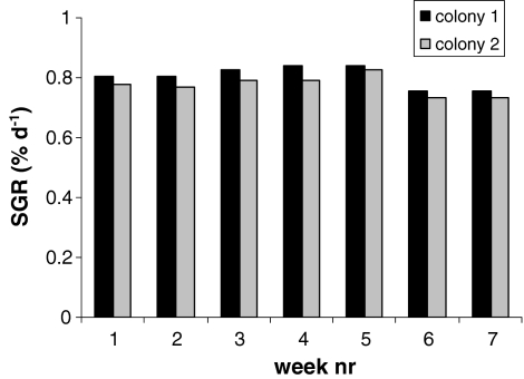
Branching corals (such as M. mirabilis in the study by Bak 1976) proliferate by continuously forming new branches that all have a similar size and shape. Therefore, these corals have a relatively constant surface to volume ratio. Their growth will be appropriately described by first order kinetics (Eq. 2), until their size has reached a point where other factors such as gravity-induced forces start to inhibit further growth. A study done in our lab on two genetically identical colonies of the branching species S. caliendrum grown under stable, controlled aquarium conditions (see “Food” and “Genotype” sections for experimental details), showed that the specific growth rate of this species remained constant throughout the monitored period (Fig. 6), thus supporting the view that branching species follow first order growth kinetics. Mass parameters such as wet mass, buoyant mass, and dry mass are suitable to monitor growth of these coral species, because they linearly correlate with other commonly used biomass estimators such as surface area, volume, or ecological volume (Rinkevich and Loya 1983).
Fig. 6.
Specific growth rate (SGR) of two colonies of S. caliendrum during seven consecutive weeks under stable aquarium conditions
Boulder-shaped corals and plate-shaped corals follow different growth kinetics. Boulder-shaped corals continuously secrete new layers of calcium carbonate upon their old skeleton, thus continuously increasing the proportion of skeletal mass to total body mass. The living tissue of these corals may either grow with a continuous rate or with a rate that slowly decreases (e.g., Montastrea annularis—Bak 1976). In case of continuous tissue growth, total coral growth can best be described by using a surface dependent growth rate constant (Eq. 3 in the paper by Sipkema et al. 2006, or similar derivatives for conically shaped objects, etc.). Growth of these corals is best determined by using surface area as an estimator for biomass. Plate-shaped corals are most likely to follow circumference-dependent growth kinetics (Eq. 4 in the paper by Sipkema et al. 2006, or derivatives thereof). Hence, growth of these corals is best determined by measuring surface area or linear extension rates.
It should be noted that these basic principles only represent a broad generalization. More sophisticated modeling approaches are needed for an exact description of species specific coral growth kinetics. For example, Crabbe (2007) reported that a 3:3 rational polynomial model described the growth of a branching species (Acropora palmata) more accurately than the simple first-order kinetics model represented by Eq. 2. Notwithstanding this, the basic principles outlined above provide a suitable tool to design coral aquaculture systems, as will be discussed in the next section.
The Economics of Coral Growth
It has been estimated that economic activities related to corals and coral reefs represent an annual turnover of 375 billion dollars worldwide (Wilkinson 1996; Bryant et al. 1998). The aquarium trade represents a growing proportion of this economic value: the trade in aquarium corals only had an estimated market size of approximately 60 million US$ per year in the period between 1997 and 2001 (Wabnitz et al. 2003), which justifies research efforts focused on optimization of coral aquaculture.
In this section, we present a case study on commercial coral breeding. Daily costs for building and maintenance of coral culture systems (Table 1) were calculated per square meter culture system surface. Calculations are based upon figures from a coral farm in a public aquarium (NAUSICAA, France), where several coral species are being bred for use in public aquaria. The figures presented here concern the branching species S. caliendrum, one of the species that is in culture at the NAUSICAA aquarium.
Table 1.
Overview of costs per category and total operational costs for coral culture systems at NAUSICAA aquarium
| Cost category | Low feed scenario | High feed scenario |
|---|---|---|
| Manpower | 6.53 | 6.53 |
| Materials | 1.42 | 1.42 |
| Energy | 0.22 | 0.22 |
| Food | 0.50 | 2.00 |
| Depreciation of system | 1.10 | 1.10 |
| Tax and insurance | 0.16 | 0.16 |
| Total costs | 9.93 | 11.43 |
All costs are given in Euros per m2 per day. Systems are depreciated in 10 years
For the commercial aquarium trade, a colony of S. caliendrum should have the size of a fist, which corresponds to a wet mass of approximately 100 g. A 1-m2 aquarium system can host up to 100 colonies of this size.
We calculated the production costs for 100 g colonies, using fragments that have an initial mass of 10 g as a starting point. Production costs were calculated for two real-life feeding scenario’s that had been applied to this coral species (see “Food” section for experimental details). Annual production of S. caliendrum biomass (in kg wet mass) and the corresponding production costs were calculated for both feeding scenarios. The growth of S. caliendrum was assumed to be exponential, following first order kinetics (see “Growth Kinetics” section; Eq. 2, Fig. 6). In Fig. 2a, it is shown that as a result of additional feeding with Artemia nauplii, the specific growth rate (μ) for S. caliendrum increased from 0.0084 to 0.0115 day−1 (i.e., a 35% increase). Both rates were used to calculate the production period, i.e., the time (t) needed for a fragment to grow from a size of 10 g (X 0) to a size of 100 g (X t):
 |
The price per colony of 100 g was calculated as follows:
It was hereby taken into account that for continuation of the culture, 10% of the harvest is needed a broodstock for the next culture.
Also, the annual production was calculated:
 |
Since stony corals are a potential resource of natural products with pharmacological properties (e.g., Alam et al. 2001), cultured corals may also be needed as biological materials for drug development studies. For this purpose, the size of the coral individuals harvested is not important. In this particular case, the best strategy for production is to continuously maintain the maximal sustained standing stock (in the example: 100 colonies of 100 g wet weight), and to harvest every day the excess growth (in the example: 0.84% and 1.15% day−1). Productivity for S. caliendrum was also calculated for this culture approach, hereby again comparing the low-feeding scenario to the high-feeding scenario.
The results of the calculations are summarized in Tables 2 and 3. This case study shows that optimization can be rewarding; as a result of optimized feeding, productivity increased with 35% while production costs increased with only 15%. The results also show that continuous harvesting reduces the production costs per kilogram coral considerably. The production costs for 100 g colonies of S. caliendrum under the two feeding scenarios (€25.40 and €30.20) are close to the average wholesale value of similarly sized aquarium corals in The Netherlands, which was €27 in the year 2010 (De Jong MarineLife, personal communication). The retail prizes for these coral colonies range between €40 and €150, depending on species and outer appearance (shape and coloration).
Table 2.
Production figures for market-size colonies of S. caliendrum, taking into account two production scenarios (low feeding and high feeding)
| Feeding regime | Production period (days) | Colonies produced per year | Price per colony (€) | Kg coral produced per year | Price per kg (€) |
|---|---|---|---|---|---|
| Low | 274 | 120 | 30.20 | 12 | 302 |
| High | 200 | 164 | 25.40 | 16.4 | 254 |
Costs per colony are provided as well as costs per kilogram for comparison with the data in Table 3
Table 3.
Production figures for continuous production of biomass of S. caliendrum under low feeding and high feeding
| Feeding regime | Production per year (kg) | Price per kg (€) |
|---|---|---|
| Low | 30.8 | 118 |
| High | 42.2 | 99 |
A practical consideration with respect to designing a coral culture in a closed aquarium setting is that semicontinuous production (for example: by renewing every week 2% of the culture) is advantageous over batch production. When operating in a semicontinuous production mode, there will be a constant standing stock inside the culture tank. This will make maintenance easier: feeding regimes and supply of calcium and carbonate do not have to be adjusted continuously to an increasing consumption by a growing standing stock.
Conclusions
Due to the adaptive flexibility of corals, their genotypic heterogeneity and the numerous factors that can potentially limit or inhibit coral growth, it is hard to give a clear-cut answer to the question “What determines coral growth?” The proposed working mechanism described in “Synopsis: What determines Coral Growth?” section of this paper implies that optimizing coral culture requires a close, genotype-specific fine-tuning between light supply, food supply, water movement, and DIC concentration. Hence, optimization can be achieved through large-scale multivariate interaction studies targeting individual species. Each species and each genotype will require a different combination of values to maximize its growth rate. Therefore, efficient high-density coral culture is best achieved by having the individual species and genotypes in separate culture systems. At present, the production costs for aquarium-cultured corals approximately equal their wholesales values. Further optimization along the lines pointed out above will make commercial culture of corals economically feasible in the near future.
Acknowledgments
This study was funded by the European Commission (project CORALZOO 012547—breeding and husbandry of stony corals in zoo’s and public aquaria) and is a joint achievement of many CORALZOO participants. Daniela Corsino and Anne Reijbroek are acknowledged for analytical support.
Open Access
This article is distributed under the terms of the Creative Commons Attribution Noncommercial License which permits any noncommercial use, distribution, and reproduction in any medium, provided the original author(s) and source are credited.
References
- Alam N, Bae BH, Hong J, Lee CO, Im KS, Jung JH. J Nat Prod. 2001;64:1059–1063. doi: 10.1021/np010148b. [DOI] [PubMed] [Google Scholar]
- Al-Horani FA, Al-Moghrabi SM, De Beer D. Mar Biol. 2003;42:419–426. [Google Scholar]
- Allemand D, Tambutté E, Girard JP, Jaubert J. J Exp Biol. 1998;201(13):2001–2009. doi: 10.1242/jeb.201.13.2001. [DOI] [PubMed] [Google Scholar]
- Allemand D, Ferrier-Pagès C, Furla P, Houlbrèque F, Puverel S, Reynaud S, Tambutté E, Tambutté S, Zoccola D. C.R. Palevol. 2004;3:453–467. [Google Scholar]
- Amar KO, Douek J, Rabinowitz C, Rinkevich B. Mar Biotechnol. 2008;10:350–357. doi: 10.1007/s10126-007-9069-2. [DOI] [PubMed] [Google Scholar]
- Anthony KRN, Fabricius KE. J Exp Mar Biol Ecol. 2000;252:221–253. doi: 10.1016/s0022-0981(00)00237-9. [DOI] [PubMed] [Google Scholar]
- Anthony KRN, Kline DI, Diaz-Pulido G, Dove S, Hoegh-Guldberg O. PNAS. 2008;105(45):17442–17446. doi: 10.1073/pnas.0804478105. [DOI] [PMC free article] [PubMed] [Google Scholar]
- Atkinson MJ, Bilger RW. Limnol Oceanogr. 1992;37(2):273–279. [Google Scholar]
- Atkinson MJ, Kotler E, Newton P. Pac Sci. 1994;48(3):296–303. [Google Scholar]
- Bak RPM. Neth J Sea Res. 1976;10(3):285–337. [Google Scholar]
- Barnes DJ, Chalker BE. In: Coral reefs: ecosystems of the world, Vol. 25. Dubinsky Z, editor. New York: Elsevier Scientific Publishing Co. Inc; 1990. pp. 109–131. [Google Scholar]
- Bongiorni L, Shafir S, Rinkevich B. Mar Pollut Bull. 2003;46:1120–1124. doi: 10.1016/S0025-326X(03)00240-6. [DOI] [PubMed] [Google Scholar]
- Bongiorni L, Shafir S, Angel D, Rinkevich B. Mar Ecol Prog Ser. 2003;253:137–144. [Google Scholar]
- Box SJ, Mumby PJ. Mar Ecol Prog Ser. 2007;342:139–149. [Google Scholar]
- Bryant D, Burke L, McManus J, Spalding M. Reefs at risk. A map-based indicator of threats to the world’s coral reefs. Washington, DC: World Resources Institute; 1998. [Google Scholar]
- Chalker BE. Mar Biol. 1981;63:135–141. [Google Scholar]
- Crabbe MJC. Comp BiolChem. 2007;31:294–297. [Google Scholar]
- Danovaro R, Bongiorni L, Corinaldesi C, Giovanelli D, Damiani E, Astolfi P, Greci L, Pusceddu A. Env Health Persp. 2008;116(4):441–447. doi: 10.1289/ehp.10966. [DOI] [PMC free article] [PubMed] [Google Scholar]
- De’ath G, Lough JM, Fabricius KE. Science. 2009;353(5910):116–119. doi: 10.1126/science.1165283. [DOI] [PubMed] [Google Scholar]
- Dennison WC, Barnes DJ. J Exp Mar Biol Ecol. 1988;115(1):67–77. [Google Scholar]
- Dubinsky Z, Jokiel PL. Pac Sci. 1994;48(3):313–324. [Google Scholar]
- Dubinsky Z, Stambler N. Glob Chang Biol. 1996;2:511–526. [Google Scholar]
- Dubinsky Z, Stambler N, Ben-Zion M, Mccloskey LR, Muscatine L, Falkowski PG. Proc R Soc Lond B Biol Sci. 1990;239(1295):231–246. [Google Scholar]
- Dullo W-C. Facies. 2005;51:33–48. [Google Scholar]
- Dustan P. Mar Biol. 1982;68:253–264. [Google Scholar]
- Enriquez S, Mendez ER, Iglesias-Prieto R. Limnol Oceanogr. 2005;50(4):1025–1032. [Google Scholar]
- Fabricius KE. Mar Pollut Bull. 2005;50:125–146. doi: 10.1016/j.marpolbul.2004.11.028. [DOI] [PubMed] [Google Scholar]
- Falkowski PG, Dubinsky Z, Muscatine L, Porter JW. Bioscience. 1984;34(11):705–709. [Google Scholar]
- Fautin DG, Buddemeier RW. Hydrobiologia. 2004;530/531:459–467. [Google Scholar]
- Ferrier-Pagès C, Gattuso J-P, Dallot S, Jaubert J. Coral Reefs. 2000;19:103–113. [Google Scholar]
- Ferrier-Pagès C, Schoelzke V, Jaubert J, Muscatine L, Hoegh-Guldberg O. J Exp Mar Biol Ecol. 2001;259(2):249–261. doi: 10.1016/s0022-0981(01)00241-6. [DOI] [PubMed] [Google Scholar]
- Ferrier-Pagès C, Witting J, Tambutté E, Sebens KP. Coral Reefs. 2003;22:229–240. [Google Scholar]
- Ferrier-Pagès C, Houlbrèque F, Wyse E, Richard C, Allemand D, Boisson F. Coral Reefs. 2005;24(4):636–645. [Google Scholar]
- Finelli CM, Helmuth BST, Pentcheff ND, Wethey DS. Water flow influences oxygen transport and photosynthetic efficiency in corals. Coral Reefs. 2006;25(1):47–57. [Google Scholar]
- Furla P, Allemand D, Shick JM, Ferrier-Pagès C, Richier S, Plantivaux A, Merle P, Tambutté S. Integr Comp Biol. 2005;45(4):595–604. doi: 10.1093/icb/45.4.595. [DOI] [PubMed] [Google Scholar]
- Fusetani N, editor. Drugs from the sea. Basel: Karger; 2000. [Google Scholar]
- Gattuso JP, Frankignoulle M, Bourge I, Romaine S, Buddemeier RW. Glob Planet Change. 1998;18:37–46. [Google Scholar]
- Gattuso JP, Allemand D, Frankignoulle M. Am Zool. 1999;39(1):160–183. [Google Scholar]
- Genin A, Lazar B, Brenner S. Nature. 1995;377(6549):507–510. [Google Scholar]
- Goreau TF. Biol Bull. 1959;116:59–75. [Google Scholar]
- Grigg RW. Coral Reefs. 1995;14(4):253–266. [Google Scholar]
- Grottoli AG. Oceanography. 2000;13(20):93–97. [Google Scholar]
- Grottoli AG, Rodrigues LJ, Palardy JE. Nature. 2006;440(7088):1186–1189. doi: 10.1038/nature04565. [DOI] [PubMed] [Google Scholar]
- Grover R, Maguer J, Reynaud-Vaganay S, Ferrier-Pagès C. Limnol Oceanogr. 2002;47(3):782–790. [Google Scholar]
- Haapkylä J, Ramade F, Salvat B. Vie Milieu. 2007;57:95–111. [Google Scholar]
- Herfort L, Thake B, Taubner I. J Phycol. 2008;44:91–98. doi: 10.1111/j.1529-8817.2007.00445.x. [DOI] [PubMed] [Google Scholar]
- Hoegh-Gulberg O, Smith GJ. J Exp Mar Biol Ecol. 1989;129:279–303. [Google Scholar]
- Houlbrèque F, Ferrier-Pagès C. Biol Rev. 2009;84:1–17. doi: 10.1111/j.1469-185X.2008.00058.x. [DOI] [PubMed] [Google Scholar]
- Houlbrèque F, Tambutté E, Ferrier-Pagès C. J Exp Mar Biol Ecol. 2003;296:145–166. [Google Scholar]
- Houlbrèque F, Tambutté E, Allemand D, Ferrier-Pagès C. J Exp Biol. 2004;207:1461–1469. doi: 10.1242/jeb.00911. [DOI] [PubMed] [Google Scholar]
- Hughes TP, Baird AH, Bellwood DR, et al. Science. 2003;301(5635):929–933. doi: 10.1126/science.1085046. [DOI] [PubMed] [Google Scholar]
- Iglesias-Prieto R, Matta JL, Robbins WA, Trench RK. PNAS. 1992;89(21):10302–10305. doi: 10.1073/pnas.89.21.10302. [DOI] [PMC free article] [PubMed] [Google Scholar]
- Jokiel PL. J Exp Mar Biol Ecol. 1978;35:87–97. [Google Scholar]
- Jokiel PL, York RH. Bull Mar Sci. 1982;32:301–315. [Google Scholar]
- Jokiel PL, Rodgers KS, Kuffner IB, Andersson AJ, Cox EF, Mackenzie FT. Coral Reefs. 2008;27:473–483. [Google Scholar]
- Jones R. Mar Pollut Bull. 2005;51:495–506. doi: 10.1016/j.marpolbul.2005.06.027. [DOI] [PubMed] [Google Scholar]
- Kaandorp JA, Kuebler JE. The algorithmic beauty of seaweeds, sponges and corals. Heidelberg: Springer; 2001. [Google Scholar]
- Kinsey DW, Davies PJ. Limnol Oceanogr. 1979;24:935–940. [Google Scholar]
- Kinzie RA, III, Takayama M, Santos SR, Coffroth MA. Biol Bull. 2001;200:51–58. doi: 10.2307/1543084. [DOI] [PubMed] [Google Scholar]
- Kleypas JA, Buddemeier RW, Archer D, Gattuso J-P, Langdon C, Opdyke BN. Science. 1999;284:118–120. doi: 10.1126/science.284.5411.118. [DOI] [PubMed] [Google Scholar]
- Knittweis L, Kraemer WE, Timm J, Kochzius M. Conserv Genet. 2009;10(1):241–249. [Google Scholar]
- Koop K, et al. Mar Pollut Bull. 2001;42:91–120. doi: 10.1016/s0025-326x(00)00181-8. [DOI] [PubMed] [Google Scholar]
- Kruszynski K, Kaandorp JA, Van Liere R. Coral Reefs. 2007;26:831–840. [Google Scholar]
- Kuffner IB. Mar Biol. 2001;138:467–476. [Google Scholar]
- Langdon C, Atkinson MJ. J Geophys Res Oceans. 2005;110:1–54. [Google Scholar]
- Lavorano S, Gili C, Muti C, Taruffi M, Corsino D, Gnone G (2008) In: Leewis RJ, Janse M (eds) Advances in coral husbandry in public aquariums. Public Aquarium Husbandry Series, vol 2., pp 19–25
- Lesser MP. Coral Reefs. 1997;16:187–192. [Google Scholar]
- Lesser MP, Weiss VM, Patterson MR, Jokiel PL. J Exp Mar Biol Ecol. 1994;178:153–179. [Google Scholar]
- Marshall AT, Clode P. J Exp Biol. 2002;205:2107–2113. doi: 10.1242/jeb.205.14.2107. [DOI] [PubMed] [Google Scholar]
- Marubini F, Davies PS. Mar Biol. 1996;127(2):319–328. [Google Scholar]
- Marubini F, Thake B. Limnol Oceanogr. 1999;44(3):716–720. [Google Scholar]
- Marubini F, Barnett H, Langdon C, Atkinson MJ. Mar Ecol Prog Ser. 2001;220:153–162. [Google Scholar]
- Marubini F, Ferrier-Pagès C, Furla P, Allemand D. Coral Reefs. 2008;27(3):491–499. [Google Scholar]
- Mass T, Genin A, Shavit U, Grinstein M, Tchernov D. Proc Nat Acad Sci. 2010;107(6):2527–2531. doi: 10.1073/pnas.0912348107. [DOI] [PMC free article] [PubMed] [Google Scholar]
- McConnaughey TA, Whelan JF. Earth Sci Rev. 1997;42:95–117. [Google Scholar]
- Meyer JL, Schultz ET. Limnol Oceanogr. 1985;30:146–156. [Google Scholar]
- Montebon ARF, Yap HT. Proc 8th Int Coral Reef Symp. 1997;2:1065–1070. [Google Scholar]
- Mucci A. Am J Sci. 1983;283:780–799. [Google Scholar]
- Muscatine L. In: Ecosystems of the world (25); coral reefs; chapter 4. Muscatine L, editor. New York: Elsevier Scientific Publishing Co. Inc; 1990. pp. 75–87. [Google Scholar]
- Muscatine L, Cernichiari E. Biol Bull. 1969;137:506–523. doi: 10.2307/1540172. [DOI] [PubMed] [Google Scholar]
- Muscatine L, Kaplan IR. Pac Sci. 1994;48:304–312. [Google Scholar]
- Nakamura T, Yamasaki H. Mar Pollut Bull. 2005;50(10):1115–1120. doi: 10.1016/j.marpolbul.2005.06.025. [DOI] [PubMed] [Google Scholar]
- Nakamura T, Van Woesik R, Yamasaki H. Mar Ecol Prog Ser. 2005;301:109–118. [Google Scholar]
- Osinga R (2008) In: Leewis RJ, Janse M (Eds.). Advances in coral husbandry in public aquariums. Public aquarium husbandry series, Vol 2, pp. 167–171
- Reynaud S, Leclercq N, Romaine-Lioud S, Ferrier-Pagés C, Jaubert J, Gattuso J-P. Glob Chang Biol. 2003;9(11):1660–1668. [Google Scholar]
- Reynaud S, Ferrier-Pagès C, Boisson F, Allemand D, Fairbanks RG. Mar Ecol Prog Ser. 2004;279:105–112. [Google Scholar]
- Rinkevich B, Loya Y. J Exp Mar Biol Ecol. 1983;73:175–184. [Google Scholar]
- Rinkevich B, Loya Y. Mar Biol. 1984;80(1):1–6. [Google Scholar]
- Schlacher TA, Stark J, Fischer ABP. Aquaculture. 2007;269:278–289. [Google Scholar]
- Schneider K, Erez J. Limnol Oceanogr. 2006;51(3):1284–1293. [Google Scholar]
- Schutter M, Van Velthoven B, Janse M, Osinga R, Janssen M, Wijffels R, Verreth J. J Exp Mar Biol Ecol. 2008;67:75–80. [Google Scholar]
- Schutter M, Crocker J, Paijmans A, Janse M, Osinga R, Verreth JAJ, Wijffels RH. Coral Reefs. 2010;29:737–748. [Google Scholar]
- Schutter M, Kranenbarg S, Wijffels RH, Verreth JAJ, Osinga R (2011) Mar Biol (in press)
- Sebens KP, Johnson AS. Hydrobiologia. 1991;226:91–101. [Google Scholar]
- Sebens KP, Witting J, Helmuth B. J Exp Mar Biol Ecol. 1997;211:1–28. [Google Scholar]
- Sebens KP, Grace SP, Helmuth B, Maney EJ, Jr, Miles JS. Mar Biol. 1998;131:347–360. [Google Scholar]
- Shafir S, Van Rijn J, Rinkevich B. Aquaculture. 2006;259:444–448. [Google Scholar]
- Shaish L, Abelson A, Rinkevich B. Dev Dyn. 2006;235:2111–2121. doi: 10.1002/dvdy.20861. [DOI] [PubMed] [Google Scholar]
- Simkiss K. Biol Rev. 1964;39:487–505. doi: 10.1111/j.1469-185x.1964.tb01166.x. [DOI] [PubMed] [Google Scholar]
- Sipkema D, Yosef NAM, Adamczewski M, Osinga R, Mendola D, Tramper J, Wijffels RH. Mar Biotechnol. 2006;8:40–51. doi: 10.1007/s10126-005-5002-8. [DOI] [PubMed] [Google Scholar]
- Snidvongs A, Kinzie RA., III Mar Biol. 1994;118:705–711. [Google Scholar]
- Sorokin YI. Limnol Oceanogr. 1973;18(3):380–385. [Google Scholar]
- Stambler N, Popper N, Dubinsky Z, Stimson J. Pac Sci. 1991;45(3):299–307. [Google Scholar]
- Stambler N, Cox EF, Vaga R. Pac Sci. 1994;48:284–290. [Google Scholar]
- Steven ADL, Broadbent AD. Proc 8th Intern Coral Reef Symp. 1997;1:867–872. [Google Scholar]
- Sunda WG. Biol Oceanogr. 1991;12:411–418. [Google Scholar]
- Tanaka Y, Miyajima T, Koike I, Hayashibara T, Ogawa H. Limnol Oceanogr. 2007;52(3):1139–1146. [Google Scholar]
- Thomas FIM, Atkinson MJ. Limnol Oceanogr. 1997;42(1):81–88. [Google Scholar]
- Titlyanov EA, Titlyanova TV. Rus J Mar Biol. 2002;28(1):S16–S31. [Google Scholar]
- Titlyanov EA, Titlyanova TV, Yamazato K, Van Woesik R. J Exp Mar Biol Ecol. 2001;263:211–225. doi: 10.1016/s0022-0981(00)00308-7. [DOI] [PubMed] [Google Scholar]
- Tomascik T. Mar Pollut Bull. 1990;21:376–381. [Google Scholar]
- Tomascik T, Sander F. Mar Biol. 1985;87:143–155. [Google Scholar]
- Torres JL. Bull Mar Sci. 2001;69(2):805–818. [Google Scholar]
- Torres JL, Armstrong RA, Corredor JE, Gilbes F. Photochem Photobiol. 2007;83:839–851. doi: 10.1562/2006-09-01-RA-1025. [DOI] [PubMed] [Google Scholar]
- Ulstrup KE, Ralph PJ, Larkum AWD, Kuhl M. Mar Biol. 2006;149:1325–1335. [Google Scholar]
- Van de Schootbrugge B, Tremolada F, Rosenthal Y, Bailey TR, Feist-Burkhardt S, Brinkhuis H, Pross J, Kent DV, Falkowski PG. Palaeogeogr Palaeoclimatol Palaeoecol. 2007;244:126–141. [Google Scholar]
- Van Katwijk MM, Meier NF, Van Loon R, Van Howe EM, Giesen WBJT, Van der Velde G. Mar Biol. 1993;117:675–683. [Google Scholar]
- Wabnitz C, Taylor M, Green E, Razak T. From ocean to aquarium. The global trade in marine ornamental species. Cambridge, UK: UNEP-WCMC report; 2003. [Google Scholar]
- Wilkinson CR. Glob Chang Biol. 1996;2:547–558. [Google Scholar]


