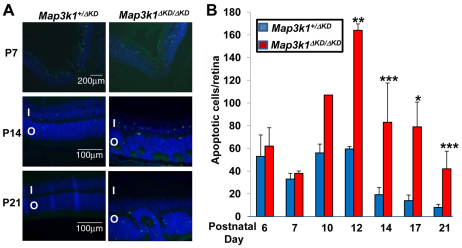
Fig. 6.
Increased apoptosis in the retinas of Map3k1ΔKD/ΔKD mice. The eyes isolated from Map3k1+/ΔKD and Map3k1ΔKD/ΔKD mice at different postnatal ages were subjected to TUNEL assays. (A) Pictures were taken under fluorescent microscopy. I, inner nuclear layer; O, outer nuclear layer. (B) The average number of apoptotic cells per retina was quantified+s.e.m.. Results represent at least four eyes at each developmental stage and genotype. Statistical analyses were carried out by comparing the numbers in different genotypes. *P<0.05, **P<0.01, ***P<0.001.

