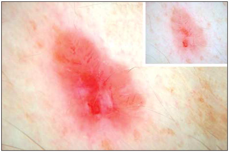
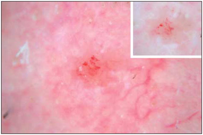
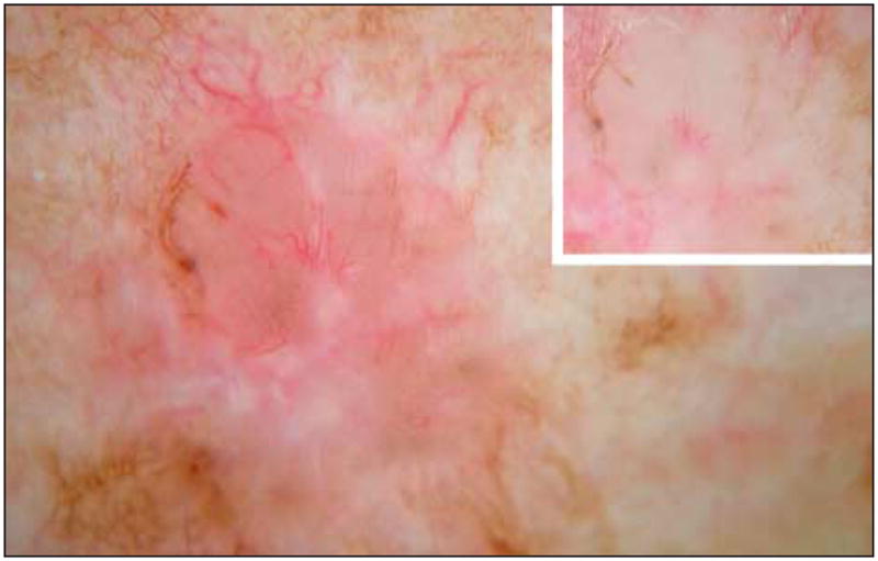
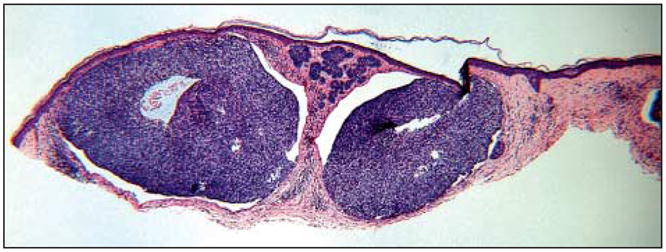
The clinical feature Semitranslucency, defined as a semiliquid or jellylike appearance, is a useful optical phenomenon for the identification of basal cell carcinoma (BCC).1 Semitranslucent areas in dermoscopic images of BCC also display the smooth, jellylike appearance of the clinical counterpart. The color ranges from reddish pink, usually seen in the thickest areas, to dull orange, often in a peripheral location (Figure 1), to gray (Figure 2 and Figure 3, insets). Figures 1 through 3 are noncontact polarized dermoscopic images of BCC, and Figure 4 is the histopathologic view of Figure 3.
Figure 1.

Figure 2.

Figure 3.

Figure 4.

Semitranslucency, which is easily appreciated without visual aids in larger BCCs, can be best appreciated with non-contact polarized dermoscopic images. Lighter colors indermoscopic images, such as those appearing with the semitranslucency phenomenon, have previously been shown to be best preserved with non contact polarized dermoscopy.2 The typical color is absent in contact polarized images, but the contrast in smoothness with the surrounding areas, the most important aspect of the jelly like phenomenon, still allows appreciation of semitranslucency in some contact polarized images (Figure 2, inset). The jellylike appearance may be lost in nonpolarized contact images (Figures 1 and 3, insets). Histopathologic examination of 10 BCCs showed that areas of semitranslucency possess basaloid tumor nodules close to the surface, with a diminished epidermal thickness and a diminished collagen layer (Figure 4). This histopathologic constellation allows transmission of light to an appreciable depth of the skin and reflection from within the tumor. Further studies of this optical phenomenon are needed to determine which histopathologic subtypes of BCC are best correlated with semitranslucency.
References
- 1.Vanker AD, Stoecker WV. An expert diagnostic program for dermatology. Comput Biomed Res. 1984;17(3):241–247. doi: 10.1016/s0010-4809(84)80015-4. [DOI] [PubMed] [Google Scholar]
- 2.Benvenuto-Andrade C, Dusza SW, Agero AL, et al. Differences between polarized light dermoscopy and immersion contact dermoscopy for the evaluation of skin lesions. Arch Dermatol. 2007;143(3):329–338. doi: 10.1001/archderm.143.3.329. [DOI] [PubMed] [Google Scholar]


