Abstract
Background
Hypothalamic glial-neuronal communications are important for the activation of luteinizing hormone releasing hormone (LHRH) secretion at the time of puberty. Since we have shown that ALC diminishes prepubertal LHRH secretion and delays puberty, we first assessed the effects of short-term ALC administration on the basal expression of a specific gene family involved in glial-neuronal communications. Secondly, since insulin like growth factor-1 (IGF-1) is a critical regulator of LHRH secretion and the pubertal process, we then assessed whether IGF-1 could induce the expression of these signaling genes and determine if ALC can block this affect.
Methods
Immature female rats were fed a liquid-diet containing ALC for 6 days beginning when 27 days old. Control animals received either the companion isocaloric liquid-diet or rat chow and water. Animals were decapitated on day 33, in the late juvenile stage of development. Medial basal hypothalamic (MBH) tissues were obtained for gene and protein analyses of glial receptor protein tyrosine phosphatase-β (RPTPβ), and the two neuronal components, contactin and contactin associated protein 1 (Caspr1). In the second experiment, IGF-1 was administered into the third ventricle (3V) and the MBH removed 6 hours after peptide delivery and the above three genes were analyzed by real-time PCR. To determine whether this action was affected by ALC, immature female rats were administered either ALC (3g/kg) or water via gastric gavage at 0900 h. At 1030 h the ALC and control groups were subdivided such that half of the animals were injected into the 3V with IGF-1 and the other half with an equal volume of saline. Rats were killed 6h after the IGF-1 injection and MBHs collected.
Results
Real-time PCR showed that when compared with control animals, ALC caused a marked decrease (p<.001) in the basal expression of the RPTPβ gene, but did not affect the expression of either contactin or Caspr1. Likewise, analysis by Western blotting demonstrated that ALC caused suppressed (p<0.001) levels of the RPTPβ protein, with the expressions of both contactin and Caspr1 proteins being unaltered. In the second experiment, results showed that only the RPTPβ gene was stimulated (p<0.05) by IGF-1 in the MBH 6 hours after peptide delivery. Assessments revealed that the IGF-1 induced increase (p< 0.01) in the expression of the RPTPβ gene was blocked by the presence of ALC.
Conclusion
Prepubertal ALC exposure is capable of interfering with hypothalamic glial-neuronal communications by suppressing the synthesis of the glial product, RPTPβ, which is required for binding to the contactin-Caspr1 complex on LHRH neuronal terminals; thus, suggesting that this action of ALC contributes to its detrimental effects on the pubertal process.
Keywords: alcohol, puberty, glia, RPTPβ, IGF-1
INTRODUCTION
It is now well accepted that prepubertal ALC exposure acts at the hypothalamic level to suppress luteinizing hormone (LH) secretion and delay pubertal development (Anderson et al., 1981; Bo et al., 1982; Dees and Skelley, 1990; Dees et al., 1995; 2000; Dissen et al., 2004; Ramaley, 1982). This action of ALC is important since elevated LH secretion at the onset of mammalian puberty is dependent upon an increase in the release of the LH-releasing hormone (LHRH) from the basal hypothalamus. At the time of puberty, an increase in the pulsatile secretion of LHRH is normally brought about by the removal of an inhibitory tone and the development of excitatory inputs. Key neurotransmitters responsible for the transsynaptic excitatory inputs controlling LHRH at puberty are IGF-1 (Danilovich et al., 1999; Hiney et al., 1991, 1996; Longo et al., 1998; Wilson, 1998; Zhen et al., 1997), kisspeptin (Hiney et al., 2009; Navarro et al., 2004; Shahab et al., 2005; Thompson et al., 2004), and neuronal glutamate (Gay and Plant 1987; Urbanski and Ojeda, 1987). The facilitative actions of each of these neurotransmitters on LHRH/LH secretion have been shown to be altered by ALC (Dissen et al., 2004; Hiney et al., 1998, 2010; Nyberg et al, 1993, 1995; Srivastava et al., 1995). In addition to these excitatory transsynaptic influences, evidence suggests that glial cells in the basal hypothalamus can also contribute to the regulation of LHRH secretion in two ways. First, secreted products of glial cells such as glutamate, IGF-1 and epidermal growth factor (EGF)-related peptides can all facilitate the synthesis/release of glial PGE2 to induce prepubertal LHRH secretion (Hiney et al., 1998, 2003; Ma et al., 1997; Mahesh et al., 2006; Ojeda and Skinner, 2006; Roth et al., 2006). Some of these actions have also been shown to be altered by ALC (Hiney et al., 1998, 2003). Second, because the LHRH neuronal terminals in the median eminence (ME) region of the basal hypothalamus are contacted by glial cell processes, it has been suggested that glial cells can also regulate LHRH secretion by contributing to plastic arrangements associated with glial-neuronal adhesiveness (Ojeda et al., 2008)
Relevant to glial-neuronal interactions and LHRH secretion at puberty is the identification of novel genes which synthesize adhesion / signaling proteins responsible for the structural integrity of bi-directional glial-neuronal communications. An example of these types of genes is the three member family consisting of neuronal contactin associated protein-1 (Caspr1), a transmembrane protein that binds to contactin on the same neuronal cell membrane. The contactin portion of this Caspr1/contactin complex is bound by a glial transmembrane protein, receptor protein tyrosine phosphatase beta (RPTPβ); thus, forming the three member assembly that can contribute to glial-neuronal adhesiveness (Peles et al., 1995; 1998). Since this system is abundant in the mammalian hypothalamus (Mungenast and Ojeda, 2005) and has cell signaling actions (Peles et al., 1997), it has been suggested that they not only provide adhesiveness to glial cell connections with LHRH neuron terminals, but also may regulate their intracellular processes (Lomniczi and Ojeda, 2008). In support of this, it has been shown that LHRH neurons express contactin and Caspr1 (Mungenast and Ojeda, 2005), the neuronal connections required for glial RPTPβ recognition and adhesiveness (Peles et al., 1997). Also, contactin has been found to participate in axonal growth, synaptogenesis and neuroendocrine function, with its expression in hypothalamic secretory neurons altered in response to changes in glial-neuronal associations (Pierre et al., 1998). Interestingly, Caspr1 appears to function as the signaling component for contactin, causing activation of intracellular signaling pathways in neurons (Peles et al., 1997). Thus, research to date indicates that this three molecule family may be acting to facilitate hypothalamic neurosecretion via changes in glial-neuronal adhesion and signaling, suggesting that changes in this arrangement could alter LHRH release and the pubertal process.
Assessing the molecules required for the development of coordinated communication networks between glia and LHRH neurons, as well as identifying substances capable of disturbing cell-cell communication systems are important. The effects of ALC on hypothalamic cell signaling systems have yet to be studied; hence, because of the known detrimental effects of ALC on LHRH secretion, we aimed in this study to first determine if chronic, prepubertal ALC exposure could alter gene expressions within the RPTPβ-contactin-Caspr1 cell adhesion / signaling system. Also, because factors that regulate this gene family are not known, and because IGF-1 plays an important role in prepubertal LHRH secretion and the timing of puberty, we aimed to determine whether prepubertal administration of this peptide could up-regulate any of these three genes, and if so, to determine whether ALC could alter that IGF-1 action.
METHODS
Adult male and female rats of the Sprague-Dawley line were purchased from Charles River (Boston, MA) and allowed to breed. Once the vaginal plug was detected, females were separated from males and allowed to deliver pups normally in the Texas A&M University lab animal facility. Female pups were weaned at twenty-one days of age and housed three per cage under controlled conditions of light (lights on, 0600h; lights off, 1800h) and temperature (23 C), with ad libitum access to food (Harland Teklad Diet, Madison, WI) and water. All procedures performed on the animals were approved by the University Animal Care and Use Committee and in accordance with the NAS-NRC Guidelines for the Care and Use of Laboratory Animals.
Experiment 1: Effects of ALC on RPTPβ, contactin and Caspr1 gene and protein expressions
Twenty-three day old rats were anethesized with 2.5% tribromoethanol (0.5ml/ 60g body weight) and surgically implanted with a permanent intragastric cannula by a procedure that has been described previously (Dees et al., 1984). All animals were allowed to recover from surgery for 4 days before the start of the experiment. At 26 days of age, rats were weighed and divided into three groups. Group 1 received a 5% ALC-liquid diet and group 2 received the companion isocaloric control liquid diet (Bioserve, Inc., Frenchtown, NJ). Group 3 served as an additional set of controls that consisted of animals that were cannulated and maintained on lab chow and water, ad libitum, throughout the experiment. On day 27, the animals began receiving their respective diet regimens as described previously (Dees and Skelley, 1990; Srivastava et al., 1995) and modified only slightly (Srivastava et al., 2009). Briefly, the liquid-diets were administered in such a manner that 6 ml of the respective diet was injected via the intragastric cannula equally dispersed over the lights-on period, and then 30 mls of diet was available ad libitum during the lights-off period. To provide an adequate food supply for these immature growing animals, the amount of diet injected via the cannula and given in the bottle at night was increased beginning on day 28 and again on days 30 and 32 (Srivastava et al., 2009). We have reported previously that this feeding regimen enabled those animals in the liquid-diet control group to grow at the same rate as the animals in the chow-fed control group (Dees and Skelley, 1990; Srivastava et. al. 2009). Additionally, this method not only ensures that all liquid diet-fed animals received approximately the same number of calories per day, but also that all of the ALC-treated animals received the same amount of ALC per day. On the morning of day 33, all animals were weighed and killed by decapitation 1.5 hours after their last gastric infusion. The animals were confirmed to be in the late juvenile stage of pubertal development as assessed by criteria described previously (Dees and Skelley, 1990; Srivastava et al., 1995). Briefly, animals in the anestrous phase had small uteri (up to 94 mg), no intraluminal fluid and closed vaginae. Animals that had entered the peripubertal period by day 33 had larger uteri (over 94 mg) with intraluminal fluid and a closed or open vagina depending on estrous phase of cycle. Since none of the ALC-treated rats had entered the peripubertal period, it was important that only late juvenile controls be used for comparisons with the ALC-treated animals; hence, the few control animals that had entered the peripubertal period by day 33 were not utilized. Trunk blood was collected and serum stored at −80 C until assayed for blood alcohol concentrations (BAC). The brains were removed and the medial basal hypothalamus (MBH) block of tissue was dissected by vertical cuts along the posterior side of the optic chiasm and the anterior edge of the mammillary bodies, with lateral cuts being made at the tuberoinfundibular sulci and along the border of the thalamus dorsally. Tissues were frozen on dry ice and stored at −80 C until further processed for RNA and protein analyses.
Experiment 2: Effect of IGF-1 on RPTPβ, contactin and Caspr1 gene expression
Twenty-two day old female rats were stereotaxically implanted with a stainless steel cannula (23 gauge) in the third ventricle of the brain. After moving 1.5 mm caudally from bregma, the cannula was lowered 8.2 mm ventrally into the third ventricle and secured with dental cement (Dees et al., 2005). After four days of recovery, the twenty-six day old rats were administered either sterile saline, or 200 ng IGF-1/3µl saline. The 200 ng dose was chosen from a previous dose response study showing the central action of IGF-1 to stimulate LH release in prepubertal female (Hiney et al., 1996). It has been shown previously that both third ventricular and systemic delivery of IGF-1 are capable of stimulating LH release in prepubertal female animals (Hiney et al., 1996). Since the site of this IGF-1 action is at the hypothalamic level (Hiney et al., 1996), the third ventricular approach is useful because it bypasses the IGF binding proteins present in blood. Rat IGF-1 was purchased from Novozymes Biopharma AU Limited, Adelaide, Australia. The peptide was delivered over a 1 minute period of time into the third ventricle at 0900 h and the animals were killed 6 h post-IGF-1/saline. The 6-hour time point was chosen based on our previous studies showing the ability of IGF-1 to induce other puberty-related genes (Dees et al., 2005; Hiney et al., 2009). Animals were killed by decapitation and proper placement of each cannula was verified. The MBH was dissected from each brain, then frozen and stored as described above.
Experiment 3: Effect of ALC on IGF-1-induced RPTPβ
Twenty-two day old female rats were implanted with third ventricular cannulae as described above. After 3 days of recovery, half of the rats were administered water (control group), and the other half received ALC (3g/kg; 1.5 ml 25% ALC /100 g rat) by gastric gavage at 0900 hours. This dose of ALC was chosen because a single intragastric injection to immature female rats yields a peak serum ALC level of approximately 160–180 mg/dl and is capable of consistently suppressing LH release (Hiney et al., 1998, 2003). The animals were left undisturbed for 90 minutes to allow time for ALC absorption. The ALC and control groups were then subdivided so that half of the rats in each group were injected into the third ventricle with IGF-1 at (200 ng/3µl) 0900h, with the other half of the animals being injected with an equal volume of saline. A second dose of ALC or water was administered gastrically at 1300 hours (4 hours after the first dose) to maintain moderately elevated serum levels of ALC over the course of the day. All animals were killed at 1630 hours, 6 hours after receiving the IGF-1 or saline. The MBHs were removed, placement of the third ventricular cannula verified, and the MBH dissected and stored frozen as described above. BACs were analyzed from tail tip blood samples collected from each animal at 1.5 and 4 hr after the first ALC injection, then from the trunk blood that was collected at the end of the experiment. The BAC reported represents the mean serum ALC level after averaging the three time points from each animal during the course of the day, then averaging the mean BACs from all of the animals to get a final BAC level for the experiment.
Isolation of total RNA
Total RNA was initially isolated from the MBH tissues by homogenizing in TRIzol Reagent (Invitrogen, CA). The homogenates were further extracted for RNA using QIAGEN RNeasy kit and treated with RNase-free DNase I according to the manufacturer’s instructions (Qiagen Inc., Valencia, CA). The integrity of the RNA was checked by the visualization of the ethidium bromide-stained 28S and 18S ribosomal RNA bands under UV light. Total RNA was quantitated spectrophotometrically by measuring its absorbance at 260 nm.
Reverse Transcription and Real-time Quantitative PCR
Total RNA (1µg) from each sample was reverse transcribed into cDNA according to the instruction manual using oligo (dT) and SuperScript III First-strand Synthesis System (Invitrogen Life Tech., CA). Real-time PCR was performed on an ABI PRISM 7500 sequence detection system as described previously (Hiney et al., 2009). Briefly, PCR reactions were performed in 25 µl reactions containing 2 µl cDNA, 500nM primer pairs and 1X SYBR green PCR master mix in 96-well plates. Primers for each gene were designed according to the guidelines of Applied Biosystems with the help of Primer Express 3.0 software (Applied Biosystems, Foster City, CA). Each primer was checked for the absence of cross-reactivity by BLAST search. Primers, specific for the house keeping gene, β-actin, were also included in all reactions separately under the same experimental conditions to normalize for the amount of RNA in the initial reverse transcription reaction. A reaction without reverse transcriptase was also carried out to ensure the specificity of the expected amplicons. The primers for the PCR reactions are as follows: rat Caspr1 [GeneBank accession NM_032061], forward, 5’-AACGCGACCTTCTTCGGTAA-3’, reverse, 5’-GCGAGCCGTAAAATGGTAGTG-3’(product size 75 bp); rat contactin [GenBank accession NM_057118], forward, 5’-AGAGCCCAGCATACCCTCAA-3’, reverse, 5’-TACGTCTGAGGGAGCCACATT-3’(product size 70 bp); rat RPTPβ [GenBank accession U09357], forward, 5’-GAACGGGCACATACATTGTACTAG-3’, reverse, 5’-TGCTCCTCTGTTTGCACCAA-3’(product size 127 bp); rat β-actin [GenBank accession NM_031144], forward, 5’-ATGCCCCGAGGCTCTCTT-3’, reverse, 5’-TGGATGCCACAGGATTCCA-3’(product size 57 bp). The thermal cycling conditions were 95°C for 10 min, followed by 40 cycles at 95 C for 15 seconds and 60 C for 1 minute. PCR product purity was confirmed by melting curve analysis for each gene at the end of the PCR reaction. Each PCR product was also electrophoresed onto 2% agarose gel containing ethidium bromide, which showed a single band of the desired size. This ruled out the possibility of any unexpected formation of primer-dimers. The relative levels of expression for each gene were determined using comparative threshold cycle method as described by Hettinger et al., (2001). The CT value for each gene was determined by subtracting the β-actin CT value for each sample from the target gene CT of each sample. Calculation of delta-delta CT in each gene involves using the highest sample delta-CT value as an arbitrary constant to subtract from all other delta-CT sample values. The changes in the relative gene expression were then determined by the formula 2−delta delta CT.
Immunoblotting
MBH tissues were homogenized in a buffer containing 1X PBS, 1% Igepal CA 630, 0.5% sodium deoxycholate, 0.1% SDS, 1mM PMSF, 0.25% protease inhibitor cocktail (Sigma Aldrich, Saint Louis, Missouri), 1 mM sodium orthovanadate at 4 C. The homogenates were incubated on ice for 30 minutes and centrifuged at 12,000Xg for 15 min. The protein concentration of the supernatants was then determined by the Bradford protein assay (Bio-Rad Laboratories, Richmond, CA) using bovine serum albumin as standard. Immunoblot analysis was performed by solubilizing equal amounts of protein (100µg) in a sample buffer containing 25 mM Tris Cl, pH 6.8, 1% SDS, 5% β-mercaptoethanol, 1mM EDTA, 4% glycerol, and 0.01% bromophenol blue and electrophores through 8% SDS-PAGE for RPTPβ and Caspr1 and 10% SDS-PAGE for contactin under reducing conditions. The separated proteins were electrophoretically transblotted onto polyvinylidene difluoride (PVDF) membranes. Membranes were blocked at room temperature in the presence of 5% nonfat dried milk/0.05% Tween 20 in PBS (pH 7.4) for 3 hr and subsequently incubated at 4 C overnight with rabbit polyclonal antibody specific for either Caspr1 (1:750; Santa Cruz Biotechnology, Inc. CA) or contactin (1:250; Santa Cruz Biotechnology, Inc.CA) and with mouse monoclonal antibody specific for RPTPβ (1:250; BD Transduction Laboratories, Franklin Lakes, NJ, USA). Following incubation, membranes were washed in PBS buffer containing 0.05% Tween-20 and then incubated with horseradish peroxidase-labeled goat anti-rabbit IgG (1:10000; Abcam Inc, Cambridge, MA) specific for Caspr1 and contactin and with goat anti-mouse IgG (1:10000; BD Pharmingen) specific for RPTPβ for 2 hr at room temperature. The specific proteins signals were detected with the enhanced chemiluminiscence method (Western Lightning Plus-ECL, Perkin Elmer, Shelton, CT) and quantified with NIH Image J software version 1.43 (National Institute of Health, Maryland). Subsequently, membranes were washed and blocked at room temperature in the presence of 5% nonfat dried milk/0.05% Tween 20 in PBS (pH 7.4) for 2 hr and reprobed with a mouse monoclonal antibody to the β-actin and goat anti-mouse secondary antibody, to normalize for the amount of sample loading. Following washing, the detection and quantitation of β-actin protein was done as described above.
Blood ALC Analysis
Blood ALC concentrations (BACs) were assessed from trunk blood samples collected at the end of Exp. 1. In Exp 3, BACs were assessed from tail tip blood samples collected from each animal at 1.5 and 4 h after the first ALC injection, and from the trunk blood collected at the end. Serum was transferred to microcentrifuge tubes for analysis of serum ALC concentrations by an enzymatic method using a diagnostic kit purchased from Genzyme, Oxford, CT. The BAC reported for Exp. 3 represents the mean serum ALC level after averaging the three time points from each animal during the course of the day, then averaging the mean BACs from all of the animals to get a final BAC level for the experiment.
Statistical Analysis
In the short-term ALC study, the differences between groups were first analyzed by ANOVA, with post hoc testing using the Student-Newman-Keuls multiple range test. Because no significant differences were detected in any of the assessments measured in the two control groups, their data were combined for presentation to simplify comparative descriptions. The differences between the combined control groups and the ALC-treated group were then analyzed using Student’s t-test. The study comparing the ability of IGF-1 vs saline to induce signal gene expression utilized the Student’s t-test. In the acute ALC study, four groups of rats were used and the differences were assessed by ANOVA, followed Student-Newman Keuls. All of these statistical tests were conducted with INSTAT software (GraphPad Software, San Diego, CA). Probability values less than 0.05 were considered to be statistically significant.
RESULTS
Effect of ALC on RPTPβ, contactin and Caspr1 gene and protein expressions
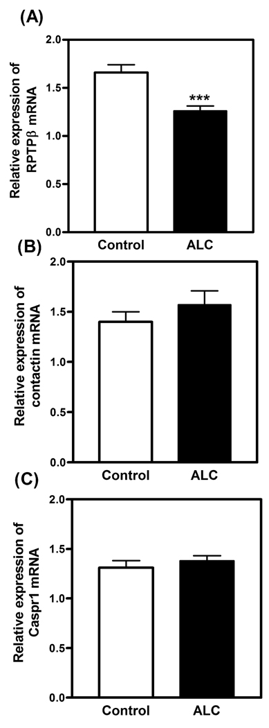
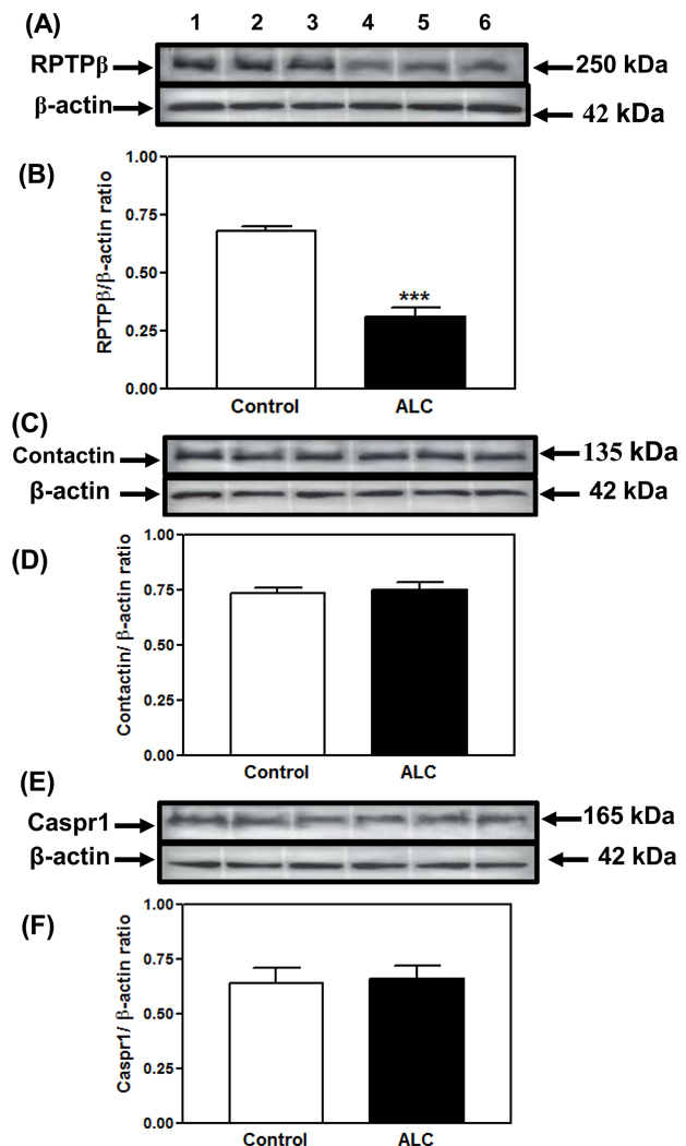
The short-term effects of ALC on the basal expression of this family of adhesion/signaling genes associated with glial-neuronal communication at the time of puberty were assessed. The blood ALC levels showed a mean± SEM of 204± 15 mg/dl, 1.5 hours after last infusion of the liquid-diet on the morning of day 6. Figure 1A shows that the basal gene expression of RPTPβ, as determined by real-time PCR, was decreased (p<0.001) in the ALC-treated rats compared with the control animals. Conversely, figures 1B&C, respectively, demonstrate that ALC did not affect the basal gene expression of either contactin or Caspr1compared with the control animals. Figure 2A depicts the representative Western immunoblot of the RPTPβ protein detected at 250 kDa. The composite graph shown in figure 2B illustrates that basal RPTPβ protein expression was markedly decreased (p<0.001) in the ALC-treated animals, an effect that paralleled the decreased expression of basal RPTPβ mRNA shown in figure 1A. The representative immunoblot of contactin protein detected at 135 kDa, as shown in figure 2C, as well as its composite graph shown in figure 2D, demonstrate no ALC-induced changes compared with control animals. Similarly, the representative immunoblot of Caspr1 protein detected at 165 kDa, shown in figure 2E, as well as its composite graph shown in figure 2F, illustrate that this protein expression was also unaffected by ALC exposure.
Figure 1.
Effect of short-term ALC exposure on basal RPTPβ (A), contactin (B) and Caspr1 (C) gene expressions in the MBH of prepubertal female rats as determined by real-time PCR. Note that ALC caused a marked decrease in basal RPTPβ gene expression compared with control animals, but did not alter the gene expression of either contactin or Caspr1. The respective bars illustrate the mean (±SEM) of an N of 12–13 per group. ***p<0.001 versus control.
Figure 2.
Effect of short-term ALC exposure on basal RPTPβ, contactin and Caspr1 protein expressions in the MBH of prepubertal rats. (A) Representative Western immunoblot of RPTPβ and β-actin proteins in the MBH isolated from control (lanes 1–3) and ALC-treated (lanes 4–6) animals. (B) Densitometric quantitation of all the bands from 2 blots assessing RPTPβ protein in the MBH. (C) Representative Western immunoblot of contactin and β-actin proteins in the MBH isolated from control (lanes 1–3) and ALC-treated (lanes 4–6) animals. (D) Densitometric quantitation of all the bands from 2 blots assessing contactin protein in the MBH. (E) Representative Western immunoblot of Caspr1 and β-actin proteins in the MBH isolated from control (lanes 1–3) and ALC-treated (lanes 4–6) animals. (F) Densitometric quantitation of all the bands from 2 blots assessing Caspr1 protein in the MBH. Note that ALC caused a marked decrease in RPTPβ protein expression compared with control animals, but did not alter the protein expression of either contactin or Caspr1.The respective bars illustrate the mean (±SEM) of an N of 10 for control and an N of 6 for ALC. All of the above data were normalized to the internal control β-actin protein. ***p<0.001 versus control.
Effects of IGF-1 on RPTPβ, contactin and Caspr1 gene expressions
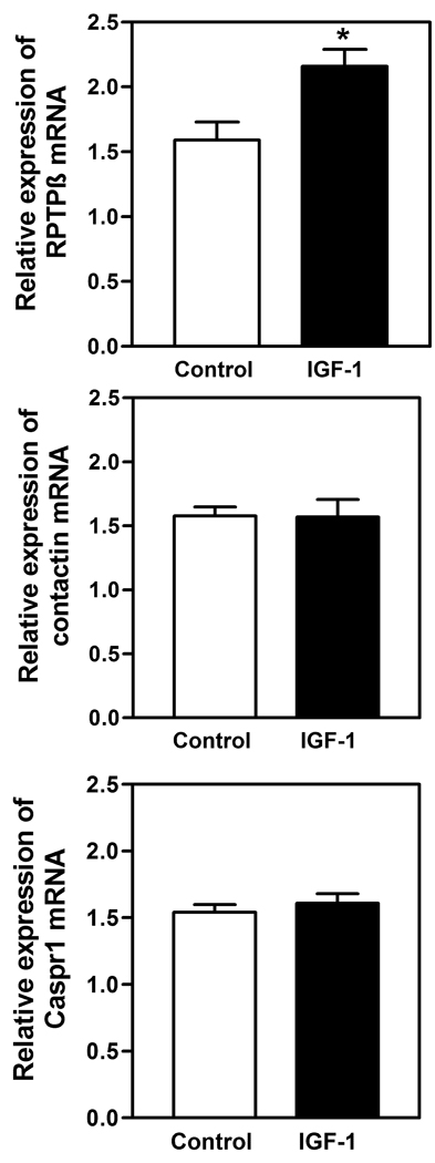
Figure 3A demonstrates the ability of IGF-1 to induce (p<0.05) the expression of the RPTPβ gene 6 hours after administration of the peptide into the third ventricle of the brain. This effect was specific for this member of the gene family, since figure 3B and C demonstrate that IGF-1 did not affect the basal expression of either contactin or Caspr1.
Figure 3.
Effect of IGF-1 on basal RPTPβ (A), contactin (B), and Caspr1 (C) gene expressions in the MBH of prepubertal female rats as determined by real-time PCR. IGF-1 (200 ng) was injected into the third ventricle and gene expressions in the MBH were assessed at 6h post-injection. Note that IGF-1 induced a significant increase in RPTPβ gene expression compared with control animals, but did not affect the expression of either contactin or Caspr1.The respective bars illustrate the mean (±SEM) of an N of 7 per group for RPTPβ and an N of 8–10 per group for contactin and Caspr1. *p<0.05 versus control.
Effect of ALC on IGF-1 induced RPTPβ gene expression
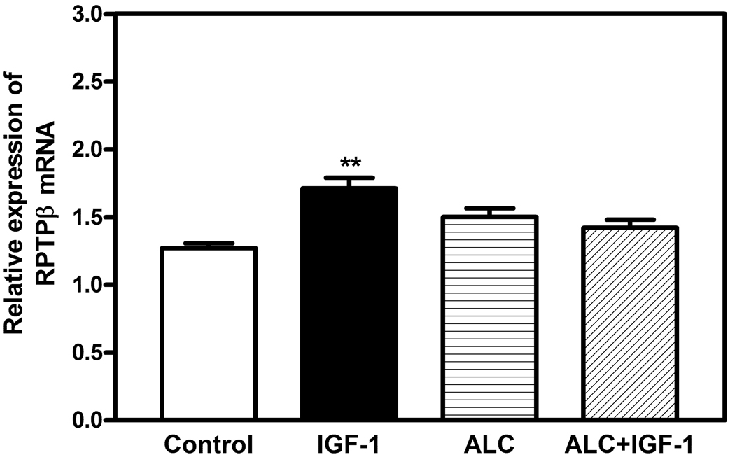
Figure 4 demonstrates again that the central administration of IGF-1 stimulated (p<0.01) the RPTPβ gene at 6 hours post-injection when compared to the control animals that were injected with saline. In addition, this figure shows that the acute presence of ALC, averaging 117± 22 level of mg/dl over the course of the day, did not alter the basal expression of the RPTPβ gene; however, the ability of IGF-1 to induce an increase in the RPTPβ expression was blocked (p<0.01) by the ALC.
Figure 4.
Effect of acute ALC exposure on basal and IGF-1 stimulated RPTPβ gene expressions in the MBH as determined by real-time PCR. The RPTPβ gene expression was assessed in the MBH at 6 h post IGF-1 injection. The IGF-1 induced an increase in the expression of the RPTPβ gene compared with the basal level shown in the controls. Note that the basal expression of the RPTPβ gene was not altered by ALC alone, but that the presence of ALC blocked the IGF-1 induced expression of the gene. The respective bars illustrate the mean (±SEM) of an N of 8–10 per group. **p<0.01 as assessed by ANOVA, with post-hoc testing using Student-Newman-Keuls.
DISCUSSION
The physiological mechanism(s) by which ALC alters LH secretion and the pubertal process has been an important area of investigation for over two decades. Compelling evidence using both rats and rhesus monkeys indicates that suppressed serum levels of LH and the resulting delay in pubertal development is due to a hypothalamic action of ALC to suppress the prepubertal secretion of LHRH (Dissen et al., 2004; Hiney and Dees, 1991; Hiney et al., 1998, 2010; Nyberg et al, 1993). This observation is important because the onset of sexual development is characterized by complex interactions within the hypothalamus that lead to the increased secretion of LHRH into the hypophyseal portal system directly from the neuron terminals in the median eminence (ME). This action prompts increased pituitary gonadotropin secretion and ovarian estradiol synthesis and secretion to drive the pubertal process to maturity. The LHRH secretory process is regulated not only by the hypothalamic actions of neuronal inputs (Hiney et al., 1991; 2009; Thompson et al., 2004; Gay and Plant 1987; Urbanski and Ojeda, 1987; Kalra and Crawley, 1992), but also by glial secreted factors (Hiney et al., 2003; Ma et al., 1997; Mahesh et al., 2006; Ojeda and Skinner, 2006; Roth et al., 2006). Additionally, in recent years there has been a growing interest with regard to contributions by other cell-cell signaling molecules that contribute to the structural organization of bi-directional glial-neuronal communications. In this regard, a novel gene family comprised of RPTPβ and the contactin-Caspr1 complex has been shown to be important for glial-neuronal adhesivness and for their cell to cell communications (Pierre et al., 1998; Peles et al.,1997; 1998). Contactin and Caspr1 provide the neuronal components, with RPTPβ being the glial partner. Once binding is complete this adhesion/signaling family can contribute to hypothalamic neuroendocrine functions (Pierre et al., 1998). Interestingly, glial-neuronal plasticity within the median eminence has been shown to change with different endocrine conditions (King and Letourneau, 1994; Prevot et al., 2000; Theodosis and Poulain, 1992), including puberty (Witkin et al., 1995), and glial cells can facilitate LHRH neuronal secretion by inducing plasticity, as well as bi-directional communications (Ojeda et al., 2000).
Because of the growing importance of adhesion/signaling molecules at puberty and because ALC alters prepubertal LHRH secretion and the pubertal process, we first assessed in this study whether ALC would affect the gene expression of any members of the RPTPβ-contactin-Caspr1 family. Our results showed that while contactin and Caspr1 gene expressions were not altered by ALC, the expression of RPTPβ was markedly decreased. Furthermore, protein expression for RPTPβ was also depressed. The fact that ALC can alter the synthesis of glial RPTPβ, which is required for binding to the neuronal contactin/Caspr1 complex, indicates its potential to disrupt the glial-neuronal adhesiveness function associated with the binding together of this three member family. Importantly, in the basal hypothalamus of immature female rhesus monkeys, glial cells were shown to express RPTPβ, and LHRH neurons were found to express both contactin and Caspr1 (Mungenast and Ojeda, 2005). Furthermore, the GT1-7 LHRH cell line has been shown to adhere to the carbonic anhydrase domain of RPTPβ in a contactin-dependent manner (Parent et al., 2007).Taken together, these results are important since they show a direct association between hypothalamic glial cells and LHRH neurons. Because glia expressing RPTPβ are densely populated within the ME, and because contactin is abundant in LHRH nerve terminals in this region, it has been suggested that these LHRH nerve terminals are a primary site of contactin dependent cell adhesion (Parent et al., 2007). Therefore, we suggest the chronic effect of ALC to decrease the synthesis of glial RPTPβ has the potential to disrupt glial-LHRH neuronal adhesive communication, and that this action, at least in part, contributes to this drugs ability to detrimentally affect the pubertal process.
The ALC-induced decrease in glial RPTPβ gene expression depicted here indicates a reduced amount of peptide available and required for binding to the contactin-Caspr1 complex on neurons, including those of the LHRH phenotype. As a result, a decreased adhesiveness between the glia and LHRH neurons in the ME would be expected, and this could contribute in several ways to altered LHRH neuronal functions observed following ALC exposure. Normally, the binding of glial RPTPβ to contactin on neuron terminals initiates the adhesion that allows for greater communication between the two cell types. Following this binding, the Caspr1 element functions as the signaling component for contactin, and subsequently, activates neuronal intracellular signaling pathways (Peles et al., 1997). The precise contribution of contactin-Caspr1 signaling and LHRH secretion remains to be determined, but previous studies have suggested that a function of the RPTPβ-contactin-Caspr1 family is to facilitate neurosecretion through changes in glial-neuronal signaling (Pierre et al., 1998; Mungenast and Ojeda, 2005). The cell adhesiveness component also promotes a more secure proximity for glial-derived secretions to bind to their receptors on the LHRH nerve terminals. Insulin-like growth factor 1 (IGF-1), transforming growth factor alpha (TGFα) and epidermal growth factor (EGF) are all puberty-related peptides that are synthesized and secreted from glia (Duenas et al., 1994; Ma et al., 1992; Ojeda et al., 2000).Once released, these peptides bind to their receptors on adjacent glial cells, thereby inducing the synthesis and secretion of PGE2. The PGE2 then binds to the PGE2-receptor on the nearby LHRH nerve terminals, facilitating the release of this peptide that controls the timing of puberty. The fact that ALC decreases prepubertal RPTPβ gene and protein expression in the hypothalamus suggests diminished glial-neuronal adhesiveness and thus, altered facilitation of LHRH release by the products secreted from the neighboring glial cells.
Another important aspect of the present study is our investigation into whether or not IGF-1 was capable of up-regulating the expression of any of the members of this adhesion/signaling gene family. We considered IGF-1 a good candidate for this because it is capable of acting at both glial and neuronal levels in association with prepubertal LHRH regulation (Dees et al., 2005; Hiney et al., 1991; 1996; Longo et al.,1998; Zhen et al.,1997). We have shown previously that IGF-1 of peripheral origin acts centrally, within the ME, to stimulate LHRH release, and that its administration accelerates the onset of female puberty in rats (Hiney et al., 1996). Subsequently, IGF-1 administration was shown to advance first ovulation in rhesus monkeys (Wilson, 1998) and IGF-1 replacement advanced puberty in GH-receptor-knockout mice that expressed very low levels of the peptide (Danilovich et al., 1999). We have now shown for the first time that the administration of IGF-1 into the third brain ventricle did not affect the gene expression of either contactin or Caspr1 within the hypothalamus; however, it induced the expression of the RPTPβ gene in this brain region at six hours post-injection. To our knowledge this is the first report of any substance capable of regulating a member of this adhesion gene family. The fact that increased circulating levels of IGF-1 at puberty can cross the blood brain barrier and enter the ME region (Hiney et al., 1996), that the peptide is also produced locally within the ME (Duenas et al., 1994), and that there is a high content of IGF-1 receptors localized to glia in this region (Lesniak et al., 1988; Marks et al., 1991), support the IGF-1/RPTPβ relationship that we have described. Additionally, this further attests to the notion that attainment of puberty is a complex interaction of events, and that glial cells through their adhesion and signal promoting capabilities, can integrate stimulatory inputs to the LHRH neuronal terminals.
As a result of our observation revealing the ability of IGF-1 to up regulate the RPTPβ gene, we assessed whether or not acute ALC administration would alter this IGF-1 action. Our results demonstrated that the presence of ALC blocked the ability of IGF-1to induce RPTPβ gene expression. This finding further suggests that this drug may affect glial-neuronal adhesiveness at this critical time of development. Based on what we know about the early positive influence of IGF-1 on LHRH release and the pubertal process (Hiney et al., 1991; 1996), we now suggest that some of the detrimental effects of ALC at puberty may be due, at least in part, to altered IGF-1 induction of adhesion signaling. This effect could be due to an ALC action at the level of the IGF-1R and/or to an altered post-receptor event. With regard to the IGF-1R, it has been shown that acute and chronic ALC administration does not alter either the gene or protein expression of this receptor (Srivastava et al. 1995; 2009), but we cannot rule out that it may affect mechanisms regulating receptor function. This suggests that ALC may target a pathway component downstream from the IGF-1R. In this regard, we have shown that chronic ALC administration suppresses the basal protein expression of phosphorylated Akt, a transduction signal activated by IGF-1 after binding to its receptor (Srivastava et al., 2009). Acutely, we have shown that ALC can block the IGF-1 induced expression of other genes involved in the pubertal process (Dees et al., 2005; Hiney et al., 2010), and that this occurs by blocking the peptide induced phosphorylation of Akt (Hiney et al., 2010). Chronic ALC administration has been shown to suppress circulating IGF-1 levels in prepubertal rats (Srivastava et al., 1995) and rhesus monkeys (Dees et al., 2000), actions associated with altered pubertal development in both species. Taken together, we suggest that decreased prepubertal levels of circulating IGF-1 available to the ME, as well as altered post-receptor transduction signals, contribute to the depressed RPTPβ gene expression that were observed following chronic ALC exposure in the present study.
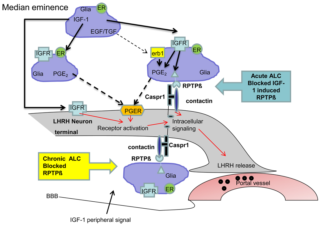
In conclusion, glial RPTPβ binding to the neuronal contactin-Caspr1 complex initiates cell adhesion and signaling communications between the glial cells and LHRH nerve terminals in the basal hypothalamus. We have shown in the present study that ALC suppresses total RPTPβ gene expression and protein levels in the MBH, suggesting a potential disruption of glial communications important for prepubertal LHRH secretory activity. Furthermore, we also revealed that IGF-1, a critical puberty-related peptide, can induce prepubertal RPTPβ gene expression in the MBH of prepubertal rats and that ALC can block this action. This glial to neuronal communication network is important for mechanisms associated with the enhanced secretion of LHRH at the time of puberty, since the glial cells integrate stimulatory influences to LHRH neuronal terminals. The schematic shown in figure 5 summarizes some important glial-glial and glial neuronal sites of intracellular communication within the median eminence, including the glial RPTPβ interaction with the LHRH neuronal contactin-Caspr1 complex.
Figure 5.
Schematic drawing showing glia-LHRH neuronal association and sites of ALC effects on RPTPβ in the median eminence of juvenile female rats. For clarity, details of other downstream pathways in this region are not shown. IGF-1, insulin-like growth factor-1; EGF, epidermal growth factor; ALC, alcohol, IGFR, Insulin-like growth factor receptor; PGE2, prostaglandin-E2; LHRH, luteinizing hormone releasing hormone; RPTPβ, receptor protein tyrosine phosphatase β; Caspr1, contactin associated protein-1; ER, estrogen receptor; TGF, transforming growth factor; BBB, blood brain barrier
Acknowledgments
This work was supported by the NIH grant AA07216 (to WLD).
REFERENCES
- Anderson RA, Willis BR, Oswald C, Gupta A, Zaneveld L. Delayed male sexual maturation induced by chronic ethanol ingestion. Fed Proc. 1981;40:825–829. [Google Scholar]
- Bo WJ, Krueger WA, Rudeen PK, Symees SK. Ethanol-induced alterations in the morphology and function of the rat ovary. Anat Rec. 1982;202:255–260. doi: 10.1002/ar.1092020210. [DOI] [PubMed] [Google Scholar]
- Danilovich N, Wernsing D, Coschigano KT, Kopchick JJ, Bartke A. Deficits in female reproductive function in GH-R-KO mice; role of IGF-1. Endocrinology. 1999;140:2637–2640. doi: 10.1210/endo.140.6.6992. [DOI] [PubMed] [Google Scholar]
- Dees WL, Dissen GA, Hiney JK, Lara F, Ojeda SR. Alcohol ingestion inhibits the increased secretion of puberty-related hormones in the developing female rhesus monkey. Endocrinology. 2000;141:1325–1331. doi: 10.1210/endo.141.4.7413. [DOI] [PubMed] [Google Scholar]
- Dees WL, Hiney JK, Nyberg CL. Effects of ethanol on the reproductive neuroendocrine axis of prepubertal and adult female rats. Chapter 14. In: Sarkar DK, Barnes CD, editors. The Reproductive Neuroendocrinology of Aging and Drug Abuse. Boca Raton, FL: CRC Press; 1995. pp. 301–334. [Google Scholar]
- Dees WL, Skelley CW. The effects of ethanol during the onset of puberty. Neuroendocrtinology. 1990;51:64–69. doi: 10.1159/000125317. [DOI] [PubMed] [Google Scholar]
- Dees WL, Skelley CW, Kozlowski GP. Intragastric cannulation as a method of ethanol administration for neruoendocrine studies. Alcohol. 1984;1:177–180. doi: 10.1016/0741-8329(84)90094-6. [DOI] [PubMed] [Google Scholar]
- Dees WL, Srivastava VK, Hiney JK. Alcohol alters IGF-1 activated Oct-2 POU gene expression in the immature female hypothalamus. J Stud Alc. 2005;666:35–45. doi: 10.15288/jsa.2005.66.35. [DOI] [PubMed] [Google Scholar]
- Dissen GA, Ojeda SR, Dearth RK, Scott H, Dees WL. Alcohol alters prepubertal luteinizing hormone (LH) secretion in female rhesus monkeys by a hypothalamic action. Endocrinology. 2004;145:4558–4564. doi: 10.1210/en.2004-0517. [DOI] [PubMed] [Google Scholar]
- Duenas M, Luquin S, Chowen JA, Torres-Aleman I, Naftolin F, Garcia-Segura LM. Gonadal hormone regulation of insulin-like growth factor-1-like immunoreactivity in hypothalamic astroglia of developing and adult rats. Neuroendocrinology. 1994;59:528–538. doi: 10.1159/000126702. [DOI] [PubMed] [Google Scholar]
- Gay VL, Plant TM. N-methyl-DL-aspartate elicits hypothalamic gonadotropin-releasing hormone release in prepubertal male rhesus monkeys. Endocrinology. 1987;120:2289–2296. doi: 10.1210/endo-120-6-2289. [DOI] [PubMed] [Google Scholar]
- Hettinger AM, Allen MR, Zhang BR, Goad DW, Malayer JR, Geisert RD. Presence of the acute phase protein, bikunin, in the endometrium of gilts during estrous cycle and early pregnancy. Biol. Reprod. 2001;65:507–513. doi: 10.1095/biolreprod65.2.507. [DOI] [PubMed] [Google Scholar]
- Hiney JK, Dearth RK, Srivastava VK, Rettori V, Dees WL. Actions of alcohol on epidermal growth factor-receptor activated luteinizing hormone secretion. J Studies Alcohol. 2003;64:809–816. doi: 10.15288/jsa.2003.64.809. [DOI] [PubMed] [Google Scholar]
- Hiney JK, Dees WL. Ethanol inhibits luteinizing hormone releasing hormone from the median eminence of prepubertal female rats in vitro: Investigation of its actions on norepinipherine and prostaglandin E2. Endocrinology. 1991;128:1404–1408. doi: 10.1210/endo-128-3-1404. [DOI] [PubMed] [Google Scholar]
- Hiney JK, Ojeda SR, Dees WL. Insulin-like growth factor-1: A possible metabolic signal involved in the regulation of female puberty. Neuroendocrinology. 1991;54:420–423. doi: 10.1159/000125924. [DOI] [PubMed] [Google Scholar]
- Hiney JK, Srivastava VK, Dees WL. Insulin like growth factor-1 stimulates hypothalamic KiSS-1 gene expression by activating Akt: Effect of alcohol. Neuroscience. 2010;166:625–632. doi: 10.1016/j.neuroscience.2009.12.030. [DOI] [PMC free article] [PubMed] [Google Scholar]
- Hiney JK, Srivastava V, Lara F, Dees WL. Ethanol blocks the central action of IGF-1 to induce luteinizing hormone secretion in the prepubertal female rat. Life Sciences. 1998;62:301–308. doi: 10.1016/s0024-3205(97)01111-9. [DOI] [PubMed] [Google Scholar]
- Hiney JK, Srivastava VK, Nyberg CL, Ojeda SR, Dees WL. Insulin like growth factor -1 of peripheral origin acts centrall to accelerate the initiation of female puberty. Endocrinology. 1996;137:3717–3828. doi: 10.1210/endo.137.9.8756538. [DOI] [PubMed] [Google Scholar]
- Hiney JK, Srivastava VK, Pine MD, Dees WL. IGF-1 activates KiSS-1 gene expression in the brain of the prepubertal female rat. Endocrinology. 2009;150:376–384. doi: 10.1210/en.2008-0954. [DOI] [PMC free article] [PubMed] [Google Scholar]
- Kalra SP, Crowley WR. Neuropeptide Y: A novel neuroendocrine peptide I the control of pituitary horone secretion and its relation to luteinizing hormone. In: Ganong WF, Martini L, editors. Front. In Neuroendocrinology. Vol. 13. NY: Raven Press; 1992. pp. 1–46. [PubMed] [Google Scholar]
- King JC, Letourneau RL. Luteinizing hormone releasing hormone terminals in the median eminence of rats undergo dynamic changes after gonadectomy, as revealed by electron microscopic image analysis. Endocrinology. 1994;134:1340–1351. doi: 10.1210/endo.134.3.8119174. [DOI] [PubMed] [Google Scholar]
- Lesniak MA, Hill JM, Kiess W, Rojeski M, Candace BP, Roth J. Receptors for insulin-like-growth factors I and II: Autoradiographic localization in rat brain and comparison to receptor for insulin. Endocrinology. 1988;123:2089–2099. doi: 10.1210/endo-123-4-2089. [DOI] [PubMed] [Google Scholar]
- Lomniczi A, Ojeda SR. A role for glial cella of the neuroendocrine brain in the central control of female sexual development. In: Purpura V, Haydon P, editors. Astrocytes in pathophysiology of the nervus system. New York, NY: Springer; 2008. [Google Scholar]
- Longo KM, Sun Y, Gore AC. Insulin like growth factor effects on gonadotropin releasing hormone biosynthesis in GT1-7 cells. Endocrinology. 1998;139:1125–1132. doi: 10.1210/endo.139.3.5852. [DOI] [PubMed] [Google Scholar]
- Ma YJ, Berg-von der Emde K, Rage F, Wetsel WC, Ojeda SR. Hypothalamic astrocytes respond to transforming growth factor alpha with secretion of neuroactive substances that stimulate the release of luteinizing hormone-releasing hormone. Endocrinology. 1997;138:19–35. doi: 10.1210/endo.138.1.4863. [DOI] [PubMed] [Google Scholar]
- Ma YJ, Junier MP, Costa ME, Ojeda SR. Transforming growth factor-α gene expression in the hypothalamus is developmentally regulated and linked to sexual maturation. Neuron. 1992;9:657–670. doi: 10.1016/0896-6273(92)90029-d. [DOI] [PubMed] [Google Scholar]
- Mahesh VB, Dhandapani KM, Brann DW. Role of astrocytes in reproduction and neuroprotection. Mol Cell Endocrinology. 2006;246:1–9. doi: 10.1016/j.mce.2005.11.017. [DOI] [PubMed] [Google Scholar]
- Marks JL, Porte D, Jr, Baskin DG. Localization of type 1 insulin-like growth factor receptor messenger RNA in the adult rat brain by in situ hybridization. Mol Endocrinol. 1991;5:1158–1168. doi: 10.1210/mend-5-8-1158. [DOI] [PubMed] [Google Scholar]
- Mungenast AE, Ojeda SR. Expression of three gene families encoding cell-cell communication molecules in the prepubertal nonhuman primate hypothalamus. J Neuroendocrinology. 2005;17:208–219. doi: 10.1111/j.1365-2826.2005.01293.x. [DOI] [PubMed] [Google Scholar]
- Navarro VM, Castellano JM, Fernandez-Fernandez R, Barriero ML, Roa J, Sanchez-Criado JE, Agilar E, Dieguez C, Pinilla L, Tena-Sempere M. Developmental and hormonally regulated mRNA expression of KiSS-1 and its putative receptor, GPR54, in rat hypothalamus and potent LH-releasing activity of KiSS-1 peptide. Endocrinology. 2004;145:4565–4574. doi: 10.1210/en.2004-0413. [DOI] [PubMed] [Google Scholar]
- Nyberg CL, Hiney JK, Minks JB, Dees WL. Ethanol alters N-methyl-DL- aspartic acid induced secretion of luteinizing hormone releasing hormone and the onset of puberty in the female rat. Neuroendocrinology. 1993;57:863–868. doi: 10.1159/000126446. [DOI] [PubMed] [Google Scholar]
- Nyberg CL, Srivastava V, Hiney JK, Lara F, Dees WL. N-methyl-D-Aspartic acid-receptor (NMDA-R) synthesis and luteinizing hormone release in immature female rats: Effects of stage of pubertal development and exposure to ethanol. Endocrinology. 1995;136:2874–2880. doi: 10.1210/endo.136.7.7789312. [DOI] [PubMed] [Google Scholar]
- Ojeda SR, Lomniczi A, Sandau US. Glial-gonadotropin hormone (GnRH) neurone interactions in the median eminence and the control of GnRH secretion. J Neuroendocrinology. 2008;20:732–742. doi: 10.1111/j.1365-2826.2008.01712.x. [DOI] [PubMed] [Google Scholar]
- Ojeda SR, Ma YJ, Lee BJ, Prevot V. Glia to neuron signaling and the neuroendocrine control of female puberty. Rec Prog Hor Res. 2000;55:197–224. [PubMed] [Google Scholar]
- Ojeda SR, Skinner MK. Puberty in the rat. In: Neill JD, editor. The Physiology of Reproduction. 3rd edn. San Diego, CA: Academic Press/Elsiever; 2006. pp. 2061–2126. [Google Scholar]
- Parent AS, Mungenast AE, Lomniczi A, Sandau US, Peles E, Bosch MA, Ronnekliev OK, Ojeda SR. A contactin-receptor-like protein tyrosine phophotase beta complex mediates adhesive communication between astroglial cell and gonadotropin releasing hormone neurons. J Neuroendocrinology. 2007;19:847–859. doi: 10.1111/j.1365-2826.2007.01597.x. [DOI] [PubMed] [Google Scholar]
- Peles E, Nativ M, Lustig M, Grumet M, Schilling J, Martinez R, Plowman GD, Schlissinger J. Identification of a novel contactin-associated transmembrane receptor with multiple domains implicated in protein-protein interactions. EMBO J. 1997;16:978–988. doi: 10.1093/emboj/16.5.978. [DOI] [PMC free article] [PubMed] [Google Scholar]
- Peles E, Nativ M, Campbell PL, Sakuria T, Martinez R, Lev S, Clary DO, Schilling J, Barnea G, Plowman GD, Grumet M, Schlessinger J. The carbonic anhydrase domain of receptor tyrosine phosphatase β is a functional ligand for the axonal cell recognition molecule contactin. Cell. 1995;82:251–260. doi: 10.1016/0092-8674(95)90312-7. [DOI] [PubMed] [Google Scholar]
- Peles E, Schlessinger J, Grumet M. Multi-ligand interactions with receptor-like protein tyrosing phosphatase β: implications for intercellular signaling. Trends Biochem Sci. 1998;23:121–124. doi: 10.1016/s0968-0004(98)01195-5. [DOI] [PubMed] [Google Scholar]
- Pierre K, Rougon G, Allard M, Bonhomme R, Gennarini G, Poulain DA, Theodosis DT. Regulated expression of the cell adhesion glucoprotein F3 in adult hypothalamic magnocellular neurons. J Neuroscience. 1998;18:5333–5343. doi: 10.1523/JNEUROSCI.18-14-05333.1998. [DOI] [PMC free article] [PubMed] [Google Scholar]
- Prevot V, Bouret S, Croix D, Alonso G, Jennes L, Mitchell V, Routtenberg A, Beayvillain JC. Growth – associated protein-43 messenger ribonucleic acid expression in gonadotropin releasing hormone neurons during the rat estrous cycle. Endocrinology. 2000;141:1648–1657. doi: 10.1210/endo.141.5.7448. [DOI] [PubMed] [Google Scholar]
- Ramaley JA. The regulation of gonadotropin secretion in immature ethanol-treated male rats. J Androl. 1982;3:248–252. [Google Scholar]
- Roth CL, McCormack AL, Lomniczi A, Mungenast AE, Ojeda SR. Quantitative proteomics identifies a major change in glial glutamate metabolism at the time of female puberty. Mol Cell Endocrinology. 2006;255:51–59. doi: 10.1016/j.mce.2006.04.017. [DOI] [PubMed] [Google Scholar]
- Shahab M, Mastronardi C, Seminara S, Crowley WF, Ojeda SR, Plant TM. Increased hypothalamic GPR54 signaling: A potential mechanism for initiation of puberty in primates. Proc Natl Acad Sci. 2005;102:2129–2134. doi: 10.1073/pnas.0409822102. [DOI] [PMC free article] [PubMed] [Google Scholar]
- Srivastava VK, Hiney JK, Dees WL. Short term alcohol administration alters KiSS-1 gene expression in the reproductive hypothalamus of prepubertal female rats. Alc. Clin. Exp. Res. 2009;33:1605–1614. doi: 10.1111/j.1530-0277.2009.00992.x. [DOI] [PMC free article] [PubMed] [Google Scholar]
- Srivastava V, Hiney JK, Nyberg CL, Dees WL. Influence of ethanol on Insulin-like growth factor I (IGF-1) and IGF-I receptor synthesis during female puberty. A correlation with serum IGF-1. Alc: Clin Exp Res. 1995;19:1467–1473. doi: 10.1111/j.1530-0277.1995.tb01009.x. [DOI] [PubMed] [Google Scholar]
- Theodosis DT, Poulain DA. Neuronal-glial and synaptic plasticity of the adult oxytocinergic system. Ann NY Acad Sci. 1992;652:303–325. doi: 10.1111/j.1749-6632.1992.tb34363.x. [DOI] [PubMed] [Google Scholar]
- Thompson EL, Patterson M, Murphy KG, Smith KL, Dhillo WS, Todd JF, Ghatei MA, Bloom SR. Central and peripheral administration of kisspeptin-10 stimulates the hypothalamic-pituitary-gonadal axis. J. Neuroendocrinology. 2004;16:850–858. doi: 10.1111/j.1365-2826.2004.01240.x. [DOI] [PubMed] [Google Scholar]
- Urbanski HF, Ojeda SR. Activation of lutenizing hormone releasing hormone release advances the onset of female puberty. Neuroendocrinology. 1987;46:273–276. doi: 10.1159/000124831. [DOI] [PubMed] [Google Scholar]
- Wilson ME. Premature elevation in serum insulin-like growth factor -1 advances first ovulation in monkeys. J Endocrinology. 1998;158:247–257. doi: 10.1677/joe.0.1580247. [DOI] [PubMed] [Google Scholar]
- Witkin JW, O’Sullivan H, Ferin M. Glial enhancement of GnRH neurons in prepubertal female rhesus monkeys. J Neuroendocrinology. 1995;7:665–671. doi: 10.1111/j.1365-2826.1995.tb00807.x. [DOI] [PubMed] [Google Scholar]
- Zhen S, Zakaria M, Wolfe A, Radovick S. Regulation of gonadotropin releasing hormone (GnRH) gene expression by IGF-1 in a cultured GnRH cell line. Molecular Endocrinology. 1997;11:1145–1155. doi: 10.1210/mend.11.8.9956. [DOI] [PubMed] [Google Scholar]


