Abstract
The diabetic Charcot foot syndrome is a serious and potentially limb-threatening lower-extremity complication of diabetes. First described in 1883, this enigmatic condition continues to challenge even the most experienced practitioners. Now considered an inflammatory syndrome, the diabetic Charcot foot is characterized by varying degrees of bone and joint disorganization secondary to underlying neuropathy, trauma, and perturbations of bone metabolism. An international task force of experts was convened by the American Diabetes Association and the American Podiatric Medical Association in January 2011 to summarize available evidence on the pathophysiology, natural history, presentations, and treatment recommendations for this entity.
The Charcot foot in diabetes poses many clinical challenges in its diagnosis and management. Despite the time that has passed since the first publication on pedal osteoarthropathy in 1883, we have much to learn about the pathophysiology, and little evidence exists on treatments of this disorder. The international task force was convened in January 2011 at the Salpêtrière Hospital in Paris, France, to review the literature and report on the definition, pathogenesis, diagnosis, and treatment of the diabetic Charcot foot. Recommendations in this report are solely the opinions of the authors and do not represent the official positions of the American Diabetes Association or the American Podiatric Medical Association.
DEFINITION
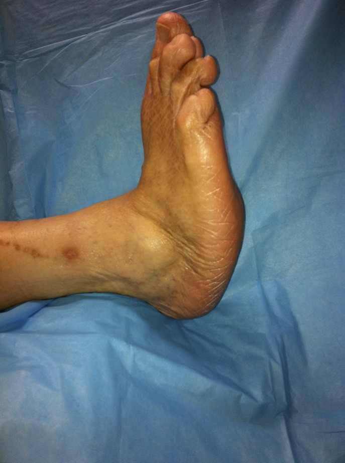
Charcot neuropathic osteoarthropathy (CN), commonly referred to as the Charcot foot, is a condition affecting the bones, joints, and soft tissues of the foot and ankle, characterized by inflammation in the earliest phase. The Charcot foot has been documented to occur as a consequence of various peripheral neuropathies; however, diabetic neuropathy has become the most common etiology. The interaction of several component factors (diabetes, sensory-motor neuropathy, autonomic neuropathy, trauma, and metabolic abnormalities of bone) results in an acute localized inflammatory condition that may lead to varying degrees and patterns of bone destruction, subluxation, dislocation, and deformity. The hallmark deformity associated with this condition is midfoot collapse, described as a “rocker-bottom” foot (Fig. 1), although the condition appears in other joints and with other presentations. Pain or discomfort may be a feature of this disorder at the active (acute) stage, but the level of pain may be significantly diminished when compared with individuals with normal sensation and equivalent degrees of injury.
Figure 1.

The typical appearance of a later-stage Charcot foot with a rocker-bottom deformity.
The set of signs and symptoms that occur together with CN qualifies this condition as a syndrome, the “Charcot foot syndrome.”
Definition and classification recommendations
Nomenclature should be standardized to CN or the Charcot foot.
Existing classifications do not provide prognostic value or direct treatment. Active or inactive should be used to describe an inflamed or stable CN, respectively. Acute and chronic can also be used in this regard, but there is no accepted measure that defines the transition point.
PATHOGENESIS
There is no singular cause for the development of the Charcot foot, but there are factors that predispose to its development, as well as a number of likely precipitating events. The current belief is that once the disease is triggered in a susceptible individual, it is mediated through a process of uncontrolled inflammation in the foot. This inflammation leads to osteolysis and is indirectly responsible for the progressive fracture and dislocation that characterizes its presentation (1). The evidence to support this hypothesis is largely circumstantial. A neurally mediated vascular reflex leading to increased peripheral blood flow and active bone resorption has been proposed as an etiological factor in the development of bone and joint destruction in neuropathic patients. However, the relationship between increased blood flow to bone and active bone resorption has not been conclusively defined.
Uncontrolled inflammation
When a bone is fractured, the release of proinflammatory cytokines including tumor necrosis factor-α and interleukin-1β leads to increased expression of the polypeptide receptor activator of nuclear factor-κB ligand (RANKL) from any of a number of local cell types. RANKL triggers the synthesis of the nuclear transcription factor nuclear factor-κβ (NF-κβ), and this in turn stimulates the maturation of osteoclasts from osteoclast precursor cells. At the same time, NF-κβ stimulates the production of the glycopeptide osteoprotegerin (OPG) from osteoblasts. This “decoy receptor” acts as an effective antagonist of RANKL (2). The fracture will also be associated with pain, and this leads to splinting of the bone, and the rise in proinflammatory cytokines is usually relatively short-lived. In the person who develops an acute Charcot foot, however, the loss of pain sensation allows for uninterrupted ambulation, with repetitive trauma. It has been suggested that this results in continual production of proinflammatory cytokines, RANKL, NF-κβ, and osteoclasts, which in turn leads to continuing local osteolysis (1). This has subsequently been shown by an increase in proinflammatory phenotypes of monocytes in those with active Charcot foot when compared with diabetic control subjects (3).
Osteoclasts generated in vitro in the presence of macrophage colony-stimulating factor and RANKL from patients with active CN have been shown to be more aggressive and exhibit an increase in their resorptive activity compared with control subjects. However, these changes are only partially inhibited by OPG, indicating that other cytokines may also be important (4).
Predisposition
Neuropathy is a universal feature of the affected limb. Although it has been suggested that people with a Charcot foot may have particular patterns of sensory loss reflecting involvement of different fibers (5,6), this is not generally accepted. Nevertheless, three groups have shown that people who have had an acute Charcot foot exhibit retention of vasodilatory reflexes in contrast to diabetic individuals with distal symmetrical neuropathy without CN (7–9).
Despite these observations, it should be noted that the syndrome might also occur in patients with a spectrum of unrelated diseases complicated by nerve damage. These include distal neuropathies caused by toxins (ethanol, drug related) and infection (leprosy), as well as diseases of the spinal cord and nerve roots (tabes dorsalis, trauma, syringomyelia) and a number of other conditions (Parkinson's disease, HIV, sarcoidosis, rheumatoid disease, and psoriasis). Although the neuroarthropathy is typically more proximal in those with disease of the spinal cord, the presentation may be otherwise indistinguishable.
Loss of protective sensation will increase the likelihood of trauma to the foot, while motor neuropathy could result in altered structure of the foot (with exaggeration of the plantar arch and clawing) and changed gait with resultant abnormal loading.
Finally, it is possible that peptides normally secreted from nerve terminals are also important in the underlying pathophysiology. Of these, calcitonin gene–related peptide (CGRP) is a likely candidate because it is known to antagonize the synthesis of RANKL. Hence, any reduction of CGRP through nerve damage will result in an increase in RANKL expression. It is of particular interest that CGRP has been reported to be necessary for the maintenance of the normal integrity of joint capsules, and it follows that any reduction in CGRP release by nerve terminals could facilitate joint dislocation (10).
Because it is not possible to identify those most likely to develop the Charcot syndrome, it is impossible to determine with any degree of confidence whether preexisting osteopenia is a significant predisposing factor. One group has, however, reported an apparent reduction in bone mineral density (BMD) of the femoral neck in the contralateral (unaffected) limb at the time of presentation. The researchers also reported an association between BMD and the relative prevalence of fracture and of dislocation in the affected foot (11).
Diabetes may be associated with osteopenia, but the available evidence suggests that reduction of BMD is a feature of type 1 diabetes more so than type 2 diabetes (12). Any reduction of BMD in type 1 diabetes may relate to loss of islet peptides such as insulin and amylin (IAPP), both of which act as growth factors for bone. Despite this, the fracture risk in type 2 diabetes may be no less than in type 1 diabetes (12), and this would explain the fact that the presentation does not appear to differ between the two types of the disease. Any associated deficiency of vitamin D—with or without renal failure and secondary hyperparathyroidism—would increase the possibility of reduced BMD in diabetes. The use of thiazolidinediones could theoretically increase the likelihood of an acute Charcot foot through an effect on bone density, but this has not yet been reported. The use of corticosteroids as immunosuppressants in people with diabetes who have had a renal and/or pancreatic transplant (13) may explain the apparent high incidence of the Charcot foot in this group.
Although the Charcot syndrome may occur in a variety of conditions, diabetes is ostensibly the most common worldwide. Diabetes may predispose to its occurrence through a number of mechanisms. Apart from the presence of neuropathy and possible osteopenia, these include the effects of advanced glycation end products, reactive oxygen species, and oxidized lipids, which may all enhance the expression of RANKL in diabetes (10). The effect of local inflammation on this pathway would similarly compound the expression of RANKL. Furthermore, a single study has reported an apparent association between two OPG-related polymorphisms in people with a history of an acute Charcot foot in diabetes (14).
Many patients recall that the onset of the condition was precipitated by trauma that is often minor in nature (15). Other cases may be triggered by different causes of local inflammation, including previous ulceration, infection, or recent foot surgery. In this respect the occurrence of an acute Charcot foot as a complication of osteomyelitis is increasingly recognized in people with diabetes. Very occasionally, the onset of an acute Charcot foot may follow successful revascularization.
DIAGNOSIS
The initial manifestations of the Charcot foot are frequently mild in nature, but can become much more pronounced with unperceived repetitive trauma. Diagnostic clinical findings include components of neurological, vascular, musculoskeletal, and radiographic abnormalities. There have been no reported cases of CN developing in the absence of neuropathy. Accordingly, peripheral sensory neuropathy associated with reduced sensation of pain is the essential predisposing condition that permits the development of the arthropathy (16–19). Because of the very presence of insensitivity, a personal history concerning antecedent trauma is often unreliable (18,20,21). Typical clinical findings include a markedly swollen, warm, and often erythematous foot with only mild to modest pain or discomfort (16,18–20,22). Acute local inflammation is often the earliest sign of underlying bone and joint injury (23). This initial clinical picture resembles cellulitis, deep vein thrombosis, or acute gout and can be misdiagnosed as such. There is most often a temperature differential between the two feet of several degrees (20,24). The affected population typically has well preserved or even exaggerated arterial blood flow in the foot. Pedal pulses are characteristically bounding unless obscured by concurrent edema. Patients with chronic deformities, however, can develop subsequent limb-threatening ischemia. Musculoskeletal deformity can be very slight or grossly evident most often due to the chronicity of the problem and the anatomical site of involvement (16,17,19,25). The classic rocker-bottom foot, with or without plantar ulceration, represents a severe chronic deformity typical for this condition (16,26,27). Radiographic and other imaging modalities can detect subtle changes consistent with active CN.
Imaging of the Charcot foot
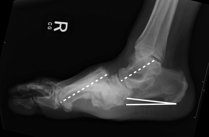
Radiographs are the primary initial imaging method for evaluation of the foot in diabetic patients. Easily available and inexpensive, they provide information on bone structure, alignment, and mineralization. X-rays may be normal or show subtle fractures and dislocations or later show more overt fractures and subluxations. In later stages, the calcaneal inclination angle is reduced and the talo-first metatarsal angle is broken (Fig. 2). Medial calcification of the arteries is present in most Charcot feet and is a frequent secondary finding on radiographs (25). However, radiographic changes of CN are typically delayed and have low sensitivity (28).
Figure 2.
Lateral X-ray of a Charcot foot deformity showing a dislocation of the tarsometatarsal joint with break in the talo-first metatarsal line (dashed lines) and a reduced calcaneal inclination angle (solid lines).
Magnetic resonance imaging (MRI) allows detection of subtle changes in the early stages of active CN when X-rays could still be normal. MRI primarily images protons in fat and water and can depict anatomy and pathology in both soft tissue and bone in great detail. Because of its unique capability of differentiating tissues with high detail, MRI has a high sensitivity and specificity for osteomyelitis and has become the test of choice for evaluation of the complicated foot in diabetic patients (29). Although not required for diagnosis when X-rays are diagnostic for Charcot bone and joint changes, MRI is very useful in making the diagnosis at its earliest onset before such changes become evident on plain films.
Nuclear medicine includes a number of exams based on the use of radioisotopic tracers. Three-phase bone scans, based on technetium-99m (99mTc), are highly sensitive for active bone pathology. However, diminished circulation can result in false-negative exams and, perhaps more importantly, uptake is not specific for osteoarthropathy. Labeled white blood cell scanning (using 111In or 99mTc) provides improved specificity for infection in the setting of neuropathic bone changes (30), but it can be difficult to differentiate soft tissue from bone. Therefore, this exam can be combined with a three-phase bone scan or sulfur colloid marrow exam when superimposed osteomyelitis is suspected (31). More recently, positron emission tomography scanning has been recognized as having potential for diagnosis of infection and differentiating the Charcot foot from osteomyelitis (32,33). However, this remains investigational at this time.
Evaluation of bone density may be useful in those with diabetes to assess onset of CN as well as fracture risk. BMD can be assessed using dual-energy X-ray absorptiometry or calcaneal ultrasound. BMD has been related to the pathological pattern of CN, whereby joint dislocation is more prevalent in those with normal mineralization versus fracture in those with diminished BMD (11).
Experts agree that radiographs are important as the first exam in virtually all settings (33,34). However, a negative result obviously should not offer any confidence regarding lack of disease. In a patient with low clinical suspicion of osteomyelitis and no sign of CN on radiographs, either three-phase bone scan or noncontrast MRI is very effective at excluding osseous disease. If the patient has an ulceration with a high likelihood of deep infection, MRI is the best diagnostic modality. Nonetheless, one test may not be adequate for full evaluation. In this setting where MRI diagnosis is indeterminate, a subsequent labeled white blood cell scan can provide more specificity and should be correlated with clinical findings. The decision of nuclear imaging versus MRI is largely based on personal preference, availability, and local experience. In general, if metal is present in the foot, nuclear medicine exams are preferred, whereas diffuse or regional ischemia makes MRI the preferred exam.
Diagnostic recommendations for active CN
The diagnosis of active Charcot foot is primarily based on history and clinical findings but should be confirmed by imaging.
Inflammation plays a key role in the pathophysiology of the Charcot foot and is the earliest clinical finding.
The occurrence of acute foot/ankle fractures or dislocations in neuropathic individuals is considered active CN because of the inflammatory process of bone healing, even in the absence of deformity.
X-rays should be the initial imaging performed, and one should look for subtle fractures or subluxations if no obvious pathology is visible.
MRI or nuclear imaging can confirm clinical suspicions in the presence of normal-appearing radiographs.
MEDICAL TREATMENT
The medical treatment of CN is aimed at offloading the foot, treating bone disease, and preventing further foot fractures (34). Because of the various etiologies of increased local bone resorption and/or secondary osteoporosis in patients with CN and limited randomized placebo-controlled trials in this area, treatment guidelines are largely based on professional opinion rather than the highest level of clinical evidence.
Offloading
Offloading at the acute active stage of the Charcot foot is the most important management strategy and could arrest the progression to deformity. Ideally, the foot should be immobilized in an irremovable total contact cast (TCC), which is initially replaced at 3 days, then checked each week. Edema reduction is often remarkable in the first few weeks of treatment. The cast should be changed frequently to avoid “pistoning” as the edema subsides. If possible, the patient should use crutches or wheelchair and should be encouraged to avoid weight bearing on the affected side. The casting is continued until the swelling has resolved and the temperature of the affected foot is within 2°C of the contralateral foot (35). An alternative device for offloading the acute active stage of CN is a prefabricated removable walking cast or “instant TCC” technique, which transforms a removable cast walker to one that is less easily removed (36,37). It is important to take into consideration that TCC may actually have unfavorable consequences on the non-Charcot limb and induce unnatural stress patterns causing ulcerations and even fractures. Furthermore, patients with CN have increased instability and risk for falling and fracture as a result of multiple comorbidities including loss of proprioception and postural hypotension. Nonetheless, it should be noted that total immobility has disadvantages in itself with a loss of muscle tone, reduction in bone density, and loss of body fitness.
Duration and aggressiveness of offloading (nonweight bearing vs. weight bearing, nonremovable vs. removable device) are guided by clinical assessment of healing of CN based on edema, erythema, and skin temperature changes (34,35). Evidence of healing on X-rays or MRI strengthens the clinical decision to transition the patient into footwear. To prevent recurrence or ulceration on subsequent deformities, various devices are recommended after an acute or active episode has resolved, including prescriptive shoes, boots, or other weight-bearing braces. Frequent monitoring is required.
Antiresorptive therapy
Treatment by antiresorptive drugs has been proposed because bone turnover in patients with active CN is excessive. However, there is little evidence to support their use, but both oral and intravenous bisphosphonates (38) have been studied in the treatment of CN in small randomized, double-blind, controlled trials (39,40) or in retrospective controlled studies (41). Some patients cannot tolerate oral bisphosphonates but may benefit from intravenous therapy using pamidronate or zoledronic acid (42). Intranasal calcitonin is another antiresorptive agent that has been studied in CN. This treatment was associated with a significantly greater reduction in cross-linked carboxy-terminal telopeptide of type I collagen and bone-specific alkaline phosphatase than standard treatment in the control group that received only calcium supplementation and offloading. Calcitonin has a safer profile in renal failure when compared with bisphosphonate therapy (43–46). However, a single dose of intravenous bisphosphonate generally does not require renal adjustment. There is no conclusive evidence for using bisphosphonates in active Charcot foot, and our understanding is evolving as more trials are currently underway.
Bone growth stimulation
There is limited evidence for the use of external bone stimulation in CN. Ultrasonic bone stimulation was reported for the treatment of CN of the ankle and for the healing of fresh fractures. Direct current electrical bone growth stimulators have been used specifically in CN patients undergoing arthrodesis and clinically tested to promote healing of fractures in the acute phase of CN in small case series. Although these findings are promising, there have been no subsequent studies to validate this method, and its use has been supported only as an adjunct therapy during the postsurgical period (45–49).
Recommendations for medical therapy
Offloading the foot and immobilization are the most important treatment recommendations in active CN and can prevent further destruction.
There is little evidence to guide the use of available pharmacological therapies to promote the healing of CN.
Protective weight bearing is required after an active episode, involving weight-bearing devices such as prescription shoes, boots, or braces.
Lifetime surveillance is advised to monitor for signs of recurrent or new CN episodes as well as other diabetic foot complications.
SURGICAL TREATMENT
Surgical treatment of Charcot arthropathy of the foot and ankle is based primarily on expert opinion. Surgery has generally been advised for resecting infected bone (osteomyelitis), removing bony prominences that could not be accommodated with therapeutic footwear or custom orthoses, or correcting deformities that could not be successfully accommodated with therapeutic footwear, custom ankle-foot orthoses, or a Charcot Restraint Orthotic Walker (50). This clinical approach is based on expert opinion and small, uncontrolled retrospective case series. There has been an increasing trend in the literature to advise earlier surgical correction of deformity and arthrodesis, based on the assumption that surgical stabilization would lead to an improved patient-perceived quality of life (51).
Several investigators have suggested that Achilles tendon lengthening combined with total contact casting has the potential to decrease the deforming forces at the midfoot and decrease the morbidity associated with CN (52–57). Exostectomy offers the potential to reduce pressure caused by bony prominences. This treatment is often combined with accommodative bracing and appears to obtain more favorable results in patients without associated ulcers (58–60).
Surgery has generally been avoided during the active inflammatory stage because of the perceived risk of wound infection or mechanical failure of fixation. Two recent case series would suggest potentially favorable outcomes with early correction of deformity combined with arthrodesis (61,62). Most case series have focused on reconstruction of the deformity by reduction and arthrodesis using standard methods of internal fixation. Because of the poor bone quality, expert opinion has advised an extended period of nonweight bearing after surgery to account for the poor bone healing and inherent weakness of the underlying osseous structures. Early surgical series showed improvement in restoring a plantigrade foot and preventing recurrence of ulceration, although nonunion, failure, and loss of initial correction were common (63–67). The concept of an internal fixation “superconstruct” that extends internal fixation beyond the zone of fusion has evolved to address these issues (68). The combination of poor bone quality and a tenuous soft tissue envelope in a relatively immune-impaired population has led many surgeons to use a modification of the external fixation method of Ilizarov to correct deformity with a limited risk for surgical-associated morbidity (69–74).
Charcot arthropathy of the ankle
Given the common failures of nonsurgical management of CN of the ankle, the task force members agree that surgical management could be considered a primary treatment. Surgical correction of deformity at the level of the ankle is likely more necessary due to the poor tolerance of deformity in the coronal plane (i.e., ankle varus or valgus) and the resultant prominence of the malleoli and their vulnerability to pressure-induced ulceration. Several small uncontrolled series have recommended augmented internal fixation followed by prolonged periods of immobilization and nonweight bearing in neuropathic patients who sustain acute ankle fractures (75–77). Acute ankle fractures in patients with complicated diabetes are associated with significantly higher rates of noninfectious complications and need for surgical revision when compared with diabetic patients without other organ system comorbidities (78). Numerous techniques have been reported without comparative effectiveness (79–82). All of the surgical studies are retrospective in nature without a control group and are based on a limited number of patients. While a strong, stable construct is required, inconclusive data exist to recommend one form of fixation over another (i.e., internal, external, or combined) in the surgical reconstruction of the foot and ankle in patients who are not infected.
Recommendations for surgical treatment
Surgical treatment is beneficial in CN cases refractory to offloading and immobilization or in the case of recalcitrant ulcers.
The initial management of acute neuropathic fractures and dislocations should not differ from other fractures.
Exostectomy is useful to relieve bony pressure that cannot be accommodated with orthotic and prosthetic means.
Lengthening of the Achilles tendon or gastrocnemius tendon reduces forefoot pressure and improves the alignment of the ankle and hindfoot to the midfoot and forefoot.
Arthrodesis can be useful in patients with instability, pain, or recurrent ulcerations that fail nonoperative treatment, despite a high rate of incomplete bony union.
For severe CN of the ankle, surgical management could be considered a primary treatment.
CONCLUSIONS
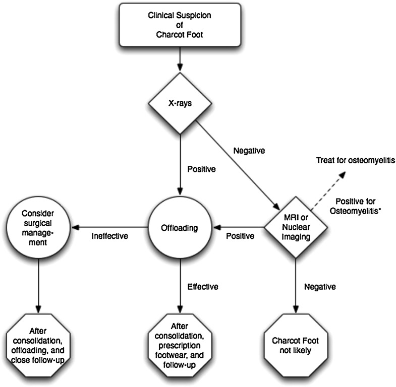
The Charcot foot syndrome is a complex complication of diabetes and neuropathy. Its destructive effects on the foot and ankle begin with a cycle of uncontrolled inflammation. The classic rocker-bottom foot deformity is a late stage of the syndrome and can be avoided by early recognition and management. Offloading is the most important initial treatment recommendation. Surgery can be helpful in early stages involving acute fractures of the foot or ankle or in later stages when offloading is ineffective. An algorithm summarizing the approach to the Charcot foot can be seen in Fig. 3.
Figure 3.
An algorithm depicting the basic approach to the Charcot foot. *Osteomyelitis can be difficult to distinguish from the Charcot foot. The reader is referred to the “Imaging of the Charcot foot” section of the article for techniques to improve specificity of various imaging modalities.
Acknowledgments
The American Diabetes Association/American Podiatric Medical Association Task Force meeting was supported by unrestricted educational grants from Small Bone Innovations, sanofi-aventis, and Integra LifeSciences. No other potential conflicts of interest relevant to this article were reported.
The authors thank Cécile Swiatek, chief librarian, from the J.-M. Charcot library at the Hospital Pitié-Salpêtrière for her assistance with the site preparation for the task force and references in this article.
References
- 1.Jeffcoate WJ, Game F, Cavanagh PR. The role of proinflammatory cytokines in the cause of neuropathic osteoarthropathy (acute Charcot foot) in diabetes. Lancet 2005;366:2058–2061 [DOI] [PubMed] [Google Scholar]
- 2.Boyce BF, Xing L. Functions of RANKL/RANK/OPG in bone modeling and remodeling. Arch Biochem Biophys 2008;473:139–146 [DOI] [PMC free article] [PubMed] [Google Scholar]
- 3.Uccioli L, Sinistro A, Almerighi C, et al. Proinflammatory modulation of the surface and cytokine phenotype of monocytes in patients with acute Charcot foot. Diabetes Care 2010;33:350–355 [DOI] [PMC free article] [PubMed] [Google Scholar]
- 4.Mabilleau G, Petrova NL, Edmonds ME, Sabokbar A. Increased osteoclastic activity in acute Charcot's osteoarthropathy: the role of receptor activator of nuclear factor-kappaB ligand. Diabetologia 2008;51:1035–1040 [DOI] [PMC free article] [PubMed] [Google Scholar]
- 5.Stevens MJ, Edmonds ME, Foster AV, Watkins PJ. Selective neuropathy and preserved vascular responses in the diabetic Charcot foot. Diabetologia 1992;35:148–154 [DOI] [PubMed] [Google Scholar]
- 6.Young MJ, Marshall A, Adams JE, Selby PL, Boulton AJ. Osteopenia, neurological dysfunction, and the development of Charcot neuroarthropathy. Diabetes Care 1995;18:34–38 [DOI] [PubMed] [Google Scholar]
- 7.Veves A, Akbari CM, Primavera J, et al. Endothelial dysfunction and the expression of endothelial nitric oxide synthetase in diabetic neuropathy, vascular disease, and foot ulceration. Diabetes 1998;47:457–463 [DOI] [PubMed] [Google Scholar]
- 8.Shapiro SA, Stansberry KB, Hill MA, et al. Normal blood flow response and vasomotion in the diabetic Charcot foot. J Diabetes Complications 1998;12:147–153 [DOI] [PubMed] [Google Scholar]
- 9.Baker N, Green A, Krishnan S, Rayman G. Microvascular and C-fiber function in diabetic Charcot neuroarthropathy and diabetic peripheral neuropathy. Diabetes Care 2007;30:3077–3079 [DOI] [PubMed] [Google Scholar]
- 10.Jeffcoate WJ, Game FL. New theories on the causes of the Charcot foot in diabetes. In The Diabetic Charcot Foot: Principles and Management. Frykberg RG, Ed. Brooklandville, MD, Data Trace Publishing Company, 2010, p. 29–44 [Google Scholar]
- 11.Herbst SA, Jones KB, Saltzman CL. Pattern of diabetic neuropathic arthropathy associated with the peripheral bone mineral density. J Bone Joint Surg Br 2004;86:378–383 [DOI] [PubMed] [Google Scholar]
- 12.Hofbauer LC, Brueck CC, Singh SK, Dobnig H. Osteoporosis in patients with diabetes mellitus. J Bone Miner Res 2007;22:1317–1328 [DOI] [PubMed] [Google Scholar]
- 13.Matricali GA, Bammens B, Kuypers D, Flour M, Mathieu C. High rate of Charcot foot attacks early after simultaneous pancreas-kidney transplantation. Transplantation 2007;83:245–246 [DOI] [PubMed] [Google Scholar]
- 14.Pitocco D, Zelano G, Gioffrè G, et al. Association between osteoprotegerin G1181C and T245G polymorphisms and diabetic Charcot neuroarthropathy: a case-control study. Diabetes Care 2009;32:1694–1697 [DOI] [PMC free article] [PubMed] [Google Scholar]
- 15.Foltz KD, Fallat LM, Schwartz S. Usefulness of a brief assessment battery for early detection of Charcot foot deformity in patients with diabetes. J Foot Ankle Surg 2004;43:87–92 [DOI] [PubMed] [Google Scholar]
- 16.Eichenholtz SN. Charcot Joints. Springfield, IL, Charles C. Thomas, 1966 [Google Scholar]
- 17.Charcot J-M, Fere C. Affections osseuses et articulaires du pied chez les tabétiques (Pied tabétique). Archives de Neurologie 1883;6:305–319 [in French] [Google Scholar]
- 18.Cofield RH, Morrison MJ, Beabout JW. Diabetic neuroarthropathy in the foot: patient characteristics and patterns of radiographic change. Foot Ankle 1983;4:15–22 [DOI] [PubMed] [Google Scholar]
- 19.Johnson JT. Neuropathic fractures and joint injuries. Pathogenesis and rationale of prevention and treatment. J Bone Joint Surg Am 1967;49:1–30 [PubMed] [Google Scholar]
- 20.Armstrong DG, Todd WF, Lavery LA, Harkless LB, Bushman TR. The natural history of acute Charcot's arthropathy in a diabetic foot specialty clinic. Diabet Med 1997;14:357–363 [DOI] [PubMed] [Google Scholar]
- 21.Henderson VE. Joint affections in tabes dorsalis. J Pathol 1905;10:211–264 [Google Scholar]
- 22.Caputo GM, Ulbrecht J, Cavanagh PR, Juliano P. The Charcot foot in diabetes: six key points. Am Fam Physician 1998;57:2705–2710 [PubMed] [Google Scholar]
- 23.Jeffcoate W. The causes of the Charcot syndrome. Clin Podiatr Med Surg 2008;25:29–42, vi [DOI] [PubMed] [Google Scholar]
- 24.McGill M, Molyneaux L, Bolton T, Ioannou K, Uren R, Yue DK. Response of Charcot's arthropathy to contact casting: assessment by quantitative techniques. Diabetologia 2000;43:481–484 [DOI] [PubMed] [Google Scholar]
- 25.Sinha S, Munichoodappa CS, Kozak GP. Neuro-arthropathy (Charcot joints) in diabetes mellitus (clinical study of 101 cases). Medicine (Baltimore) 1972;51:191–210 [DOI] [PubMed] [Google Scholar]
- 26.Sanders LJ, Frykberg RG. The Charcot foot (Pied de Charcot). In Levin and O'Neal's The Diabetic Foot. 7th ed Bowker JH, Pfeifer MA, Eds. Philadelphia, Mosby Elsevier, 2007, p. 257–283 [Google Scholar]
- 27.Morrison WB, Shortt CP, Ting AYI. Imaging of the Charcot foot. In The Diabetic Charcot Foot: Principles and Management. Frykberg RG, Ed. Brooklandville, MD, Data Trace Publishing Company, 2010, p. 65–84 [Google Scholar]
- 28.Morrison WB, Ledermann HP. Work-up of the diabetic foot. Radiol Clin North Am 2002;40:1171–1192 [DOI] [PubMed] [Google Scholar]
- 29.Morrison WB, Ledermann HP, Schweitzer ME. MR imaging of the diabetic foot. Magn Reson Imaging Clin N Am 2001;9:603–613, xi [PubMed] [Google Scholar]
- 30.Larcos G, Brown ML, Sutton RT. Diagnosis of osteomyelitis of the foot in diabetic patients: value of 111In-leukocyte scintigraphy. AJR Am J Roentgenol 1991;157:527–531 [DOI] [PubMed] [Google Scholar]
- 31.Palestro CJ, Mehta HH, Patel M, et al. Marrow versus infection in the Charcot joint: indium-111 leukocyte and technetium-99m sulfur colloid scintigraphy. J Nucl Med 1998;39:346–350 [PubMed] [Google Scholar]
- 32.Keidar Z, Militianu D, Melamed E, Bar-Shalom R, Israel O. The diabetic foot: initial experience with 18F-FDG PET/CT. J Nucl Med 2005;46:444–449 [PubMed] [Google Scholar]
- 33.Hopfner S, Krolak C, Kessler S, Tiling R. Preoperative imaging of Charcot neuroarthropathy. Does the additional application of 18F-FDG-PET make sense? Nuklearmedizin 2005;45:15-20 [DOI] [PubMed] [Google Scholar]
- 34.Frykberg RG, Eneroth M. Principles of conservative management. In The Diabetic Charcot Foot: Principles and Management. Frykberg RG, Ed. Brooklandville, MD, Data Trace Publishing Company, 2010, p. 93–116 [Google Scholar]
- 35.Armstrong DG, Lavery LA. Monitoring healing of acute Charcot's arthropathy with infrared dermal thermometry. J Rehabil Res Dev 1997;34:317–321 [PubMed] [Google Scholar]
- 36.Armstrong DG, Lavery LA, Wu S, Boulton AJ. Evaluation of removable and irremovable cast walkers in the healing of diabetic foot wounds: a randomized controlled trial. Diabetes Care 2005;28:551–554 [DOI] [PubMed] [Google Scholar]
- 37.Armstrong DG, Short B, Espensen EH, Abu-Rumman PL, Nixon BP, Boulton AJ. Technique for fabrication of an “instant total-contact cast” for treatment of neuropathic diabetic foot ulcers. J Am Podiatr Med Assoc 2002;92:405–408 [DOI] [PubMed] [Google Scholar]
- 38.Selby PL, Young MJ, Boulton AJ. Bisphosphonates: a new treatment for diabetic Charcot neuroarthropathy? Diabet Med 1994;11:28–31 [DOI] [PubMed] [Google Scholar]
- 39.Jude EB, Selby PL, Burgess J, et al. Bisphosphonates in the treatment of Charcot neuroarthropathy: a double-blind randomised controlled trial. Diabetologia 2001;44:2032–2037 [DOI] [PubMed] [Google Scholar]
- 40.Pitocco D, Ruotolo V, Caputo S, et al. Six-month treatment with alendronate in acute Charcot neuroarthropathy: a randomized controlled trial. Diabetes Care 2005;28:1214–1215 [DOI] [PubMed] [Google Scholar]
- 41.Anderson JJ, Woelffer KE, Holtzman JJ, Jacobs AM. Bisphosphonates for the treatment of Charcot neuroarthropathy. J Foot Ankle Surg 2004;43:285–289 [DOI] [PubMed] [Google Scholar]
- 42.Hofbauer LC, Hamann C, Ebeling PR. Approach to the patient with secondary osteoporosis. Eur J Endocrinol 2010;162:1009–1020 [DOI] [PubMed] [Google Scholar]
- 43.Bem R, Jirkovská A, Fejfarová V, Skibová J, Jude EB. Intranasal calcitonin in the treatment of acute Charcot neuroosteoarthropathy: a randomized controlled trial. Diabetes Care 2006;29:1392–1394 [DOI] [PubMed] [Google Scholar]
- 44.Molines L, Darmon P, Raccah D. Charcot's foot: newest findings on its pathophysiology, diagnosis and treatment. Diabetes Metab 2010;36:251–255 [DOI] [PubMed] [Google Scholar]
- 45.Ulbrecht JS, Wukich DK. The Charcot foot: medical and surgical therapy. Curr Diab Rep 2008;8:444–451 [DOI] [PubMed] [Google Scholar]
- 46.Wukich DK, Sung W. Charcot arthropathy of the foot and ankle: modern concepts and management review. J Diabetes Complications 2009;23:409–426 [DOI] [PubMed] [Google Scholar]
- 47.Strauss E, Gonya G. Adjunct low intensity ultrasound in Charcot neuroarthropathy. Clin Orthop Relat Res 1998;(349):132–138 [DOI] [PubMed] [Google Scholar]
- 48.Hockenbury RT, Gruttadauria M, McKinney I. Use of implantable bone growth stimulation in Charcot ankle arthrodesis. Foot Ankle Int 2007;28:971–976 [DOI] [PubMed] [Google Scholar]
- 49.Petrisor B, Lau JT. Electrical bone stimulation: an overview and its use in high risk and Charcot foot and ankle reconstructions. Foot Ankle Clin 2005;10:609–620, vii–viii [DOI] [PubMed] [Google Scholar]
- 50.Pinzur MS. Surgical management: history and general principles. In The Diabetic Charcot Foot: Principles and Management. Frykberg RG, Ed. Brooklandville, MD, Data Trace Publishing Company, 2010, p. 165–188 [Google Scholar]
- 51.Dhawan V, Spratt KF, Pinzur MS, Baumhauer J, Rudicel S, Saltzman CL. Reliability of AOFAS diabetic foot questionnaire in Charcot arthropathy: stability, internal consistency, and measurable difference. Foot Ankle Int 2005;26:717–731 [DOI] [PubMed] [Google Scholar]
- 52.Lavery LA, Armstrong DG, Boulton AJ; Diabetex Research Group. Ankle equinus deformity and its relationship to high plantar pressure in a large population with diabetes mellitus. J Am Podiatr Med Assoc 2002;92:479–482 [DOI] [PubMed] [Google Scholar]
- 53.Armstrong DG, Lavery LA. Elevated peak plantar pressures in patients who have Charcot arthropathy. J Bone Joint Surg Am 1998;80:365–369 [DOI] [PubMed] [Google Scholar]
- 54.Hastings MK, Mueller MJ, Sinacore DR, Salsich GB, Engsberg JR, Johnson JE. Effects of a tendo-Achilles lengthening procedure on muscle function and gait characteristics in a patient with diabetes mellitus. J Orthop Sports Phys Ther 2000;30:85–90 [DOI] [PubMed] [Google Scholar]
- 55.Maluf KS, Mueller MJ, Strube MJ, Engsberg JR, Johnson JE. Tendon Achilles lengthening for the treatment of neuropathic ulcers causes a temporary reduction in forefoot pressure associated with changes in plantar flexor power rather than ankle motion during gait. J Biomech 2004;37:897–906 [DOI] [PubMed] [Google Scholar]
- 56.Holstein P, Lohmann M, Bitsch M, Jørgensen B. Achilles tendon lengthening, the panacea for plantar forefoot ulceration? Diabetes Metab Res Rev 2004;20(Suppl. 1):S37–S40 [DOI] [PubMed] [Google Scholar]
- 57.Mueller MJ, Sinacore DR, Hastings MK, Lott DJ, Strube MJ, Johnson JE. Impact of Achilles tendon lengthening on functional limitations and perceived disability in people with a neuropathic plantar ulcer. Diabetes Care 2004;27:1559–1564 [DOI] [PubMed] [Google Scholar]
- 58.Catanzariti AR, Mendicino R, Haverstock B. Ostectomy for diabetic neuroarthropathy involving the midfoot. J Foot Ankle Surg 2000;39:291–300 [DOI] [PubMed] [Google Scholar]
- 59.Rosenblum BI, Giurini JM, Miller LB, Chrzan JS, Habershaw GM. Neuropathic ulcerations plantar to the lateral column in patients with Charcot foot deformity: a flexible approach to limb salvage. J Foot Ankle Surg 1997;36:360–363 [DOI] [PubMed] [Google Scholar]
- 60.Laurinaviciene R, Kirketerp-Moeller K, Holstein PE. Exostectomy for chronic midfoot plantar ulcer in Charcot deformity. J Wound Care 2008;17:53–55, 57–58 [DOI] [PubMed] [Google Scholar]
- 61.Mittlmeier T, Klaue K, Haar P, Beck M. Should one consider primary surgical reconstruction in Charcot arthropathy of the feet? Clin Orthop Relat Res 2010;468:1002–1011 [DOI] [PMC free article] [PubMed] [Google Scholar]
- 62.Simon SR, Tejwani SG, Wilson DL, Santner TJ, Denniston NL. Arthrodesis as an early alternative to nonoperative management of Charcot arthropathy of the diabetic foot. J Bone Joint Surg Am 2000;82-A:939–950 [DOI] [PubMed] [Google Scholar]
- 63.Papa J, Myerson M, Girard P. Salvage, with arthrodesis, in intractable diabetic neuropathic arthropathy of the foot and ankle. J Bone Joint Surg Am 1993;75:1056–1066 [DOI] [PubMed] [Google Scholar]
- 64.Pakarinen TK, Laine HJ, Honkonen SE, Peltonen J, Oksala H, Lahtela J. Charcot arthropathy of the diabetic foot. Current concepts and review of 36 cases. Scand J Surg 2002;91:195–201 [DOI] [PubMed] [Google Scholar]
- 65.Stone NC, Daniels TR. Midfoot and hindfoot arthrodeses in diabetic Charcot arthropathy. Can J Surg 2000;43:449–455 [PMC free article] [PubMed] [Google Scholar]
- 66.Pinzur M. Surgical versus accommodative treatment for Charcot arthropathy of the midfoot. Foot Ankle Int 2004;25:545–549 [DOI] [PubMed] [Google Scholar]
- 67.Garapati R, Weinfeld SB. Complex reconstruction of the diabetic foot and ankle. Am J Surg 2004;187(5A):81S–86S [DOI] [PubMed] [Google Scholar]
- 68.Assal M, Stern R. Realignment and extended fusion with use of a medial column screw for midfoot deformities secondary to diabetic neuropathy. J Bone Joint Surg Am 2009;91:812–820 [DOI] [PubMed] [Google Scholar]
- 69.Pinzur MS. Neutral ring fixation for high-risk nonplantigrade Charcot midfoot deformity. Foot Ankle Int 2007;28:961–966 [DOI] [PubMed] [Google Scholar]
- 70.Farber DC, Juliano PJ, Cavanagh PR, Ulbrecht J, Caputo G. Single stage correction with external fixation of the ulcerated foot in individuals with Charcot neuroarthropathy. Foot Ankle Int 2002;23:130–134 [DOI] [PubMed] [Google Scholar]
- 71.Fabrin J, Larsen K, Holstein PE. Arthrodesis with external fixation in the unstable or misaligned Charcot ankle in patients with diabetes mellitus. Int J Low Extrem Wounds 2007;6:102–107 [DOI] [PubMed] [Google Scholar]
- 72.Wukich DK, Belczyk RJ, Burns PR, Frykberg RG. Complications encountered with circular ring fixation in persons with diabetes mellitus. Foot Ankle Int 2008;29:994–1000 [DOI] [PubMed] [Google Scholar]
- 73.Bevilacqua NJ, Rogers LC. Surgical management of Charcot midfoot deformities. Clin Podiatr Med Surg 2008;25:81–94, vii [DOI] [PubMed] [Google Scholar]
- 74.Rogers LC, Bevilacqua NJ, Frykberg RG, Armstrong DG. Predictors of postoperative complications of Ilizarov external ring fixators in the foot and ankle. J Foot Ankle Surg 2007;46:372–375 [DOI] [PubMed] [Google Scholar]
- 75.Connolly JF, Csencsitz TA. Limb threatening neuropathic complications from ankle fractures in patients with diabetes. Clin Orthop Relat Res 1998;(348):212–219 [PubMed] [Google Scholar]
- 76.Jani MM, Ricci WM, Borrelli J, Jr, Barrett SE, Johnson JE. A protocol for treatment of unstable ankle fractures using transarticular fixation in patients with diabetes mellitus and loss of protective sensibility. Foot Ankle Int 2003;24:838–844 [DOI] [PubMed] [Google Scholar]
- 77.Perry MD, Taranow WS, Manoli A, 2nd, Carr JB. Salvage of failed neuropathic ankle fractures: use of large-fragment fibular plating and multiple syndesmotic screws. J Surg Orthop Adv 2005;14:85–91 [PubMed] [Google Scholar]
- 78.Wukich DK, Joseph A, Ryan M, Ramirez C, Irrgang JJ. Outcomes of ankle fractures in patients with uncomplicated versus complicated diabetes. Foot Ankle Int 2011;32:120–130 [DOI] [PubMed] [Google Scholar]
- 79.Ayoub MA. Ankle fractures in diabetic neuropathic arthropathy: can tibiotalar arthrodesis salvage the limb? J Bone Joint Surg Br 2008;90:906–914 [DOI] [PubMed] [Google Scholar]
- 80.Dalla Paola L, Volpe A, Varotto D, et al. Use of a retrograde nail for ankle arthrodesis in Charcot neuroarthropathy: a limb salvage procedure. Foot Ankle Int 2007;28:967–970 [DOI] [PubMed] [Google Scholar]
- 81.Caravaggi C, Cimmino M, Caruso S, Dalla Noce S. Intramedullary compressive nail fixation for the treatment of severe Charcot deformity of the ankle and rear foot. J Foot Ankle Surg 2006;45:20–24 [DOI] [PubMed] [Google Scholar]
- 82.Pinzur MS, Noonan T. Ankle arthrodesis with a retrograde femoral nail for Charcot ankle arthropathy. Foot Ankle Int 2005;26:545–549 [DOI] [PubMed] [Google Scholar]


