Abstract
Blunt neck trauma with an associated laryngotracheal injury is rare. We report a patient with blunt neck trauma who came to the emergency room and was sent to ward without realizing the seriousness of the situation. He presented later with respiratory distress and an anesthesiologist was called in for emergency airway management. Airway management in such a situation is described in this report.
Keywords: Airway management, blunt neck trauma, blunt tracheal trauma, pneumomediastinum, pneumothorax
Introduction
Blunt and penetrating trauma to the neck can result in acute or overt airway compromise with life-threatening implications. Acute isolated laryngotracheal trauma is uncommon (incidence 0.03%) and therefore early assessment, recognition, and prompt appropriate management are of paramount importance to reduce morbidity and mortality.[1] In this report, we describe how the delay in diagnosis contributed to significant morbidity in a case of blunt tracheal trauma.
Case Report
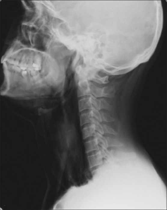
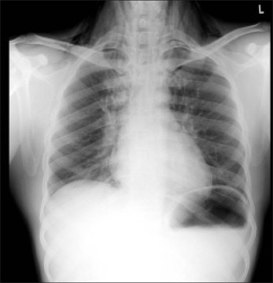
A 40-year-old man sustained a blunt injury to his neck on falling off a stool. He presented soon after the incident in the emergency room with swelling of the face and neck with the change in voice. He was not comfortable lying down and preferred to sit up. There was no evidence of external injury to the neck. He was hemodynamically stable and maintaining oxygen saturation (SpO2) of 96-98% on room air. The anterior-posterior and lateral X-rays of the neck showed a rightward deviation of the trachea, evidence of soft tissue injury, and surgical emphysema [Figure 1]. The chest X-ray did not reveal any evidence of pneumothorax [Figure 2]. A computerized tomography scan of the neck could not be done as he was unable to lie down.
Figure 1.

The anterior–posterior and lateral X-rays of the neck showing rightward deviation of the trachea, evidence of soft tissue injury, and surgical emphysema
Figure 2.

Chest X--ray: A–P view, normal
He was evaluated by the ENT resident who admitted him for observation in the general ward. Six hours later, the patient complained of difficulty in breathing and worsening of the surgical emphysema of the face and neck was noted. The SpO2 was 95% despite oxygen supplementation using a venture face mask set at FiO2 0.6. He was hemodynamically stable, alert, and talking. The anesthesiologist on call was requested to evaluate the patient. He immediately recognized the gravity of the situation and the possibility of blunt tracheal injury and decided that any further assessment and airway management was to be performed in the operating room (OR). The patient was shifted to the OR in the upright position on oxygen and a senior anesthesiologist was called in to help. The anesthesiologist's plan was to perform awake fiberoptic intubation or tracheostomy followed by an endoscopic evaluation of the extent of the tracheal injury.
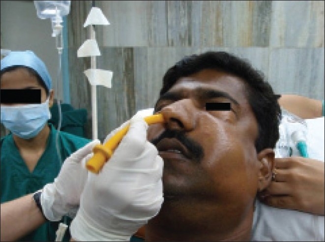
In the OR, with the patient supported with pillows in the sitting position [Figure 3], the airway was anesthetized with 4% lignocaine aerosol using a nebulizer, lignocaine viscous gargles and 1% lignocaine, 10 mL, injected slowly at the base of the tongue by the drip technique. Nasal pledgets soaked in 4% lignocaine and oxymetazoline drops were applied. Oxygen was supplemented initially using a face mask and later through the fiberoptic bronchoscope. He remained hemodynamically stable during this period. Awake nasal fibreoptic intubation was performed with ease. A blood-tinged streak/tear was noted in the anterior subglottic region, a 7.0-mm ID endotracheal tube was passed beyond the tear and the cuff inflated. In view of the patient's condition and the likelihood of further deterioration, the need to evaluate the tracheal tear and perform tracheostomy was discussed with the ENT surgeons. They opined to wait and observe the patient in the intensive care unit (ICU).
Figure 3.

Patient positioned in a sitting position with pillows under the neck
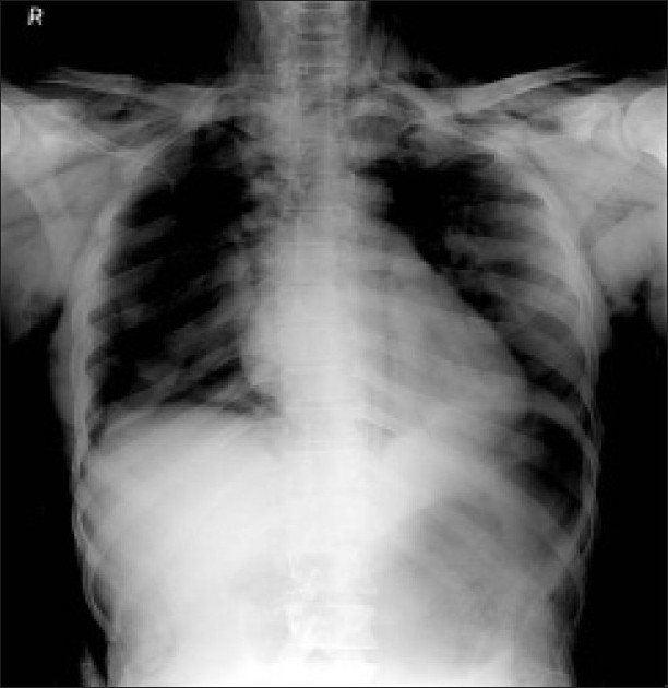
In the ICU, his condition rapidly deteriorated, surgical emphysema worsened (extended to the entire face, neck, chest wall, and upper limbs up to the fingers), and respiratory distress developed with SpO2 falling to 80%. Bilateral pneumothorax was now evident on the chest X-ray [Figure 4]. Bilateral intercostal drainage tubes were inserted and the ENT surgeons decided to perform a tracheostomy in the OR. A fiberoptic bronchoscope was passed via a swivel connector and oxygen administered with a breathing circuit to evaluate the tracheal trauma. The tip of the tracheal tube and Murphy's eye were found to be completely obstructed by edematous tracheal mucosa. The tracheostomy was performed under local infiltration supplemented by incremental doses of midazolam and propofol. Sevoflurane in 100% oxygen on spontaneous respiration was also administered.
Figure 4.

Chest X-ray showing bilateral pneumothorax
Hypopharyngoscopy and microlaryngoscopy following the tracheostomy revealed gross edema of the ventricular bands, vocal cords, and a large left ventricular air cyst. An anterior subglottic mucosal laceration with the displacement of the cricoid cartilage was visualized. No further surgical intervention was done. The patient was observed in the ICU for 24 h and the surgical emphysema dramatically reduced. Two weeks later, he was evaluated under anesthesia and the tracheal tear was found to have healed well. The tracheostomy was decannulated prior to discharge.
Discussion
Laryngotracheal injuries are uncommon and can be very deceptive. Serious injury to laryngotracheal anatomy could exist even in the absence of any visible external injury. Hoarseness of voice, pain, dyspnea, dysphagia, subcutaneous emphysema and hemoptysis are some of the clinical signs and symptoms which point toward an internal injury to the airway.[2] It is not uncommon for a laryngeal fracture to be missed initially.[2] In the case reported, blunt trauma to the neck, change in voice, and inability to lie down should have alerted the primary physicians of the possibility of a significant injury and he should have been observed in a high dependency unit after admission to the hospital. Ideally, an evaluation of the extent of injury should have been performed immediately.[3]
Once the anesthesiologist had been informed, they were cautious about “not attempting any airway management” in the ward. It was important to have an experienced anesthesiologist present for further management.[3] In the OR, after securing the airway, the extent of the injury should have been evaluated and tracheostomy performed if required, but this decision is not a very easy to take.[2] If the internal injury is minor, tracheostomy can possibly be avoided and conservative management would suffice. In the presence of massive surgical emphysema, we should have had a high index of suspicion for pneumomediastinum and perhaps inserted intercostal tubes prophylactically to avoid its progression to pneumothorax.
To conclude, laryngeal trauma even in the absence of signs of an external injury can be life threatening. Prompt diagnosis of such injuries requires a high index of suspicion. Securing the airway early in an appropriate setting is crucial and a top priority.
Footnotes
Source of Support: Nil
Conflict of Interest: None declared.
References
- 1.Cicala RS, Kudsk KA, Butts A, Nguyen H, Fabian TC. Initial evaluation and management of upper airway injuries in trauma patients. J Clin Anesth. 1991;3:91–8. doi: 10.1016/0952-8180(91)90003-6. [DOI] [PubMed] [Google Scholar]
- 2.Hwang SY, Yeak SC. Management dilemmas in laryngeal trauma. J Laryngol Otol. 2004;118:325–8. doi: 10.1258/002221504323086471. [DOI] [PubMed] [Google Scholar]
- 3.Pierre EJ, McNeer RR, Shamir MY. Early management of the traumatized airway. Anesthesiol Clin. 2007;25:1–11. doi: 10.1016/j.anclin.2006.11.001. [DOI] [PubMed] [Google Scholar]


