Abstract
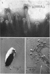
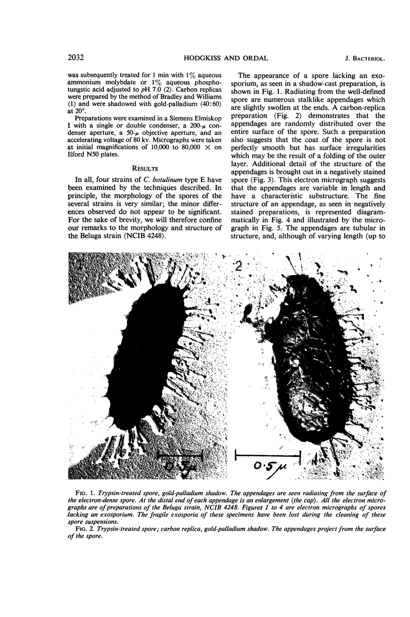
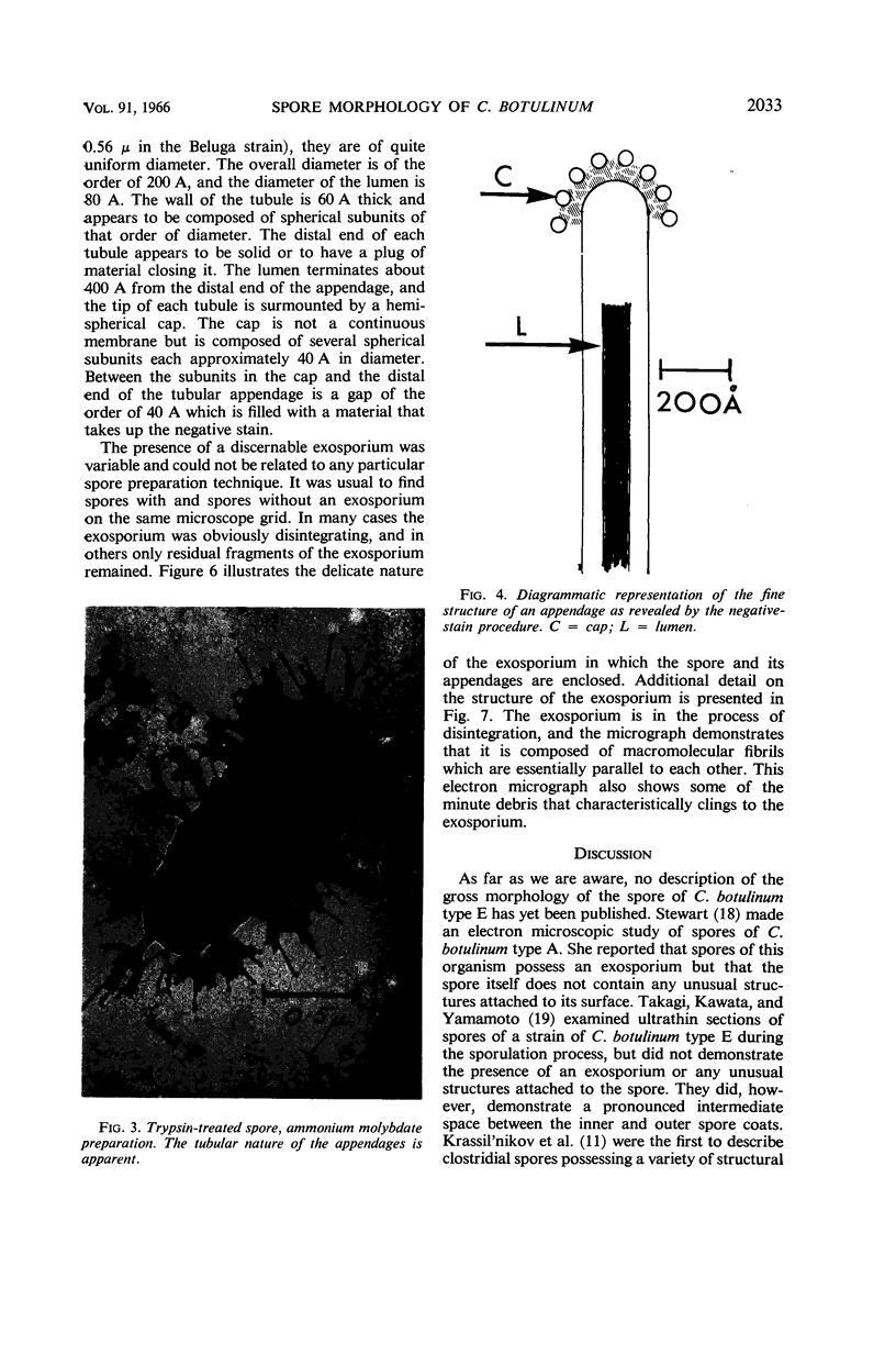
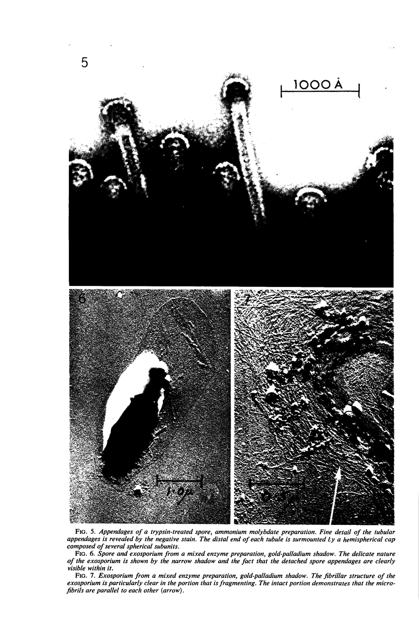
Hodgkiss, W. (Torry Research Station, Aberdeen, Scotland), and Z. John Ordal. Morphology of the spore of some strains of Clostridium botulinum type E. J. Bacteriol. 91:2031–2036. 1966.—The spores of four strains of C. botulinum type E show an unusual and elaborate morphology. Numerous tubular appendages radiate from the surface of the spore. The spore and its appendages are enclosed in a delicate exosporium. The electron microscopic morphology of the spores, as seen in metal-shadowed and negatively stained preparations, and by the carbon-replica technique, is described.
Full text
PDF
Images in this article
Selected References
These references are in PubMed. This may not be the complete list of references from this article.
- ANINGA S., VAN DEN HOOFF A. An electron microscope study on the shape of the spores of Bacillus polymyxa. Antonie Van Leeuwenhoek. 1956;22(4):327–330. doi: 10.1007/BF02538345. [DOI] [PubMed] [Google Scholar]
- BRADLEY D. E., WILLIAMS D. J. An electron microscope study of the spores of some species of the genus Bacillus using carbon replicas. J Gen Microbiol. 1957 Aug;17(1):75–79. doi: 10.1099/00221287-17-1-75. [DOI] [PubMed] [Google Scholar]
- BRENNER S., HORNE R. W. A negative staining method for high resolution electron microscopy of viruses. Biochim Biophys Acta. 1959 Jul;34:103–110. doi: 10.1016/0006-3002(59)90237-9. [DOI] [PubMed] [Google Scholar]
- Brinton C. C., Jr The structure, function, synthesis and genetic control of bacterial pili and a molecular model for DNA and RNA transport in gram negative bacteria. Trans N Y Acad Sci. 1965 Jun;27(8):1003–1054. doi: 10.1111/j.2164-0947.1965.tb02342.x. [DOI] [PubMed] [Google Scholar]
- DUGUID J. P. Fimbriae and adhesive properties in Klebsiella strains. J Gen Microbiol. 1959 Aug;21:271–286. doi: 10.1099/00221287-21-1-271. [DOI] [PubMed] [Google Scholar]
- DUGUID J. P., SMITH I. W., DEMPSTER G., EDMUNDS P. N. Non-flagellar filamentous appendages (fimbriae) and haemagglutinating activity in Bacterium coli. J Pathol Bacteriol. 1955 Oct;70(2):335–348. doi: 10.1002/path.1700700210. [DOI] [PubMed] [Google Scholar]
- GERHARDT P., RIBI E. ULTRASTRUCTURE OF THE EXOSPORIUM ENVELOPING SPORES OF BACILLUS CEREUS. J Bacteriol. 1964 Dec;88:1774–1789. doi: 10.1128/jb.88.6.1774-1789.1964. [DOI] [PMC free article] [PubMed] [Google Scholar]
- HOUWINK A. L., van ITERSON W. Electron microscopical observations on bacterial cytology; a study on flagellation. Biochim Biophys Acta. 1950 Mar;5(1):10–44. doi: 10.1016/0006-3002(50)90144-2. [DOI] [PubMed] [Google Scholar]
- MANTON I. Observations on the microanatomy of the spermatozoid of the bracken fern (Pteridium aquilinum). J Biophys Biochem Cytol. 1959 Dec;6:413–418. doi: 10.1083/jcb.6.3.413. [DOI] [PMC free article] [PubMed] [Google Scholar]
- TAKAGI A., KAWATA T., YAMAMOTO S. Electron microscope studies on ultrathin sections of spores of the Clostridium group, with special reference to the sporulation and germination process. J Bacteriol. 1960 Jul;80:37–46. doi: 10.1128/jb.80.1.37-46.1960. [DOI] [PMC free article] [PubMed] [Google Scholar]
- THORNLEY M. J., HORNE R. W. Electron microscope observations on the structure of fimbriae, with particular reference to Klebsiella strains, by the use of the negative staining technique. J Gen Microbiol. 1962 Apr;28:51–56. doi: 10.1099/00221287-28-1-51. [DOI] [PubMed] [Google Scholar]
- TRESNER H. D., DAVIES M. C., BACKUS E. J. Electron microscopy of Streptomyces spore morphology and its role in species differentiation. J Bacteriol. 1961 Jan;81:70–80. doi: 10.1128/jb.81.1.70-80.1961. [DOI] [PMC free article] [PubMed] [Google Scholar]


