

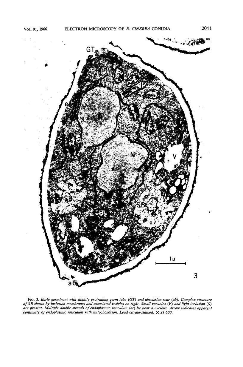
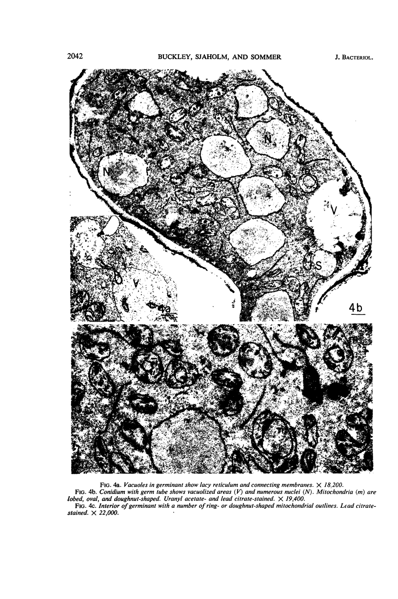
Abstract
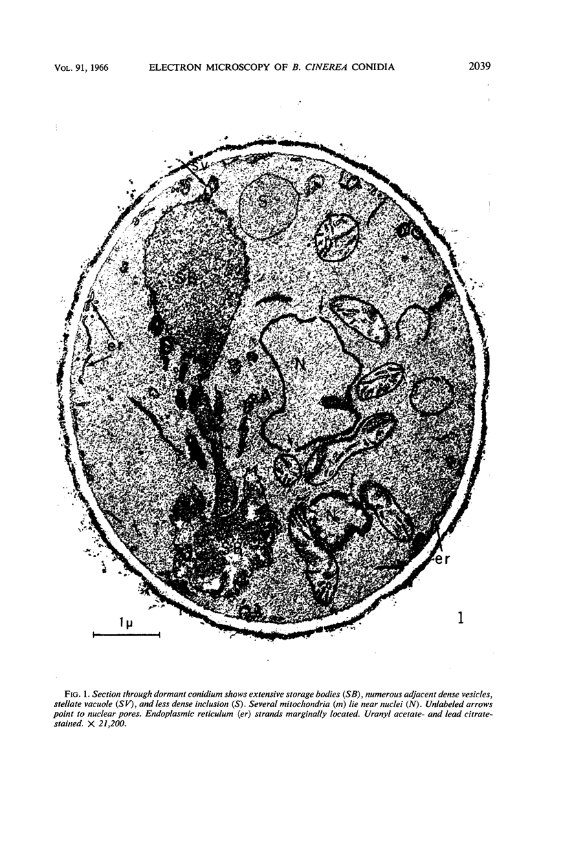
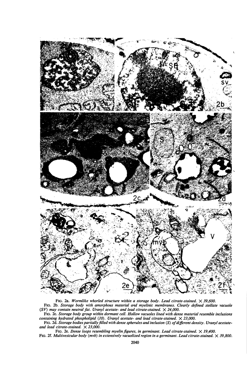
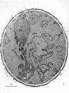
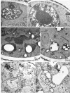
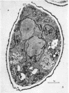
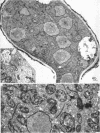
Buckley, Patricia M. (University of California, Davis), Virginia E. Sjaholm, and N. F. Sommer. Electron microscopy of Botrytis cinerea conidia. J. Bacteriol. 91:2037–2044. 1966.—Sections of germinating and nongerminating Botrytis cinerea conidia were examined with an electron microscope. Uranyl acetate or lead citrate provided contrast between membranes and cytoplasm. Membrane-bounded, dense inclusions previously unreported in dormant spores were termed “storage bodies.” Whorled structures, spherules, granules, and membrane loops were seen within these inclusions. The various forms assumed by the enclosed materials closely resemble phospholipid inclusions described for other cells. It is suggested that the inclusions provide material for the assembly of membranous organelles during germination. Utilization of the stored material apparently results in extensive vacuolization in advanced germinants.
Full text
PDF
Images in this article
Selected References
These references are in PubMed. This may not be the complete list of references from this article.
- CHRISTENSEN A. K., CHAPMAN G. B. Cup-shaped mitochondria in interstitial cells of the albino rat testis. Exp Cell Res. 1959 Nov;18:576–579. doi: 10.1016/0014-4827(59)90323-4. [DOI] [PubMed] [Google Scholar]
- HAWKER L. E., HENDY R. J. AN ELECTRON-MICROSCOPE STUDY OF GERMINATION OF CONIDIA OF BOTRYTIS CINEREA. J Gen Microbiol. 1963 Oct;33:43–46. doi: 10.1099/00221287-33-1-43. [DOI] [PubMed] [Google Scholar]
- Hohl H. R. Nature and Development of Membrane Systems in Food Vacuoles of Cellular Slime Molds Predatory upon Bacteria. J Bacteriol. 1965 Sep;90(3):755–765. doi: 10.1128/jb.90.3.755-765.1965. [DOI] [PMC free article] [PubMed] [Google Scholar]
- LARSON D. A. FINE-STRUCTURAL CHANGES IN THE CYTOPLASM OF GERMINATING POLLEN. Am J Bot. 1965 Feb;52:139–154. [PubMed] [Google Scholar]
- NECAS O., HAVELKOVA M., SOUDEK D. SUBMICROSCOPIC MORPHOLOGY OF RHIZOPUS NIGRICANS. Folia Microbiol (Praha) 1963 Sep;40:290–292. doi: 10.1007/BF02868772. [DOI] [PubMed] [Google Scholar]
- REVEL J. P., ITO S., FAWCETT D. W. Electron micrographs of myelin figures of phospholipide simulating intracellular membranes. J Biophys Biochem Cytol. 1958 Jul 25;4(4):495–498. doi: 10.1083/jcb.4.4.495. [DOI] [PMC free article] [PubMed] [Google Scholar]
- RICHARDSON S. H., HULTIN H. O., GREEN D. E. STRUCTURAL PROTEINS OF MEMBRANE SYSTEMS. Proc Natl Acad Sci U S A. 1963 Nov;50:821–827. doi: 10.1073/pnas.50.5.821. [DOI] [PMC free article] [PubMed] [Google Scholar]
- SOMMER N. F., CREASY M., ROMANI R. J., MAXIE E. C. AN OXYGEN-DEPENDENT POSTIRRADIATION RESTORATION OF RHIZOPUS STOLONIFER SPORANGIOSPORES. Radiat Res. 1964 May;22:21–28. [PubMed] [Google Scholar]
- STOECKENIUS W. An electron microscope study of myelin figures. J Biophys Biochem Cytol. 1959 May 25;5(3):491–500. doi: 10.1083/jcb.5.3.491. [DOI] [PMC free article] [PubMed] [Google Scholar]