

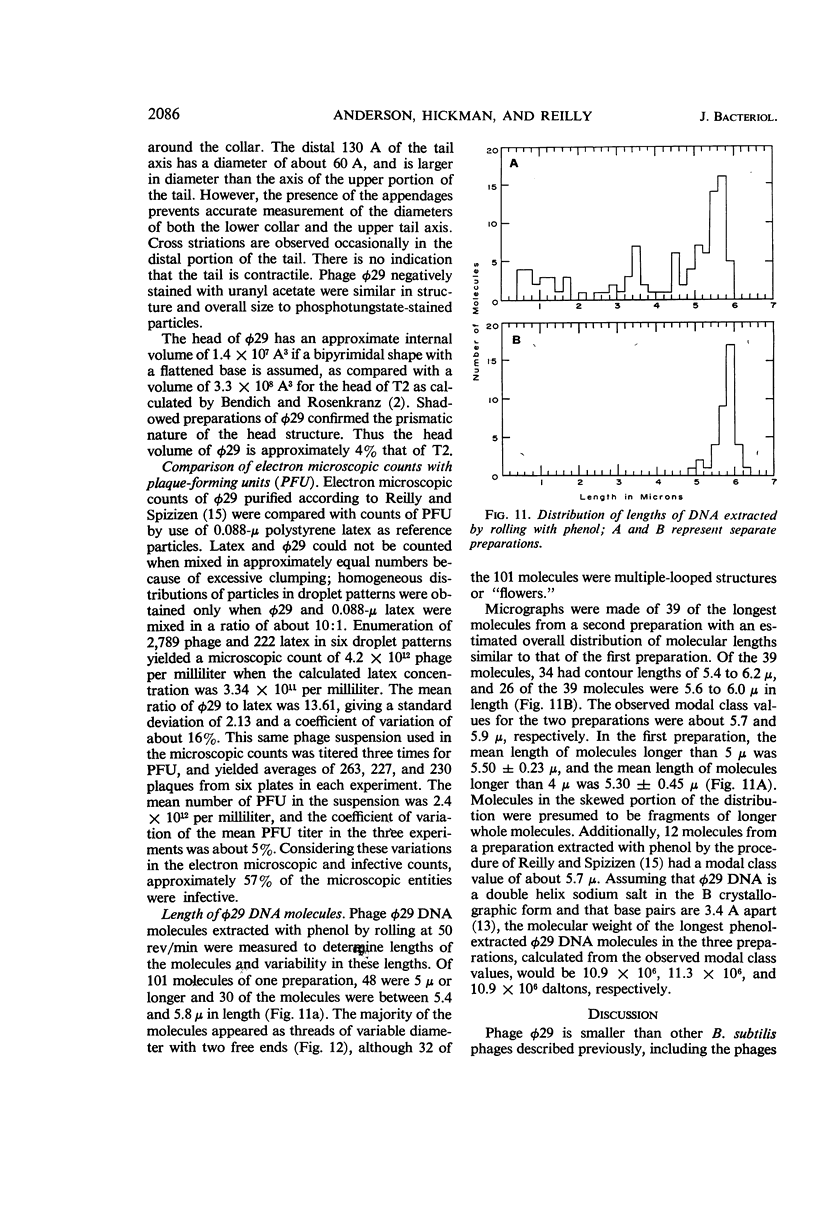
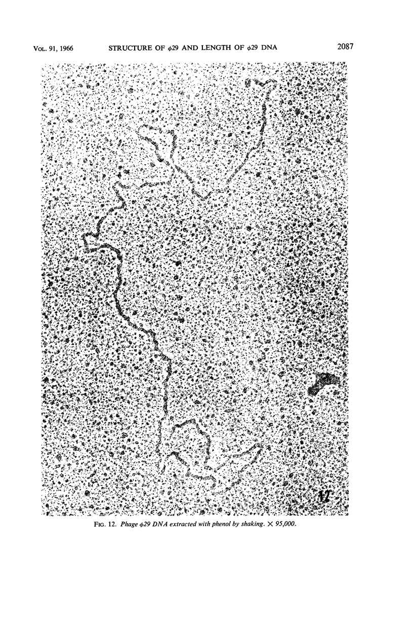
Abstract
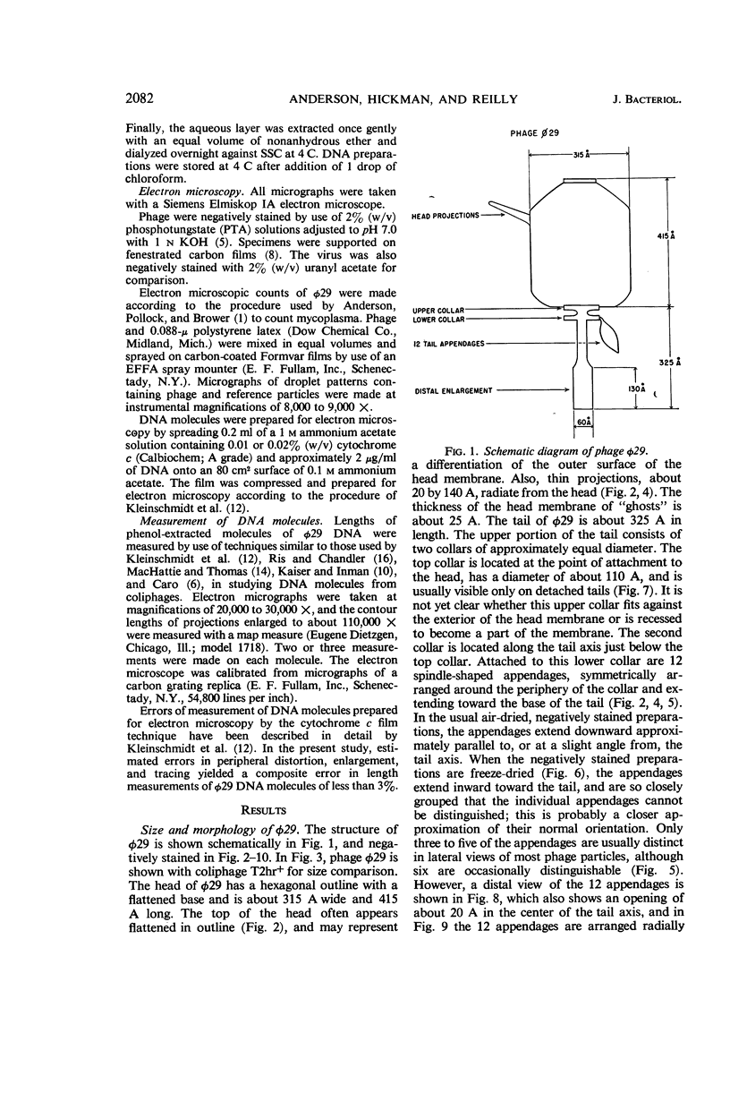
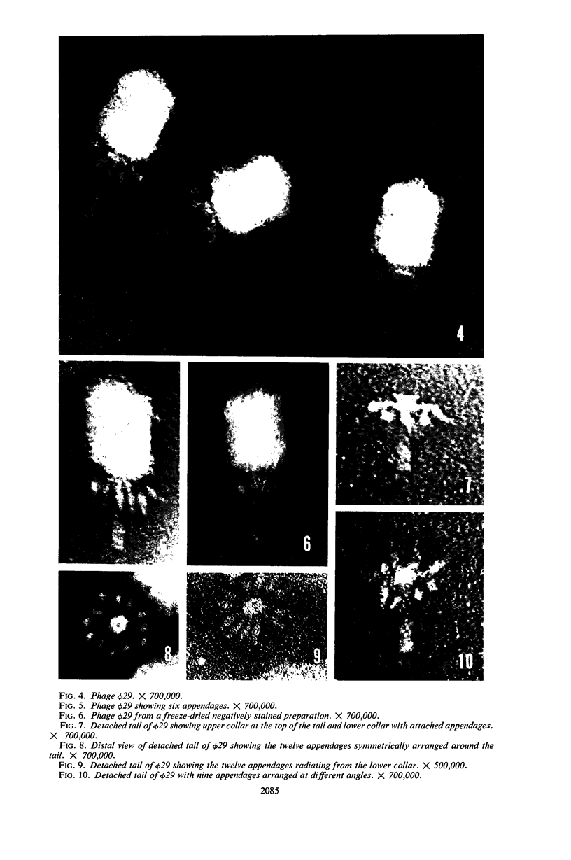
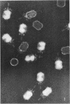
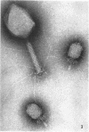
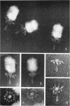
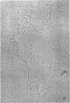
Anderson, D. L. (University of Minnesota, Minneapolis), D. D. Hickman, and B. E. Reilly. Structure of Bacillus subtilis bacteriophage φ29 and the length of φ29 deoxyribonucleic acid. J. Bacteriol. 91:2081–2089. 1966—Bacillus subtilis bacteriophage φ29 were negatively stained with phosphotungstic acid. The head of φ29 has a hexagonal outline with a flattened base, and is about 315 A wide and 415 A in length. The virus has an intricate tail about 325 A in length. Twelve spindle-shaped appendages are attached to the lower of two collars which comprise the proximal portion of the tail. The distal 130 A of the tail axis has a diameter of about 60 A and is larger in diameter than the axis of the upper portion of the tail. Comparison of electron microscopic counts of φ29 with plaque-forming units indicated that about 50% of the microscopic entities were infective. Phenol-extracted φ29 deoxyribonucleic acid (DNA) molecules were prepared for electron microscopy by the cytochrome c film technique of Kleinschmidt et al. Measurement of contour lengths of DNA molecules from three preparations gave skewed distributions of lengths with observed modal class values ranging from 5.7 to 5.9 μ. Assuming that φ29 DNA is a double helix in the B form, the corresponding molecular weights would be 10.9 × 10⁶ to 11.3 × 10⁶ daltons. The largest DNA molecules would have a volume of 1.9 × 10⁷ A3 which is about 25% greater than the estimated 1.4 × 10⁷ A3 internal volume of the phage head.
Full text
PDF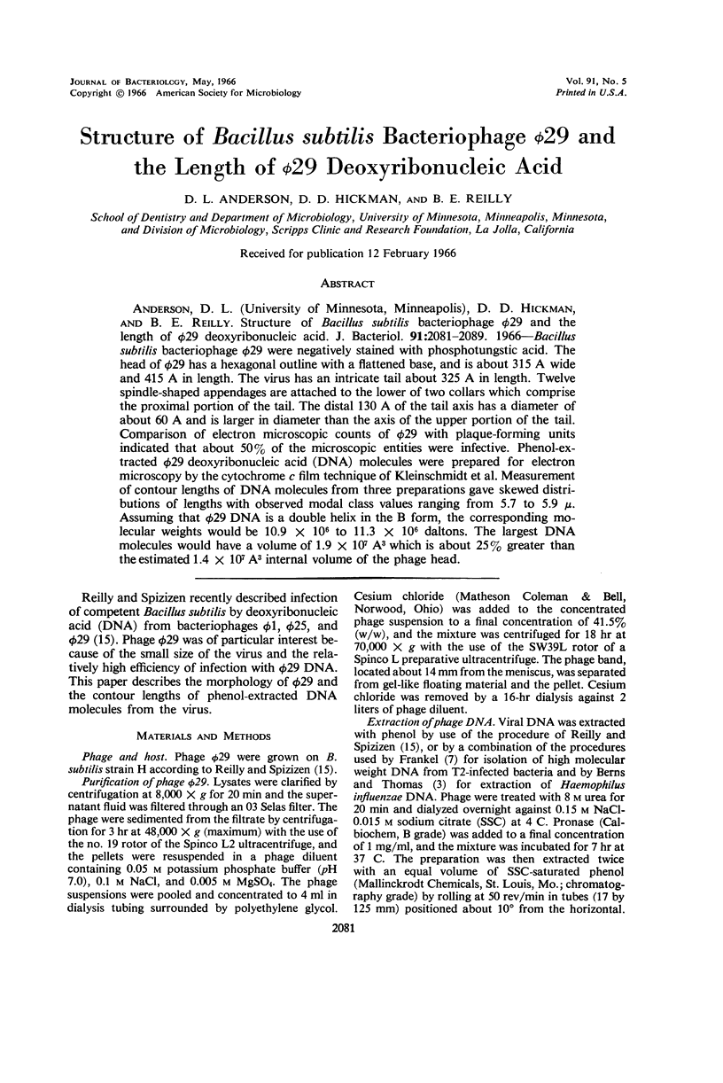

Images in this article
Selected References
These references are in PubMed. This may not be the complete list of references from this article.
- Anderson D. L., Pollock M. E., Brower L. F. Morphology of Mycoplasma laidlawii type A. I. Comparison of electron microscopic counts with colony-forming units. J Bacteriol. 1965 Dec;90(6):1764–1767. doi: 10.1128/jb.90.6.1764-1767.1965. [DOI] [PMC free article] [PubMed] [Google Scholar]
- BERNS K. I., THOMAS C. A., Jr ISOLATION OF HIGH MOLECULAR WEIGHT DNA FROM HEMOPHILUS INFLUENZAE. J Mol Biol. 1965 Mar;11:476–490. doi: 10.1016/s0022-2836(65)80004-3. [DOI] [PubMed] [Google Scholar]
- BRENNER S., HORNE R. W. A negative staining method for high resolution electron microscopy of viruses. Biochim Biophys Acta. 1959 Jul;34:103–110. doi: 10.1016/0006-3002(59)90237-9. [DOI] [PubMed] [Google Scholar]
- Bradley D. E. The isolation and morphology of some new bacteriophages specific for Bacillus and Acetobacter species. J Gen Microbiol. 1965 Nov;41(2):233–241. doi: 10.1099/00221287-41-2-233. [DOI] [PubMed] [Google Scholar]
- CARO L. G. THE MOLECULAR WEIGHT OF LAMBDA DNA. Virology. 1965 Feb;25:226–236. doi: 10.1016/0042-6822(65)90201-1. [DOI] [PubMed] [Google Scholar]
- FRANKEL F. R. An unusual DNA extracted from bacteria infected with phage T2. Proc Natl Acad Sci U S A. 1963 Mar 15;49:366–372. doi: 10.1073/pnas.49.3.366. [DOI] [PMC free article] [PubMed] [Google Scholar]
- IONESCO H., RYTER A., SCHAEFFER P. SUR UN BACT'ERIOPHAGE H'EBERG'E PAR LA SOUCHE MARBURG DE BACILLUS SUBTILIS. Ann Inst Pasteur (Paris) 1964 Dec;107:764–776. [PubMed] [Google Scholar]
- KLEINSCHMIDT A. K., LANG D., JACHERTS D., ZAHN R. K. [Preparation and length measurements of the total desoxyribonucleic acid content of T2 bacteriophages]. Biochim Biophys Acta. 1962 Dec 31;61:857–864. [PubMed] [Google Scholar]
- Kaiser A. D., Inman R. B. Cohesion and the biological activity of bacteriophage lambda DNA. J Mol Biol. 1965 Aug;13(1):78–91. doi: 10.1016/s0022-2836(65)80081-x. [DOI] [PubMed] [Google Scholar]
- MACHATTIE L. A., THOMAS C. A., Jr DNA FROM BACTERIOPHAGE LAMBDA: MOLECULAR LENGTH AND CONFORMATION. Science. 1964 May 29;144(3622):1142–1144. doi: 10.1126/science.144.3622.1142. [DOI] [PubMed] [Google Scholar]
- REILLY B. E., SPIZIZEN J. BACTERIOPHAGE DEOXYRIBONUCLEATE INFECTION OF COMPETENT BACILLUS SUBTILIS. J Bacteriol. 1965 Mar;89:782–790. doi: 10.1128/jb.89.3.782-790.1965. [DOI] [PMC free article] [PubMed] [Google Scholar]
- SEAMAN E., TARMY E., MARMUR J. INDUCIBLE PHAGES OF BACILLUS SUBTILIS. Biochemistry. 1964 May;3:607–613. doi: 10.1021/bi00893a001. [DOI] [PubMed] [Google Scholar]