Abstract
The two-stage exchange arthroplasty (one- or two-stage) is believed to be the gold standard for the management of infections following total knee arthroplasty. We herein report a novel two-stage exchange arthroplasty technique using an antibiotic-impregnated cement intramedullary nail, which can be easily prepared during surgery using a straight thoracic tube and a Steinmann pin, and may provide additional stability to the knee to maintain normal mechanical axis. In addition, there is less pain between the period of prosthesis removal and subsequent reimplantation. Less soft tissue contracture, less scar adhesion, easy removal of the cement intramedullary nail, and successful infection control are the advantages of this technique.
Keywords: Total knee arthroplasty, Infection, Two-stage reimplantation, Static-spacer, Antibiotic-impregnated cement, Cement rod
Deep prosthetic infection following total knee arthroplasty (TKA) is an uncommon yet undesirable clinical and economical outcome for patients and orthopedic surgeons. Many treatment options have been reported and the optimal method varies by the extent of infection and the patient's underlying medical condition.1,2) Among the existing methods, two-stage reimplantation with intravenous antibiotics for the time interval has an excellent success rate and is currently the most commonly accepted standard management.3,4) A cement spacer can be used in this technique to maintain knee stability, to prevent shortening of the extensor mechanism and the ligament, capsular retraction and to reduce pain. A static or mobile articulating spacer is used for these purposes.
Each spacer has advantages and disadvantages. In maintaining a stable knee, the static spacer reduces the level of pain during the period between the two procedures (interval period). In addition, the static spacer technique is easier to complete than the mobile spacer technique. The static spacer is unstable, however, and maintains the limb in a malaligned position, which causes bone loss and lower range of motion following reimplantation.5) The advantage of a mobile articulating spacer is preservation of knee motion before reimplantation. Several reports showed improved range of knee motion following reimplantation. The mobile spacer technique is costly and complicated however, and causes intra-operative sterilization challenges as it uses the previous prosthesis as a cement template and produces cement wear debris from the cement-cement surface.6-10)
We report the application of the static spacer technique using a novel antibiotic-impregnated cement rod for the treatment of infected TKA. The technique conferred better rigidity and stability and less bone loss compared to the conventional static spacer. Normal anatomic alignment of the lower extremity and reduced soft tissue adhesion were observed.
SURGICAL TECHNIQUE
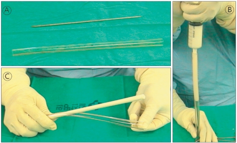
The following section describes the treatment procedure for patients with deep infection following TKA using modified static spacers. The original prosthesis was removed, followed by intensive irrigation and wide debridement of the infected soft tissue. A 36 Fr-diameter straight thoracic catheter (Mallinckrodt Medical, Athlone, Ireland) and a Steinmann pin measuring 3.0 mm in diameter and 22 cm in length were used to prepare the antibiotic-impregnated cement rod. Vancomycin (2 g) was then added to the Gentamicin bone cement (DePuy, Warsaw, IN, USA). At the late liquid stage of the cement, antibiotic-impregnated cement was poured into 50 mL of enema syringe. The Steinmann pin was inserted into the tube, and the prepared cement was delivered through the tube. During this procedure, the other end of the tube was partially sealed with a clamping device. The tube was rolled gently to straighten the rod. After the cement hardened, the tube was removed from the cement rod using a surgical knife. Finally, a cement rod measuring 9 mm in diameter and 22 cm in length was formed (Fig. 1).
Fig. 1.
(A) Preparation of the Steinmann pin and 36 Fr-diameter silicone tube. (B) Filling of the 50 mL enema syringe with the antibiotic-imprgnated cement. (C) Peeling off the tube.
An entry hole was created at the center of the distal femur and proximal tibia for the insertion of the cement rod. The rod was then inserted into the femur and tibia through this hole. During insertion, it was important to place the center of the cement rod on an imaginary joint line. The proximal medullary canal of the tibia was filled with antibiotic-impregnated cement up to the surface of the proximal tibia, while the surgical assistant maintained proper anatomic alignment and joint space. Subsequently, the same procedure was performed for the distal femur. The space between the cement of the distal femur and the proximal tibia was filled with more antibiotic-impregnated cement. Finally, the suprapatellar pouch and medial and lateral gutter space were filled with antibiotic-impregnated cement to reduce soft tissue adhesion (Fig. 2). Cylinder splint immobilization was required for three days after the operation, after which a fixed-angle knee brace was used and toe-touching ambulation was allowed until the reimplantation surgery.
Fig. 2.
Intramedullary nailing technique with the cement-impregnated rod. (A) Insertion of the cement-impregnated nail into the medullary canals of the femur and tibia, and approximation of the joint line and space with a previously marked line on the nail. (B) During this procedure, an assistant is needed to maintain sufficient joint space and anatomic alignment. (C) Covering the proximal tibia and distal femur with antibiotic-impregnated cement. (D) Filling the joint space with cement around the nail. (E) Filling the suprapatellar pouch, medial and lateral gutter space with cement to reduce soft tissue adhesion.
DISCUSSION
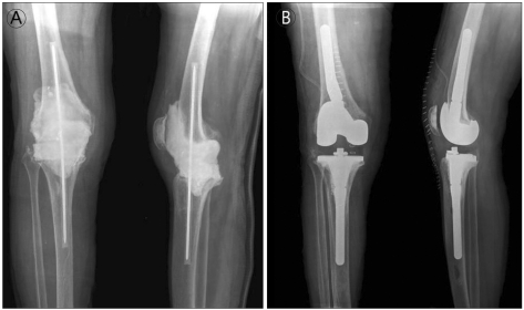
From April to November 2007, the authors performed the static technique in four patients using the novel antibiotic-impregnated cement rod for the treatment of infected TKA (culture: staphylococcus in three cases and no bacteria in one case). After the implant removal surgery, six weeks of intravenous antibiotic treatment was administered. Follow-up laboratory studies, including erythrocyte sedimentation rate and C-reactive protein, culture study via knee aspiration and a frozen biopsy from the second-stage operation (< 5 polymorpho-leukocytes/high power field) were performed to confirm successful eradication of the infection. The second-stage reimplantation was performed when all the criteria for the validation of infection control were met (Fig. 3). The second-stage reimplantation was performed using the rectus snipping approach, and more than 90° of flexion was obtained intra-operatively. In all four patients, no re-infection was evident after two and a half years of follow-up. The range of motion of the knee joints were respectively improved from 50 to 80, 95, and 100, and the knee society scores were 70, 86, 65, and 84, respectively, in the last follow-up evaluation.
Fig. 3.
A 76-year-old female with chronic deep infection 18 months after primary total knee arthroplasty. (A) Immediate postoperative plain radiograph at the first-stage operation with the antibiotic-impregnated cement intramedullary nailing in situ. Good anatomic alignment and sufficient joint space were achieved. (B) Plain radiograph at the immediate postoperative period of the second-stage reimplantation with stem extension.
Fehring et al.8) emphasized the importance of resting the joint in septic joint conditions. Others also reported that the static spacer technique provides more stability than the mobile spacer technique in patients with severe bone loss.5,8)
The main advantage of this technique is the maintenance of a normally aligned lower limb during the interval period. This maintains knee stability in combination with early muscle strengthening exercises, including quadriceps setting exercise, and enables the patient to comfortably dress and manage him/herself during the intervening period. The cement rod and static spacer provide a stable gap between the femur and tibia, thereby minimizing soft tissue contracture and shortening of the lower limb. In addition, symmetric maintenance of the soft tissue of both medial and lateral gutters requires no additional soft tissue balancing at the second-stage reimplantation.
A metal intramedullary nail can also be used to stabilize the knee joint. The additional cost of a metal nail, however, and the difficulty in infection control due to biofilm formation around the metal nail can be troublesome. In addition, a metal nail cannot be removed easily at the second stage reimplantation due to its hardness, and soft tissue adhesion of the femoral or tibial medullary canals. On the other hand, an antibiotic-impregnated cement rod enjoys advantages, such as inexpensive antibiotic delivery to the marrow spaces and easy removal by using a Hercules cutter. In addition, a static spacer is anchored to the cement rod, which can prevent spacer migration and bone erosion. This feature is believed to generate less cement wear debris than with the conventional static spacer or mobile articulating spacer technique. During the conduct of the antibiotic-impregnated cement rod technique, antibiotic-impregnated cement was applied to the proximal tibia, distal femur, joint gap space, suprapatellar pouch, and both gutters in a stepwise manner. This technique enables easy removal of the cement and reduced soft tissue adhesion at second-stage reimplantation.
Footnotes
No potential conflict of interest relevant to this article was reported.
References
- 1.Blom AW, Brown J, Taylor AH, Pattison G, Whitehouse S, Bannister GC. Infection after total knee arthroplasty. J Bone Joint Surg Br. 2004;86(5):688–691. doi: 10.1302/0301-620x.86b5.14887. [DOI] [PubMed] [Google Scholar]
- 2.Hanssen AD, Rand JA. Evaluation and treatment of infection at the site of a total hip or knee arthroplasty. Instr Course Lect. 1999;48:111–122. [PubMed] [Google Scholar]
- 3.Goldman RT, Scuderi GR, Insall JN. 2-stage reimplantation for infected total knee replacement. Clin Orthop Relat Res. 1996;(331):118–124. doi: 10.1097/00003086-199610000-00016. [DOI] [PubMed] [Google Scholar]
- 4.Windsor RE, Insall JN, Urs WK, Miller DV, Brause BD. Two-stage reimplantation for the salvage of total knee arthroplasty complicated by infection: further follow-up and refinement of indications. J Bone Joint Surg Am. 1990;72(2):272–278. [PubMed] [Google Scholar]
- 5.Emerson RH, Jr, Muncie M, Tarbox TR, Higgins LL. Comparison of a static with a mobile spacer in total knee infection. Clin Orthop Relat Res. 2002;(404):132–138. doi: 10.1097/00003086-200211000-00023. [DOI] [PubMed] [Google Scholar]
- 6.Hofmann AA, Kane KR, Tkach TK, Plaster RL, Camargo MP. Treatment of infected total knee arthroplasty using an articulating spacer. Clin Orthop Relat Res. 1995;(321):45–54. [PubMed] [Google Scholar]
- 7.Leone JM, Hanssen AD. Management of infection at the site of a total knee arthroplasty. Instr Course Lect. 2006;55:449–461. [PubMed] [Google Scholar]
- 8.Fehring TK, Odum S, Calton TF, Mason JB. Articulating versus static spacers in revision total knee arthroplasty for sepsis: the Ranawat Award. Clin Orthop Relat Res. 2000;(380):9–16. doi: 10.1097/00003086-200011000-00003. [DOI] [PubMed] [Google Scholar]
- 9.Goldstein WM, Kopplin M, Wall R, Berland K. Temporary articulating methylmethacrylate antibiotic spacer (TAMMAS): a new method of intraoperative manufacturing of a custom articulating spacer. J Bone Joint Surg Am. 2001;83-A(Suppl 2 Pt 2):92–97. [PubMed] [Google Scholar]
- 10.Haddad FS, Masri BA, Campbell D, McGraw RW, Beauchamp CP, Duncan CP. The PROSTALAC functional spacer in two-stage revision for infected knee replacements: prosthesis of antibiotic-loaded acrylic cement. J Bone Joint Surg Br. 2000;82(6):807–812. doi: 10.1302/0301-620x.82b6.10486. [DOI] [PubMed] [Google Scholar]


