Abstract
Transforming growth factor beta (TGF-β) is an important regulator of cell growth, and loss of TGF-β signaling is a hallmark of carcinogenesis. The Smad3/4 adaptor protein β2-spectrin (β2SP) is emerging as a potent regulator of tumorigenesis through its ability to modulate the tumor suppressor function of TGF-β. However, to date the role of the TGF-β signaling pathway at specific stages of the development of hepatocellular carcinoma (HCC), particularly in relation to the activation of other oncogenic pathways remains poorly delineated. Here, we identify a mechanism by which β2SP, a crucial Smad3 adaptor, modulates CDK4, cell cycle progression, and suppression of HCC. Increased expression of β2SP inhibits phosphorylation of the retinoblastoma gene product (Rb) and markedly reduces CDK4 expression to a far greater extent than other CDKs and cyclins. Furthermore, suppression of CDK4 by β2SP efficiently restores Rb hypophosphorylation and cell cycle arrest in G1. We further demonstrate that β2SP interacts with CDK4 and Smad3 in a competitive and TGF-β-dependent manner. In addition, haploinsufficiency of cdk4 in β2sp+/− mice results in a dramatic decline in HCC formation compared to that observed in β2sp+/− mice.
Conclusions
Thus, β2SP deficiency leads to CDK4 activation and contributes to dysregulation of the cell cycle, cellular proliferation, oncogene overexpression, and the formation of HCCs. Our data highlight CDK4 as an attractive target for the pharmacologic inhibition of HCC and demonstrate the importance of β2sp+/− mice as a model of pre-clinical efficacy in the treatment of HCC.
Keywords: β2-spectrin, CDK4, TGF-β, Hepatocellular Cancer
INTRODUCTION
The transforming growth factor β (TGF-β) signaling pathway is involved in multiple cellular processes, including cell growth, differentiation, adhesion, migration, and apoptosis. TGF-β is particularly active as an anti-mitogenic cytokine, functioning as a profound tumor suppressor by inhibiting cell cycle progression and arresting cells in early G1 phase. TGF-β signaling is mediated by type I and type II transmembrane serine/threonine kinase receptors (TβRI and TβRII) and such intracellular mediators as the Smad proteins (1, 2). TGF-β ligand binding to TβRII results in phosphorylation at glycine-serine repeats in the cytoplasmic tail domain of TβRI by TβRII. TβRI in turn phosphorylates the C-terminal serines of Smad2, and Smad3. This activity facilitates the dissociation of Smad2 and Smad3 from the microtubule cytoskeleton, and enables their association with Smad4. The heteromeric Smad2/3 and Smad4 complex is then able to translocate to the nucleus where it binds directly to Smad Binding Elements (SBEs), as well as to a number of co-activators to directly modulate TGF-β-regulated gene expression. TGF-β possesses oncogenic potential, which contributes to tumor progression later in carcinogenesis, but TGF-β also acts as a tumor suppressor at the early stages of tumor development by inhibiting proliferation and inducing apoptosis (2, 3). Importantly, inactivation of TGF-β signaling is thought to play a role in the development of a number of cancers (4). For example, the expression of Smad4 is lost in half of all pancreatic adenocarcinomas and one-third of all colon cancers. In addition, mutations in TβRII have been demonstrated in a subset of colonic and gastric cancers due to microsatellite instability (5, 6).
Recent studies have described a negative feedback control of Smad activity by CDK4 and CDK2 (7). Smad3 is a physiological target of these two kinases and mutation of the CDK4/CDK2 phosphorylation sites on Smad3 results in an enhancement of Smad3 transcriptional activity. This suggests that CDK4 and CDK2 negatively regulate the transcriptional activity and antiproliferative function of Smad3. Most human cancers appear to have lost their growth-inhibitory response to TGF-β. Interestingly, only about 10% of tumors appear to exhibit loss of expression of the TGF-β receptors or Smad family members, suggesting that other mechanisms such as loss of expression of scaffolding proteins, or amplification and over-expression of cell cycle regulatory proteins such as cyclin D1 and/or CDK loci may account for the loss of TGF-β signaling in human tumors.
We previously demonstrated that β2-spectrin (β2SP, or SPTBN1; Gene ID: 6711, MIM ID: 182790) is essential for normal TGF-β signaling by facilitating complex formation and the nuclear translocation of Smad3/4 (8). We previously found that mice containing a β2sp haploinsufficiency (β2sp+/−) mice spontaneously develop HCC, and that 11% of human HCC cancer cell lines exhibited a splice site mutation in β2SP exon 15 (9, 10). In addition, most cases of human HCC, gastric cancer, and lung cancer demonstrate significant reductions in β2SP expression (11–13). These results suggest that β2SP acts as a tumor suppressor and that the inhibition of β2SP function is a critical mechanism by which normal cells can escape from the regulation of proliferation in carcinogenesis. However, the exact mechanisms by which β2SP regulates cellular proliferation and suppression of liver carcinogenesis are unclear. We previously reported that the introduction of β2SP decreases CDK4 expression and results in the accumulation of cells in G1 phase (13). In contrast, a β2sp null mutation in mouse embryonic fibroblasts (MEF) results in increased levels of CDK4, while the siRNA-mediated knockdown of β2SP results in hyperphosphorylation of the retinoblastoma gene product Rb in HepG2 and CPAE cells (12, 14). These results imply that CDK4 is a strong mediator of the TGF-β-β2SP signaling pathway and its regulation of the cell cycle. To address the relationship between β2SP and CDK4, we examined the effect of changes in β2SP and CDK4 expression on progression through the cell cycle. We identified a TGF-β- and Smad3-dependent interaction between β2SP and CDK4. We also found that the decreased levels of CDK4 in β2sp+/− mice crossed with cdk4+/− mice efficiently prevented the spontaneous development of HCC seen in β2sp+/− mice. Thus, our investigation provides evidence that CDK4 activation is a critical step in the dysregulation of cellular proliferation due to alterations in β2SP expression. CDK4 may thus be an attractive therapeutic target in β2SP-deficient HCCs.
EXPERIMENTAL PROCEDURES
Mice
Animals were cared for in accordance with Institutional guidelines using approved protocols. β2sp+/− and cdk4+/− mice, and intercrosses were maintained and genotyped by PCR as described previously (8)(15).
Immunological Analysis
Antibodies against the following proteins were used in this study: CDK2, CDK6, cyclin B, cyclin D1, cyclin E, c-myc, Rb, β-actin, α-tubulin (Santa Cruz Biotechnology), phospho-RbSer807/811 (Cell Signaling Technology), CDK4 (Santa Cruz Biotechnology or Cell Signaling Technology), Ki-67 (Novus), V5 (Invitrogen), FLAG (Sigma), and β2SP (Santa Cruz Biotechnology or VA-1 (16)).
RESULTS
Changing of CDK4 in response to changes in β2SP expression
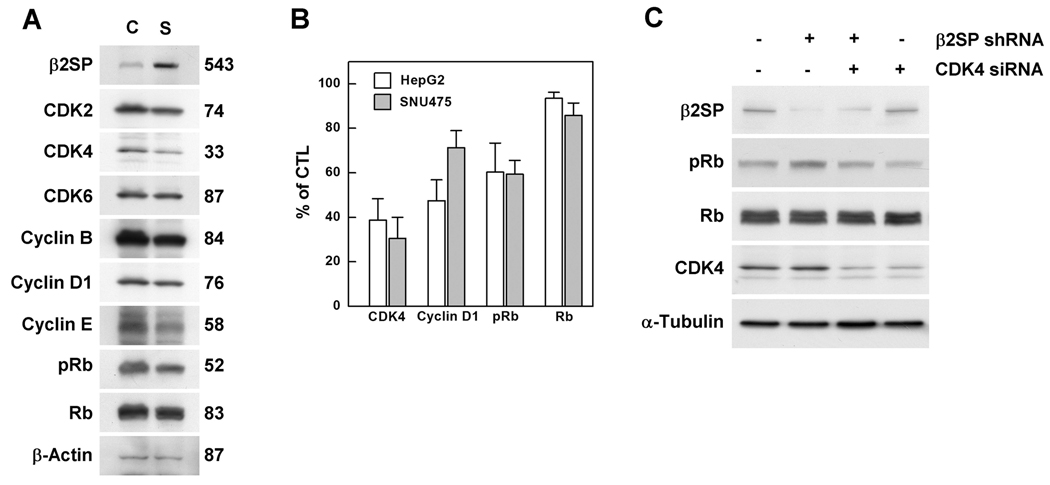
β2SP expression is tightly related to the levels of CDK4, and the G1/S transition suggesting the role of β2SP in the expression of CDK4, and cell cycle progression (12, 13). However, it is not yet clear whether CDK4 is the sole partner of β2SP in the TGF-β-β2SP-mediated signaling pathway. Thus, we examined the expression of several cell cycle regulatory proteins responsible for Rb phosphorylation upon the overexpression of β2SP in SNU-475 HCC cells. As shown in Fig. 1A, β2SP overexpression decreased the expression of several proteins responsible for cell cycle regulation including phosphorylated Rb (pRb). Comparing protein expression from identical preparations, the most dramatic reduction in expression was for CDK4 (33% of control) suggesting that CDK4 is a downstream effector in cell cycle regulation mediated by β2SP signaling. Then, we further compared the expression levels of CDK4, cyclin D1, pRb, and Rb upon transfection of β2SP in HepG2 and SNU475 cells in three independent experiments. The most remarkable reductions of CDK4 were shown in HepG2 (39%) and SNU475 (31%) cells (Fig. 1B). However, it was not clear that the change in CDK4 due to the loss of β2SP was sufficient to disrupt the cell cycle. Thus, we tested whether the increase in Rb phosphorylation was due to the down-regulation of β2SP or activation of CDK4. We inhibited β2SP expression in SNU-475 cells by the infection of a lentivirus containing shRNA against β2SP and then analyzed Rb phosphorylation. β2SP expression was decreased by 44% after lentiviral infection and Rb phosphorylation was increased by 55%, while the levels of Rb were unchanged (Fig. 1C). To determine whether CDK4 is responsible for Rb phosphorylation due to the down-regulation of β2SP, we inhibited CDK4 expression by siRNA in SNU-475 cells infected with β2SP shRNA. CDK4 siRNA in the presence of β2SP shRNA restored Rb phosphorylation to basal levels (Fig. 1C). These results suggest that CDK4 is a key regulator of Rb phosphorylation affected by β2SP expression.
Fig. 1. β2SP affects CDK4 expression.
(A) SNU-475 cells were transfected with V5-β2SP (S) or empty vector (C). Two days later, cell cycle regulatory proteins were analyzed by Western blotting from identical membrane. The numbers indicate relative amounts of each protein (%) after β2SP overexpression. (B) Expression levels of CDK4, cyclin D1, pRb, and Rb were measured in HepG2 and SNU475 cells upon transfection of β2SP. Means±standard errors from at least three independent experiments are given. (C) SNU-475 cells were infected with empty or β2SP shRNA lentivirus, followed by puromycin selection. Infected cells were then transfected with scrambled or CDK4 siRNA. The cells were analyzed by Western blotting.
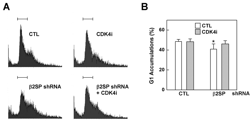
We then determined whether the induction of CDK4 expression due to the down-regulation of β2SP accelerates cell cycle progression. SNU-475 cells were infected with the β2SP shRNA lentivirus followed by treatment with a CDK4 inhibitor, and then analyzed cell cycle phases by FACS with PI staining. The number of cells in G1 phase was significantly decreased from 46 to 35% upon the knock down of β2SP (Fig. 2A and 2B). Additional treatment with CDK inhibitor (200 nM) in β2SP shRNA-treated cells returned 43% of the cells to G1. However, we did not detect the additional accumulation of cells in G1 phase in control lentiviral-treated cells exposed to the same CDK4 inhibitor. Taken together, these data demonstrate that dysregulation of the cell cycle resulting from the disruption of β2SP expression is mediated by CDK4 activation and Rb phosphorylation.
Fig. 2. CDK4 activation is responsible for the G1/S transition following β2SP shRNA infection.
SNU-475 cells were infected with empty (CTL) or β2SP shRNA lentivirus followed by puromycin selection. Then, the cells were treated with a 200 nM CDK4 inhibitor, and analyzed by flow cytometry with PI staining. (A) Representative histograms and (B) distribution of SNU-475 cells in G1 phase from three independent experiments. Statistically significant differences (P < 0.05) were determined by paired t-tests and indicated with asterisks.
Interactions between β2SP and CDK4
We further investigated the mechanism by which β2SP modulates CDK4 by examining interactions between these proteins. Recent reports indicate that CDK4 phosphorylates Smad3 to inhibit its transcriptional activity and anti-proliferative functions (7). Thus, we sought to determine whether CDK4 phosphorylates β2SP as it does Smad3. We incubated purified CDK4-cyclin D1 complex with β2SP in the presence of [γ32P] ATP, separated the proteins by SDS-PAGE, and then performed autoradiography. Using this method, β2SP phosphorylation by CDK4 was not detected (data not shown).
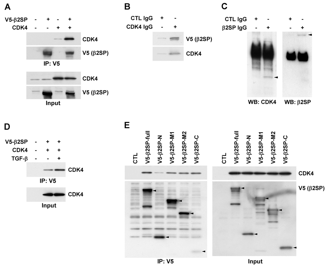
We continued to examine the interactions between β2SP and CDK4 by the immunoprecipitation of V5 epitope-tagged β2SP (V5-β2SP) and CDK4 expressed in 293T cells. As shown in Fig. 3A, β2SP and CDK4 were successfully expressed and an interaction between β2SP and CDK4 was observed. In addition, our reverse-immunoprecipitaion analysis further confirmed the interaction of these proteins (Fig. 3B). To know whether the interactions between β2SP and CDK4 might occur under physiological condition, we tested the interaction in 293T cells with endogenous level of protein. The results showed that CDK4 was associated in the anti-β2SP antibody precipitated complex but not in normal IgG precipitated (Fig. 3C). Next, we tested whether the interaction between β2SP and CDK4 is influenced by TGF-β. The interaction of β2SP with CDK4 became more than two-fold stronger (225%) upon treatment with TGF-β (Fig. 3D). To further delineate the binding region(s) for CDK4 on β2SP, we tested the ability of overexpressed CDK4 to capture β2SP fragments generated from the full-length protein. Studies of four protein fragments spanning the length of β2SP revealed that CDK4 interacts to 17-repeat middle region with ankyrin binding motif and the C-terminal fragment with abundant phosphorylation residues while not to the well known actin binding N-terminal fragment (Fig. 3E). Our data indicate that β2SP interacts with CDK4 in a TGF-β-dependent manner, and that the 17-repeat region and C-terminal domain of β2SP are required for the interaction.
Fig. 3. β2SP interacts with CDK4.
(A) 293T cells were transfected with V5-β2SP and/or CDK4. Two days later, the cells were immunoprecipitated with anti-V5 antibody-conjugated agarose and subjected to Western blotting using the indicated antibodies. (B) V5-β2SP and CDK4 transfected 293T cells were immunoprecipitated with control or anti-CDK4 rabbit IgG, and then analyzed by the Western blotting using anti-V5 and anti-CDK4 mouse IgG. (C) Untransfected 293T cells were immunoprecipitated with control or anti-β2SP rabbit IgG, and subjected to Western blotting using anti-CDK4 and anti-β2SP mouse IgG. Protein and rabbit IgG complexes were precipitated with protein-A agarose. (D) V5-β2SP and CDK4 transfected 293T cells were treated with TGF-β (100 pM) for 1 day, and immunoprecipitated as above. (E) Full-length or fragments of β2SP, and CDK4 transfected 293T cells were immunoprecipitated, and analyzed. The inputs represent 5% of the protein extracts without immunoprecipitation.
Smad3 prevents the interaction of CDK4 with β2SP
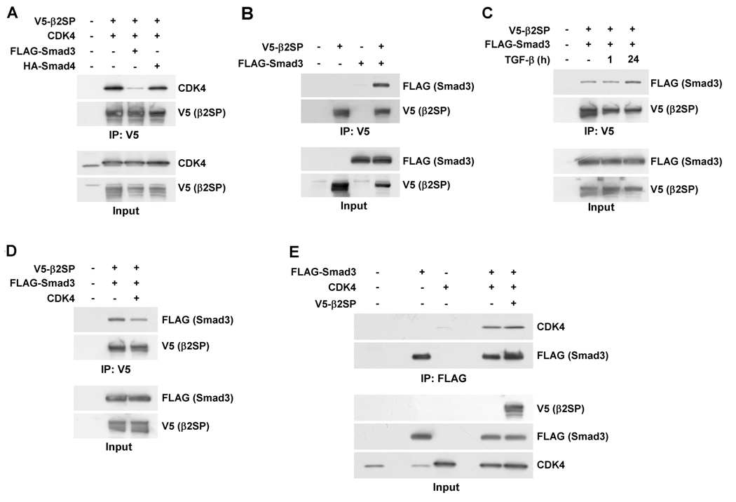
It has been reported that CDK4 phosphorylates Smad3, which inhibits the transcriptional activity and anti-proliferative functions of Smad3 (7). Based on this report, we tested whether the presence of Smad3 or 4 influences the interaction of β2SP with CDK4. Surprisingly, the addition of Smad3 prevented the interaction between β2SP and CDK4, while the introduction of Smad4 had no effect on the coupling of β2SP with CDK4 (Fig. 4A). To further analyze the effect of Smad3 on the binding ability of β2SP, we tested the interaction of β2SP with Smad3 in 293T cells. As shown in Fig. 4B and 4C, the interaction between β2SP and Smad3 was confirmed and increased upon treatment with TGF-β. To further substantiate the β2SP-CDK4 and β2SP-Smad3 interactions, we performed the reverse immunoprecipitation experiment (i.e., we tested the effect of CDK4 on the β2SP-Smad3 interaction). CDK4 decreased the binding of β2SP with Smad3 (Fig. 4D). Thus, the binding of β2SP to CDK4 and Smad3 can be competitively inhibited by the addition of Smad3 or CDK4, respectively. We also examined whether Smad3 interacts with CDK4 and whether this interaction is influenced by the presence of β2SP. Immunoprecipitation assays revealed that CDK4 interacted with Smad3 (Fig. 4E). In addition, in the presence of β2SP, the binding of Smad3 with CDK4 was unchanged. These findings suggest that β2SP, Smad3, and CDK4 form a complex and that the Smad3-CDK4 interaction is stronger than that of β2SP with Smad3 or CDK4. However, we cannot rule out the possibility that additional protein(s) are required for complex formation.
Fig. 4. Smad3 competes with CDK4 for binding to β2SP.
(A) Smad3 but not Smad4 competes with CDK4 for binding to β2SP. 293T cells were transfected with V5-β2SP and CDK4 in the presence of Smad3 or Smad4. (B and C) β2SP interacts with Smad3 in a TGF-β-dependent manner. 293T cells were transfected with V5-β2SP and FLAG-Smad3 in the absence and presence of TGF-β (100 pM; B and C, respectively). (D) CDK4 inhibits the β2SP-Smad3 interaction. 293T cells were transfected with V5-β2SP and FLAG-Smad3 in the absence or presence of CDK4. (E) The CDK4-Smad3 interaction is not affected by β2SP. 293T cells were transfected with CDK4 and FLAG-Smad3 in the absence or presence of β2SP. Extracts prepared from the transfected cells were immunoprecipitated by anti-V5 (A-D) or anti-FLAG (E) antibody-conjugated agarose. The inputs represent 5% of the protein extracts without immunoprecipitation.
Haploinsufficiency of CDK4 prevents HCC in β2SP+/− mice
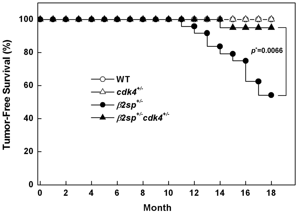
We previously showed that β2sp+/− mice spontaneously developed the HCC formation with elevated CDK4 function (17). To examine the contribution of CDK4 to HCC formation due to the alteration of β2SP, we generated double-heterozygous mutant mice by crossing β2sp+/− and cdk4+/− mice and followed cohorts of wild-type, β2sp+/−, cdk4+/−, and β2sp+/− cdk4+/− animals. The mice of each genotype were healthy and could not be easily distinguished. None of the mice exhibited abnormalities until twelve months. At thirteen months of age, the β2sp+/− mutant mice exhibited HCC with a substantially increased incidence of HCC up to 46% (11 out of 24) until 18 months of age. In contrast, only one out of 20 (5%) of the β2sp+/− cdk4+/− mice showed HCC during same period. By 18 months of age, none of the wild-type or cdk4+/− animals showed any sign of neoplasia, including HCC. Thus, although one out of 20 β2sp+/− cdk4+/− mice exhibited HCC, the lifespan and incidence of HCC in the β2sp+/− cdk4+/− animals was remarkably improved compared to the β2sp+/− mice. When we compared the survival of β2sp+/− cdk4+/− mice to β2sp+/− mice, the survival was significantly improved according to the log-rank test (p=0.0066). These results suggest that the reduction of CDK4 in β2sp+/− mice efficiently prevented HCC formation.
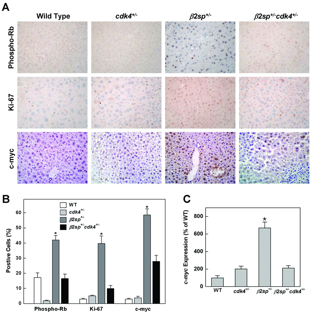
To examine the molecular events occurring after the reduction in CDK4 in the β2sp+/− mice, we performed the immunohistochemical analysis of pre-cancerous normal liver tissue to determine whether cellular proliferation-related molecular markers were altered (Fig. 6A). Statically significant upregulation of pRb, and Ki-67 staining were identified in liver sections from the β2sp+/− mice but not in liver tissues from the wild-type or cdk4+/− mice. Notably, statically significant reductions were identified in the nuclei of hepatocytes from the β2sp+/− cdk4+/− mice, suggesting that the inhibition of CDK4 could restore the dysregulated cell cycle and hyperproliferation caused by the disruption of β2SP (Fig. 6B).
Fig. 6. Reduced proliferation and down-regulation of CDK4 by c-myc in β2sp+/− mice.
Normal livers from 18-month-old mice were prepared for immunohistochemistry or RT-PCR. (A and B) Detection of pRb, Ki-67, and c-myc in liver sections (40X magnification). (C) Expression of c-myc mRNA in liver tissues (n=3) using 18S rRNA as a control. Means±standard errors are given. The asterisks indicate P < 0.01.
Transduction of the TGF-β signal suppresses oncogenic signals by preventing the transcription of c-myc (18). In this study, we found that liver carcinogenesis due to changes in β2SP expression also affects c-myc expression. c-myc-positive hepatocytes were abundant in liver sections from β2sp+/− mice but not in those from wild-type or cdk4+/− mice. However, in the β2sp+/− cdk4+/− mice, c-myc levels were significantly reduced after the down-regulation of CDK4. We performed quantitative RT-PCR to directly compare c-myc expression in liver tissues from these mice. A statistically significant increase of c-myc transcription was detected in the β2sp+/− mice (670% compared to wild type) but suppressed by the down-regulation of CDK4 in β2sp+/− cdk4+/− mice (202% compared to wild type) (Fig. 6C). Together, these observations indicate that the activation of CDK4 caused by β2SP disruption results not only in dysregulation of the cell cycle and hyperproliferation but also activates oncogenic signals that facilitate HCC formation.
DISCUSSION
TGF-β is a multifunctional regulatory polypeptide affecting multiple cellular functions, including proliferation, differentiation, and apoptosis. TGF-β inhibits cell cycle progression during G1 through the control of CDKs. In mammalian cells, tightly regulated cyclins and CDKs act sequentially during the G1/S transition and are required for cell cycle progression. The mechanisms whereby TGF-β arrests the cell cycle have been studied primarily in epithelial cells with emphasis on the regulation of G1 cyclin-dependent kinases. In mink lung epithelial cells, TGF-β treatment induces the inhibition of CDK4 synthesis and CDK2 inactivation with a subsequent G1 arrest. In human HaCaT keratinocytes, TGF-β induces a growth arrest through the down-regulation of cell-cycle regulators, including cyclin E, cyclin A, CDK2, and CDK4. In fact, germline transmission of activated cdk4 (R24C) mutation in mice results in spontaneous tumor formation, and facilitated tumorigenesis in an oncogenic background (15, 19). Several cyclin-dependent kinase inhibitors have been implicated in the TGF-β-induced cell-cycle arrest. TGF-β induces the up-regulation of the CDK inhibitor p15INK4B, which specifically inhibits the enzymatic activities of CDK4 and CDK6, thereby preventing progression through G1 phase of the cell cycle. However, because multiple cell-cycle regulators are involved in TGF-β signaling, this raises several questions related to their actual roles in specific cell types. Among the regulatory proteins responsible for cell cycle progression, CDK4 is essential for the progression from early to mid-G1, at which cells are believed to commit to DNA synthesis and eventually mitosis. CDK4-cyclin D1 phosphorylates Rb. This enables E2F release from Rb, resulting in the transcription of a number of genes that are necessary for DNA synthesis and cell cycle progression.
Previously, the only known substrate of CDK4 was Rb; however, Matsuura et al. (2004) demonstrated that CDK4 phosphorylates Smad3 and inhibits Smad3-mediated TGF-β signaling. A loss of TGF-β responsiveness results in dysregulated cell growth and is believed to be a crucial step in the development of various tumors, including liver cancer. Most tumors exhibit a loss of responsiveness to TGF-β signaling, and the expression of cyclins and CDKs is often enhanced in tumor cells (20). We previously demonstrated that β2SP is a critical mediator of the TGF-β signaling pathway and acts as a tumor suppressor (8, 11, 21). β2SP interacts and facilitates the nuclear translocation of Smad3 and Smad4, which enables proper TGF-β signaling. β2SP expression is significantly decreased or absent in lung, gastric, liver, and colon tumors. Moreover, β2sp+/− mice developed spontaneous HCC whereas β2sp+/− smad4+/− mice exhibited enhanced formation of spontaneous gastric cancers. In addition, the expression of CDK4 and cyclin D1 is significantly increased in gastric cancers and HCCs from β2sp altered mice (12, 13, 17, 21).
Notably, our data revealed that the activation of CDK4 is an essential step in HCC formation due to alterations in β2SP. First, significant reductions in CDK4 and phosphorylated Rb were observed upon the overexpression of β2SP in the SNU-475 HCC cells. Next, the reduction of CDK4 by siRNA transfection restored β2SP-mediated increases in phosphorylated Rb to basal levels. In addition, the role of CDK4 in the G1/S transition was tested following the alteration of β2SP expression whereupon the chemical inhibition of CDK4 rescued the abnormal G1/S transition caused by infection of the β2SP-shRNA. We further found that β2SP interacts with CDK4 and Smad3 in a competitive and TGF-β-dependent manner. Finally, the genetic inhibition of CDK4 in mice produced by crossing cdk4+/− with β2sp+/− mice efficiently prevented HCC formation compared to that in β2sp+/− mice, and was accompanied by decreased proliferation and oncogene expression in the liver. Taken together, these results imply that CDK4 activation is required for dysregulation of the cell cycle and that the inhibition of CDK4 prevents abnormal G1/S transition and HCC formation due to alterations in β2SP expression.
In addition to cell cycle regulation, we examined the expression of c-myc as a reflection of TGF-β signaling and oncogenic stimulation in β2sp mutant mice, which provided us with insight into the hepatocarcinogenic mechanisms caused by alterations in TGF-β-β2SP signaling. By RT-PCR and histological analysis of aged normal livers, the expression of c-myc was dramatically increased in β2sp+/− mice and returned to normal upon the down-regulation of CDK4 in β2sp+/− cdk4+/− mice. Moreover, it has been reported that the phosphorylation of Smad3 by CDK4 and CDK2 inhibits its transcriptional activity and anti-proliferative function (7). Because cancer cells often exhibit high levels of CDK activity, lowering Smad3 activity via the phosphorylation of CDK may contribute to tumorigenesis and TGF-β resistance in cancer patients. Recently, it is suggested that CDK4, together with JNK, alters tumor-suppressive TGF-β signaling to malignant characteristics by transcriptional activation of c-Myc in later stages of human colorectal cancer. These results suggest that the activation of CDK4 due to changes in β2SP expression stimulates the expression of c-myc, which could cause pre-cancerous tissue to progress to malignancy.
Finally, we previously demonstrated that the majority of human HCCs exhibited reduced β2SP expression (17). In response to β2SP deficiency, the activation of CDK4 contributes to the phosphorylation of Rb, facilitation of the G1/S transition, and induction of the oncogene c-myc, leading to liver malignancy. We also found that the activation of CDK4 does not merely increase the proliferative activity of liver tissue, but actually transforms normal tissue into pre-cancerous tissue by suppressing the inhibitory functions of TGF-β. Our data highlight CDK4 as an attractive target for pharmacologic inhibition and demonstrate the importance of β2sp+/− mice as a model of pre-clinical efficacy in the treatment of HCC due to β2SP alterations. Thus, our work greatly underscores the potential for targeting CDK4 in the treatment and prevention of cancer, specifically HCC, and studies are currently ongoing to assess the efficacy of the tumor-specific inhibition of CDK4 in cancer patients (22).
Fig. 5. Kaplan-Meier curves of HCC survival.
The wild-type (n=24), and cdk4+/− (n=20) mice were normal and did not develop cancer. By contrast, 46% of the 18-month-old β2sp+/− (n=24) mice spontaneously developed HCC. Remarkably, only one out of 20 β2sp+/− cdk4+/− mice developed HCC at the same age. Statistically significant difference (p=0.0066) was determined by log-rank test in comparison of the tumor-free survival between β2sp+/− and β2sp+/− cdk4+/− mice.
Acknowledgments
Financial Supports:
This study was supported by National Institute of Health, RO1-CA106614 (LM), RO1-CA042857 (LM), RO1-DK58637 (BM), PO1-CA130821 (LM), RC2-AA019392, VA Merit Award (LM), R. Robert and Sally D. Funderburg Research Scholar (LM), the Ben Orr Award (LM), National Cancer Center, Korea, NCC-0910020 (SSK), Korea Healthcare Technology R & D Project, A090095 (SSK), and National Research Foundation, Korea, 2010-0021822 (SSK).
Abbreviations
- TGF-β
transforming growth factor-β
- HCC
hepatocellular cancer
- β2SP
β2-spectrin
REFERENCES
- 1.Massague J. G1 cell-cycle control and cancer. Nature. 2004;432:298–306. doi: 10.1038/nature03094. [DOI] [PubMed] [Google Scholar]
- 2.Mishra L, Derynck R, Mishra B. Transforming growth factor-beta signaling in stem cells and cancer. Science. 2005;310:68–71. doi: 10.1126/science.1118389. [DOI] [PubMed] [Google Scholar]
- 3.Bierie B, Moses HL. Tumour microenvironment: TGFbeta: the molecular Jekyll and Hyde of cancer. Nat Rev Cancer. 2006;6:506–520. doi: 10.1038/nrc1926. [DOI] [PubMed] [Google Scholar]
- 4.Derynck R, Zhang YE. Smad-dependent and Smad-independent pathways in TGF-beta family signalling. Nature. 2003;425:577–584. doi: 10.1038/nature02006. [DOI] [PubMed] [Google Scholar]
- 5.Markowitz S, Wang J, Myeroff L, Parsons R, Sun L, Lutterbaugh J, Fan RS, et al. Inactivation of the type II TGF-beta receptor in colon cancer cells with microsatellite instability. Science. 1995;268:1336–1338. doi: 10.1126/science.7761852. [DOI] [PubMed] [Google Scholar]
- 6.Grady WM, Myeroff LL, Swinler SE, Rajput A, Thiagalingam S, Lutterbaugh JD, Neumann A, et al. Mutational inactivation of transforming growth factor beta receptor type II in microsatellite stable colon cancers. Cancer Res. 1999;59:320–324. [PubMed] [Google Scholar]
- 7.Matsuura I, Denissova NG, Wang G, He D, Long J, Liu F. Cyclin-dependent kinases regulate the antiproliferative function of Smads. Nature. 2004;430:226–231. doi: 10.1038/nature02650. [DOI] [PubMed] [Google Scholar]
- 8.Tang Y, Katuri V, Dillner A, Mishra B, Deng CX, Mishra L. Disruption of transforming growth factor-beta signaling in ELF beta-spectrin-deficient mice. Science. 2003;299:574–577. doi: 10.1126/science.1075994. [DOI] [PubMed] [Google Scholar]
- 9.Katuri V, Tang Y, Li C, Jogunoori W, Deng CX, Rashid A, Sidawy AN, et al. Critical interactions between TGF-beta signaling/ELF, and E-cadherin/beta-catenin mediated tumor suppression. Oncogene. 2006;25:1871–1886. doi: 10.1038/sj.onc.1209211. [DOI] [PMC free article] [PubMed] [Google Scholar]
- 10.Yao ZX, Jogunoori WS, Choufani S, Rashid A, Blake T, Yao W, Kreishman P, et al. Epigenetic silencing of {beta}-spectrin a TGF-{beta} signaling/scaffolding protein in a human cancer stem cell disorder: Beckwith-Wiedemann syndrome. J Biol Chem. 2010 doi: 10.1074/jbc.M110.162347. [DOI] [PMC free article] [PubMed] [Google Scholar]
- 11.Katuri V, Tang Y, Marshall B, Rashid A, Jogunoori W, Volpe EA, Sidawy AN, et al. Inactivation of ELF/TGF-beta signaling in human gastrointestinal cancer. Oncogene. 2005;24:8012–8024. doi: 10.1038/sj.onc.1208946. [DOI] [PubMed] [Google Scholar]
- 12.Kim SS, Shetty K, Katuri V, Kitisin K, Baek HJ, Tang Y, Marshall B, et al. TGF-beta signaling pathway inactivation and cell cycle deregulation in the development of gastric cancer: role of the beta-spectrin, ELF. Biochem Biophys Res Commun. 2006;344:1216–1223. doi: 10.1016/j.bbrc.2006.03.236. [DOI] [PMC free article] [PubMed] [Google Scholar]
- 13.Baek HJ, Kim SS, da Silva FM, Volpe EA, Evans S, Mishra B, Mishra L, et al. Inactivation of TGF-beta signaling in lung cancer results in increased CDK4 activity that can be rescued by ELF. Biochem Biophys Res Commun. 2006;346:1150–1157. doi: 10.1016/j.bbrc.2006.05.195. [DOI] [PubMed] [Google Scholar]
- 14.Baek HJ, Lim SC, Kitisin K, Jogunoori W, Tang Y, Marshall MB, Mishra B, et al. Hepatocellular cancer arises from loss of transforming growth factor beta signaling adaptor protein embryonic liver fodrin through abnormal angiogenesis. Hepatology. 2008;48:1128–1137. doi: 10.1002/hep.22460. [DOI] [PMC free article] [PubMed] [Google Scholar]
- 15.Rane SG, Dubus P, Mettus RV, Galbreath EJ, Boden G, Reddy EP, Barbacid M. Loss of Cdk4 expression causes insulin-deficient diabetes and Cdk4 activation results in beta-islet cell hyperplasia. Nat Genet. 1999;22:44–52. doi: 10.1038/8751. [DOI] [PubMed] [Google Scholar]
- 16.Mishra L, Cai T, Yu P, Monga SP, Mishra B. Elf3 encodes a novel 200-kD beta-spectrin: role in liver development. Oncogene. 1999;18:353–364. doi: 10.1038/sj.onc.1202313. [DOI] [PubMed] [Google Scholar]
- 17.Kitisin K, Ganesan N, Tang Y, Jogunoori W, Volpe EA, Kim SS, Katuri V, et al. Disruption of transforming growth factor-beta signaling through beta-spectrin ELF leads to hepatocellular cancer through cyclin D1 activation. Oncogene. 2007;26:7103–7110. doi: 10.1038/sj.onc.1210513. [DOI] [PMC free article] [PubMed] [Google Scholar]
- 18.Chen CR, Kang Y, Massague J. Defective repression of c-myc in breast cancer cells: A loss at the core of the transforming growth factor beta growth arrest program. Proc Natl Acad Sci U S A. 2001;98:992–999. doi: 10.1073/pnas.98.3.992. [DOI] [PMC free article] [PubMed] [Google Scholar]
- 19.Rane SG, Cosenza SC, Mettus RV, Reddy EP. Germ line transmission of the Cdk4(R24C) mutation facilitates tumorigenesis and escape from cellular senescence. Mol Cell Biol. 2002;22:644–656. doi: 10.1128/MCB.22.2.644-656.2002. [DOI] [PMC free article] [PubMed] [Google Scholar]
- 20.Ito Y, Matsuura N, Sakon M, Miyoshi E, Noda K, Takeda T, Umeshita K, et al. Expression and prognostic roles of the G1-S modulators in hepatocellular carcinoma: p27 independently predicts the recurrence. Hepatology. 1999;30:90–99. doi: 10.1002/hep.510300114. [DOI] [PubMed] [Google Scholar]
- 21.Tang Y, Katuri V, Srinivasan R, Fogt F, Redman R, Anand G, Said A, et al. Transforming growth factor-beta suppresses nonmetastatic colon cancer through Smad4 and adaptor protein ELF at an early stage of tumorigenesis. Cancer Res. 2005;65:4228–4237. doi: 10.1158/0008-5472.CAN-04-4585. [DOI] [PubMed] [Google Scholar]
- 22.Johnson SM, Torrice CD, Bell JF, Monahan KB, Jiang Q, Wang Y, Ramsey MR, et al. Mitigation of hematologic radiation toxicity in mice through pharmacological quiescence induced by CDK4/6 inhibition. J Clin Invest. 2010;120:2528–2536. doi: 10.1172/JCI41402. [DOI] [PMC free article] [PubMed] [Google Scholar]


