

Abstract
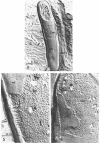
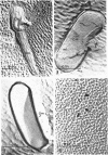
Remsen, C. C. (Swiss Federation Institute of Technology, Zurich, Switzerland), and D. G. Lundgren. Electron microscopy of the cell envelope of Ferrobacillus ferrooxidans prepared by freeze-etching and chemical fixation techniques. J. Bacteriol. 92:1765–1771. 1966.—A comparison was made of the fine structure of the cell envelope of the gram-negative bacterium Ferrobacillus ferrooxidans when cells were prepared for microscopy by freeze-etching and chemical fixation techniques. Cell envelopes of chemically fixed cells appeared as five separate layers distinguishable by their location and electron density. Frozen-etched cells showed a three-layered complex with each layer measuring approximately 100 A in thickness. The latter technique is considered to be “artifact-free” and, as a technique, yields purely morphological information on the natural state. The three layers revealed by freeze-etching are: the outer layer, a lipoprotein-lipopolysaccharide layer; the middle layer, a layer composed of globular protein attached to fibrillar mucopeptide; and the innermost layer, the cytoplasmic membrane. The latter was covered with 100 to 120 A particles. The relationship of the aforementioned layers to those seen in chemically fixed cells is discussed.
Full text
PDF


Images in this article


Selected References
These references are in PubMed. This may not be the complete list of references from this article.
- BROWN A. D., SHOREY C. D. THE CELL ENVELOPES OF TWO EXTREMELY HALOPHILIC BACTERIA. J Cell Biol. 1963 Sep;18:681–689. doi: 10.1083/jcb.18.3.681. [DOI] [PMC free article] [PubMed] [Google Scholar]
- DEPETRIS S. ULTRASTRUCTURE OF THE CELL WALL OF ESCHERICHIA COLI. J Ultrastruct Res. 1965 Apr;12:247–262. doi: 10.1016/s0022-5320(65)80098-3. [DOI] [PubMed] [Google Scholar]
- DUGAN P. R., LUNDGREN D. G. ENERGY SUPPLY FOR THE CHEMOAUTOTROPH FERROBACILLUS FERROOXIDANS. J Bacteriol. 1965 Mar;89:825–834. doi: 10.1128/jb.89.3.825-834.1965. [DOI] [PMC free article] [PubMed] [Google Scholar]
- KELLENBERGER E., RYTER A., SECHAUD J. Electron microscope study of DNA-containing plasms. II. Vegetative and mature phage DNA as compared with normal bacterial nucleoids in different physiological states. J Biophys Biochem Cytol. 1958 Nov 25;4(6):671–678. doi: 10.1083/jcb.4.6.671. [DOI] [PMC free article] [PubMed] [Google Scholar]
- MOOR H. DIE GEFRIER-FIXATION LEBENDER ZELLEN UND IHRE ANWENDUNG IN DER ELEKTRONENMIKROSKOPIE. Z Zellforsch Mikrosk Anat. 1964 Apr 28;62:546–580. [PubMed] [Google Scholar]
- MOOR H., MUHLETHALER K., WALDNER H., FREY-WYSSLING A. A new freezing-ultramicrotome. J Biophys Biochem Cytol. 1961 May;10:1–13. doi: 10.1083/jcb.10.1.1. [DOI] [PMC free article] [PubMed] [Google Scholar]
- MURRAY R. G., STEED P., ELSON H. E. THE LOCATION OF THE MUCOPEPTIDE IN SECTIONS OF THE CELL WALL OF ESCHERICHIA COLI AND OTHER GRAM-NEGATIVE BACTERIA. Can J Microbiol. 1965 Jun;11:547–560. doi: 10.1139/m65-072. [DOI] [PubMed] [Google Scholar]
- Mahoney R. P., Edwards M. R. Fine Structure of Thiobacillus thiooxidans. J Bacteriol. 1966 Aug;92(2):487–495. doi: 10.1128/jb.92.2.487-495.1966. [DOI] [PMC free article] [PubMed] [Google Scholar]
- OGURA M. High resolution electron microscopy on the surface structure of Escherichia coli. J Ultrastruct Res. 1963 Apr;8:251–263. doi: 10.1016/s0022-5320(63)90006-6. [DOI] [PubMed] [Google Scholar]
- REYNOLDS E. S. The use of lead citrate at high pH as an electron-opaque stain in electron microscopy. J Cell Biol. 1963 Apr;17:208–212. doi: 10.1083/jcb.17.1.208. [DOI] [PMC free article] [PubMed] [Google Scholar]
- SILVERMAN M. P., LUNDGREN D. G. Studies on the chemoautotrophic iron bacterium Ferrobacillus ferrooxidans. I. An improved medium and a harvesting procedure for securing high cell yields. J Bacteriol. 1959 May;77(5):642–647. doi: 10.1128/jb.77.5.642-647.1959. [DOI] [PMC free article] [PubMed] [Google Scholar]
- STEERE R. L. Electron microscopy of structural detail in frozen biological specimens. J Biophys Biochem Cytol. 1957 Jan 25;3(1):45–60. doi: 10.1083/jcb.3.1.45. [DOI] [PMC free article] [PubMed] [Google Scholar]
- WEIDEL W., FRANK H., LEUTGEB W. Autolytic enzymes as a source of error in the preparation and study of gram-negative cell walls. J Gen Microbiol. 1963 Jan;30:127–130. doi: 10.1099/00221287-30-1-127. [DOI] [PubMed] [Google Scholar]
- WEIDEL W., FRANK H., MARTIN H. H. The rigid layer of the cell wall of Escherichia coli strain B. J Gen Microbiol. 1960 Feb;22:158–166. doi: 10.1099/00221287-22-1-158. [DOI] [PubMed] [Google Scholar]