Abstract
Purpose
Reports of dissimilar stereothresholds for contour and random-dot (RD) targets may reflect differences in stimulus properties or differences between local and global stereoscopic processing mechanisms. In this study, we evaluated whether the stereothresholds obtained using low- and high-density RD stimuli are consistent with a distinction between local and global disparity processing.
Methods
Stereothresholds were measured in eight normal subjects for a small disparate line segment superimposed on RD surrounds with densities that ranged between 0.07% and 28.3%.
Results
Stereothresholds averaged 0.23 arc min for a RD density of 0.39% and approximately doubled for lower and higher densities. The increase in stereothresholds at low densities is likely due to the increased spacing between elements, which reduces their usefulness as a reference for relative disparity judgments. The increase in stereothresholds at high densities is attributed to a crowding effect.
Conclusions
Because the stereothresholds measured with RD stimuli of low and high density are limited by different constraints, they can be considered to be different types of stereotargets. However, because the stereothresholds measured for RD targets of varying densities are similar to those determined previously for a local, two-rod stereotarget, it is likely that all of these stimuli are processed by a single disparity-processing mechanism.
Keywords: stereothreshold, local stereopsis, global stereopsis, random dot stereogram, dot density
Stereograms consist of two-dimensional monocular half-images that are combined by the brain into a three-dimensional depth percept. The two half-images contain reference elements that are in the same relative locations and test elements that are offset in their relative spatial locations in the two eyes. This offset, or relative binocular disparity, causes a perception of depth. The stereothreshold is the smallest relative binocular disparity that yields a perception of depth. Stereopsis has been categorized as “local” and “global,” but the definitions of these terms are ambiguous.
Julesz1 distinguished between local and global stereograms based on the number of dots contained within the stereogram. This definition was adopted by Westheimer & McKee2 who formed a stereogram that they considered “local” because it only contained 49 dots. In support of the distinction made by Julesz, Pressmar & Hasse3 reported phenomenologically different responses to low density and high density random dot stereograms (RDSs), which the authors considered to be local and global stereograms, respectively.
Some researchers describe local stereograms as stimuli composed of individual features or contours seen in depth, such as line stereograms, and global stereograms as complex, patterned stimuli composed of many features, such as RDSs.4,5, 6 This definition is also consistent with the distinction between the stereogram types based on element density, because RDSs that contain very few dots are essentially isolated contours seen in depth; whereas RDSs that contain a high density of dots comprise textures with many features.
Clinically, stereopsis is assessed commonly using local contour stereograms (e.g. the Titmus circles or the Randot stereotest for fine disparities),7, 8 although global random-dot based tests also are available (e.g., Preschool Randot test and TNO Stereo Test). Discrepant results for stereothresholds assessed with local and global stereograms have been reported.8–10 In contrast, Fawcett7 only found differences between the thresholds for local and global targets in patients with abnormal binocularity, although the similar thresholds obtained for normal subjects could reflect a floor effect because the tests used in this study could not measure stereothresholds lower than 40 arc seconds. However, Stevenson, Cormack, & Schor11 reported similar average thresholds on the order of a few arc seconds using global RDSs and the local Howard Dolman Apparatus. Disagreement about the similarity between thresholds using local and global stereotargets may be explained by differences in the properties of the targets,12,11 such as the spacing between the constituent elements,8, 13, 14 size,15–17 contrast,18–22 viewing duration,9, 22, 23 and spatial frequency content,24–26 or, alternatively, by differences in the neural mechanisms that underlie the processing of local and global stereograms.27
Consistent with the second alternative, Westheimer28 distinguished between local processes that ascribe depth values to individual features and global processes that ascribe depth values to the overall configuration and are relatively unaffected by the depth values of the constituent internal features. The perception of depth in a dense RDS presumably would reflect the operation of the latter, global process.
Other authors distinguish between local and global processing mechanisms based on the extent of the correspondence problem – the matching ambiguity that exists in global stimuli.1, 29–32
Attributing their definition to Julesz, Bruce, Green, & Georgeson33 define local stereopsis as the mechanism that processes edge or dot stimuli, and global stereopsis as the mechanism that processes complex pattern stimuli that involve the evaluation of many possible local matches. Similarly, global stereopsis has been described as the integration across multiple pattern elements to solve the correspondence problem by selecting the disparity for which the maximum number of elements in the two half views are matched.32, 34
Still other authors claim that local and global processing mechanisms share a common disparity-processing stage, followed by a subsequent, global level of analysis to resolve the ambiguity that is produced when multiple possible stereoscopic matches exist.1, 35
To determine whether different neural mechanisms process local and global stimuli, it is necessary to form local and global stereograms that have similar properties. Based on the definition that a small number of dots should appear as local, isolated targets in depth and that many dots should appear as a random-dot texture and not as individual dots in depth,1 it should be possible to obtain both local and global stereograms by varying the density of the same RDS. Although previous studies examined changes in correlation thresholds36 and stereoscopic efficiency37 as a function of RD density, to our knowledge, the effect of RD density on stereothresholds for depth discrimination has not been evaluated systematically.
Following Tyler,27 we provisionally define a local disparity mechanism as one that processes the disparity at one location in the field without reference to the disparities at other regions of the field, and a global disparity mechanism as one that involves interactions between local disparity mechanisms. One outcome of this interaction may be the assignment of depth information to empty regions between texture elements to form a disparity-defined surface, which Wilcox & Duke38 found to occur when approximately 10% of the stereogram area is covered by texture elements. In addition, densely spaced texture elements in a stereogram undergo disparity averaging11, 39, 40 and result in a reduced sensitivity to differences in disparity.2, 41 For example, Tsirlin et al.42 reported that the disambiguation of two (or more) stereoscopic surfaces separated by 1.9 arc minutes of disparity worsens with increasing RD density.
To evaluate whether the stereothresholds obtained using low- and high-density RDSs are consistent with the distinction made above between local and global targets, in this study we superimposed a small disparate line segment (i.e. a single feature) within RDS surrounds of various densities. If the RDS stimulus is processed by a local disparity mechanism, then the disparities of the random dots and the line segment should be determined independently and the depth value of the line segment should remain conspicuous regardless of the RD density. However, if the stimulus is processed by a global disparity mechanism, then the depth of the line segment should be less easily discriminated as its disparity is averaged, at least to some extent, with the disparity of the surrounding dots. A hierarchical processing scheme, in which local disparity extraction is followed by global interactions between local disparity mechanisms27 also is consistent with a decrease in the ease of discrimination of the disparate line segment. We therefore expected that the stereothreshold for the line segment would increase when it was embedded in RDSs of high density, if the processing of high-density stereotargets reflects the operation of a global disparity mechanism.*
METHODS
Subjects
Eight healthy, fully corrected subjects between the ages of 24 and 58, including the two authors, participated in the experiment. Each subject had a stereothreshold of at least 40 arc seconds for both crossed and uncrossed disparities, as measured with the Titmus Stereo Test (Stereo Optical Co., Chicago, IL, USA). The research adhered to the tenets of the Declaration of Helsinki, and the experimental protocol was reviewed by the University of Houston Committee for the Protection of Human Subjects. Informed consent was obtained before the experiments were conducted.
Stimuli
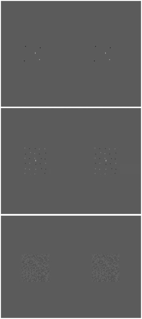
The stimuli were created in Matlab (The MathWorks; Natick, MA) with the Psychophysics toolbox 43, 44 and displayed on a gamma-corrected, 29-cm wide LCD monitor of 1024 X 768 pixels. Each pixel is 0.03 cm in size and subtends 0.85 arc minutes at the viewing distance of 114 cm. As illustrated in Figure 1, the stimuli comprised two identical half images of static 171 arc minutes X 171 arc minutes RDSs that were presented side-by-side on the laptop monitor. To ensure that the mean luminance of the display did not vary with dot density, each dot contained within the RDS was randomly either darker or lighter than the gray background.
Figure 1.
Stimuli to assess stereothresholds. One frame of two identical half images of (a) 0.07%, (b) 0.92%, and (c) 28.3% density 171 arc minutes X 171 arc minutes RDSs, presented side-by-side on the laptop monitor, with a superimposed line segment. In the experiment, the contrast of the RDs and the line segments shown in each panel were adjusted to the same multiple of the observers’ contrast detection thresholds.
To measure stereothresholds for dots that are offset in the stereogram by less than one pixel,45 each 1-pixel dot was convolved with a Gaussian envelope (SD= 1.75 pixels), to yield dots with a full width at half height (corresponding to ±1.15 standard deviations) of approximately 4 pixels (3.5 arc minutes). Each RDS was constructed from a regularly spaced array of Gaussian dots, to which horizontal and vertical position jitter was added with a SD equal to 15% of the average dot-to-dot spacing. To produce relative disparities, the Gaussian dots (but not the superimposed line segment) were offset horizontally by half of the required positional offset in opposite directions in the two half images. Eight densities of the RDS background were selected, ranging from 0.07% to 28.3%. We define the area of each dot as the proportion of its Gaussian amplitude that is either 50% of the maximum value above or 50% of the maximum value below the mean luminance. Because the dot diameter corresponded to 4 pixels, the area of each dot was 4π, or 12.57 square pixels. The RDS density is defined as the percentage of the total stereogram area that is covered by dots.
A single 1 X 13 pixel bright line segment was convolved with the same Gaussian envelope, and transparently pasted at the center of the RDS, plus or minus 1.8 arc minutes (SD) of horizontal and vertical position jitter. The transparent paste operation inserted the line into the RD image without obscuring the neighboring background dots and prevented wrap around of the luminance values*.
During each stimulus presentation, one of 15 possible arrangements of dots for each density was randomly selected and presented for 500 ms.
Stimulus Presentation
Experiments were performed in a dark room. A mirror haploscope and an opaque divider mounted between the haploscope mirrors in the median plane provided full separation between the half images presented to each eye.46,47 Prior to the experiment, the positions of the haploscope mirrors were adjusted for each subject to produce perceived alignment of a pair of vertically separated flashed Nonius lines, in order to minimize horizontal fixation disparity.
Variation of Stereothreshold with RD Density
To match the detectability of the vertical line segment that was superimposed on RDSs with different densities, contrast-detection thresholds for both the line segment and the RDS images were measured prior to the experimental sessions. For each subject, the RDS dot and line contrasts were adjusted for equal visibility across dot densities (see the Appendix - available at [LWW insert link]).
Although retinal image disparities were introduced into the background of RDs and the superimposed line segment always had zero disparity, subjects judged whether the line segment was in front or behind the RD surround. Prior to the experiment, subjects ran a set of approximately 20 practice trials at each RD density to become familiar with the task. No additional practice was deemed necessary, as the subjects were familiar with the feedback sounds and the use of the auxiliary button box from the preceding contrast-detection experiments.
In each session, one of eight RD densities was tested. A session consisted of a total of 70 trials, in which 7 disparities (3 crossed, 3 uncrossed, and 0 disparity) were presented in a random order using the method of constant stimuli. After each response, audio feedback was provided to indicate if it was correct or incorrect. Within a session, the range of disparities was selected individually to obtain a suitable psychometric function, which plotted the percentage of “near” responses as a function of disparity. The psychometric function was fit with a cumulative Gaussian and the threshold was estimated as the semi-interquartile range (=0.675 X SD) of the fitted cumulative Gaussian function. Stereothresholds were measured at least twice for each observer, in a random order.
RESULTS
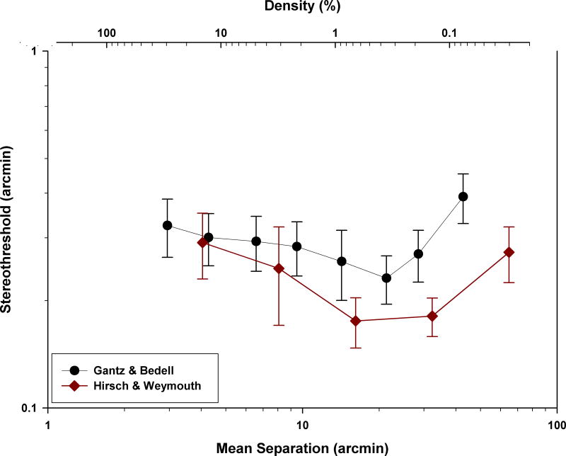
Stereothresholds for depth discrimination of a line superimposed on RDSs with different dot densities average approximately 0.23 arc minutes, corresponding to 13.8 arc seconds, for a RD density of 0.39% and approximately double for both lower and higher RD densities (Figure 2, upper axis). The effect of dot density on the stereothresholds is significant (Repeated Measures ANOVA, Fdf=7,49=3.01, p=0.03, Huynh-Feldt corrected). Pairwise comparisons indicated that the stereothreshold measured with 0.07% density differs significantly from all but the two highest RD densities (p values range from 0.002 to 0.038, Huynh-Feldt corrected) and that the stereothreshold measured with 0.39% density differs significantly from that for 28.28% density (Fdf=1,49= 5.87, p= 0.035, Huynh-Feldt corrected).
Figure 2.
Mean stereothresholds for depth discrimination of a line superimposed on RDSs with various dot densities (black circles) are plotted as a function of the mean line-to-dot separation, equal to one half of the mean RD separation (lower axis) and the RD density (upper axis). Error bars represent the standard error of the mean. Stereothresholds are approximately 0.23 arc minutes for a RD density of 0.39%, and increase at both lower and higher densities. For comparison, data are shown from a previous study (filled diamonds) that measured stereothresholds for two rods that varied in their horizontal separation.
DISCUSSION
Stereothresholds are lowest at a background RD density of 0.39% (mean line-to-dot separation of 21.4 arc minutes) and increase for lower and higher RD densities. For small mean line-to-dot separations (high RD densities), the results are consistent with a study by Westheimer and McKee2 that measured stereothresholds with a 49-element random-dot stimulus with various inter-element separations. They found that stereothresholds increase for small inter-element separations (i.e., at high densities), starting when the separation reaches approximately 10 arc minutes. Because they varied the element separation by scaling the entire target’s size, a change in the number of dots can not account for the increase in stereothreshold at high element densities. Instead, they and other authors attributed the increase in stereothreshold that occurs at small inter-element separations to a crowding effect – an interference in localizing one element produced by the presence of neighboring elements.2, 48, 49 Disparity averaging also is reported to occur for dense stereoscopic stimuli11, 39, 40, 42 and has been suggested to contribute to the crowding effect for stereothresholds.49 In support of this suggestion, studies that examined crowding for orientation discrimination and letter acuity found that the features and locations of the surrounded and flanking targets undergo averaging, even when observers can clearly differentiate the individual component stimuli.50, 51
Although the study by Westheimer & McKee2 did not measure stereothresholds for inter-element separations larger than 15 arc minutes, other studies demonstrated an increase in stereothreshold with a further increase in element separation.46, 52 Large separations between the test and reference elements may degrade the usefulness of the reference target for determining the relative disparity of the test elements. As illustrated by the filled diamonds in Figure 2, Hirsch & Weymouth52 also reported that stereothresholds increase at both small and large target separations, using a target that consisted of two horizontally separated rods. The similar trend shown for these local rod targets and for the small line segment superimposed on our static RD targets suggests that both types of targets are processed by the same disparity-processing mechanism. This suggestion is consistent with the results of a recent perceptual-learning experiment, in which the improvement of stereothreshold produced by training observers with sparse RD targets transferred to dense RD targets, and vice versa.47
The goal of this study was to examine which RD densities could be considered to represent local and global stereograms. Under the assumption that local and global stereotargets are processed by different mechanisms we hypothesized that stereothresholds for an embedded line segment would be best for low densities of the background RDS, when individual elements are processed separately by local disparity mechanisms, and would be worse for high RDS densities, when the disparities of the line and nearby RDs can interact within a global disparity mechanism. However, we found that stereothresholds for static RDSs are optimal for 0.39% density RDSs, and worsen for both lower and higher densities. For low densities, these findings are consistent with those of other authors, who attribute the results to the increased spacing between elements, which reduces the usefulness of the reference target for relative disparity judgments. For high densities, our findings are consistent with an elevation of stereothresholds because ofcrowding, which may be attributable to disparity averaging.11, 39, 40, 42 and occurs regardless of whether the individual elements in the stereogram are distinguishable.2, 41, 48 Because the stereothresholds measured with RDSs of low and high density are limited by different constraints, they can be considered to be different types of stereotargets. Low density RDSs (<0.39%) may be considered “local” stereotargets, and high density RDSs (>0.39%) may be considered “global” stereotargets. However, because of the similarity between the stereothresholds measured using a range of RD densities and the stereothresholds measured with a two-rod stereotarget (a “local” stereotarget), it is likely that all of these stimuli are processed by a single disparity-processing mechanism.
Supplementary Material
Acknowledgments
We thank Hope Queener, Scott Stevenson, and Girish Kumar for assistance with programming. Support was provided by Core Center grant P30 EY 007551 and a University of Houston Vision Research Student Grant.
APPENDIX
The appendix is available at [LWW insert link].
Footnotes
For very low densities, the separation between dots in the RD background and test line is expected to become too large for the RD background to provide an optimal disparity reference. Regardless of the disparity processing mechanism involved, stereothresholds for depth discrimination of the test line are expected to increase for very low RD densities, or large target separations (e.g. Hirsch & Weymouth52).
The transparent paste operation was implemented as follows. Within the region where the line segment was pasted, the luminance of each pixel in the background RD pattern was scaled to the inverse of the luminance of the line. The luminance values of the line and the scaled background were then added. The whole image was rescaled to obtain luminance pixel values that ranged between 0 and 255 and the range of the rescaled pixel values was reduced in the screen color look-up table to produce contrasts less than 100%.
References
- 1.Julesz B. Foundations of Cyclopean Perception. Chicago: The University of Chicago Press; 1971. [Google Scholar]
- 2.Westheimer G, McKee SP. Stereogram design for testing local stereopsis. Invest Ophthalmol Vis Sci. 1980;19:802–9. [PubMed] [Google Scholar]
- 3.Pressmar SB, Haase W. Effects on depth perception and pattern recognition in random dot stereograms by changing the matrix dot density. Ophthalmologe. 2001;98:955–9. doi: 10.1007/s003470170043. [DOI] [PubMed] [Google Scholar]
- 4.Harwerth RS, Fredenburg PM, Smith EL., III Temporal integration for stereoscopic vision. Vision Res. 2003;43:505–17. doi: 10.1016/s0042-6989(02)00653-3. [DOI] [PubMed] [Google Scholar]
- 5.Rose D, Price E. Functional separation of global and local stereopsis investigated by cross-adaptation. Neuropsychologia. 1995;33:269–74. doi: 10.1016/0028-3932(94)00122-6. [DOI] [PubMed] [Google Scholar]
- 6.Harwerth RS, Schor CM. Binocular Vision. In: Kaufman PL, Alm A, Adler FH, editors. Adler’s Physiology of the Eye: Clinical Application. 10. St. Louis, MO: Mosby; 2002. pp. 484–510. [Google Scholar]
- 7.Fawcett SL. An evaluation of the agreement between contour-based circles and random dot-based near stereoacuity tests. J AAPOS. 2005;9:572–8. doi: 10.1016/j.jaapos.2005.06.006. [DOI] [PubMed] [Google Scholar]
- 8.Saladin JJ. Stereopsis from a performance perspective. Optom Vis Sci. 2005;82:186–205. doi: 10.1097/01.opx.0000156320.71949.9d. [DOI] [PubMed] [Google Scholar]
- 9.Harwerth RS, Rawlings SC. Viewing time and stereoscopic threshold with random-dot stereograms. Am J Optom Physiol Opt. 1977;54:452–7. doi: 10.1097/00006324-197707000-00004. [DOI] [PubMed] [Google Scholar]
- 10.Frisby JP, Mein J, Saye A, Stanworth A. Use of random-dot sterograms in the clinical assessment of strabismic patients. Br J Ophthalmol. 1975;59:545–52. doi: 10.1136/bjo.59.10.545. [DOI] [PMC free article] [PubMed] [Google Scholar]
- 11.Stevenson SB, Cormack LK, Schor CM. Hyperacuity, superresolution and gap resolution in human stereopsis. Vision Res. 1989;29:1597–605. doi: 10.1016/0042-6989(89)90141-7. [DOI] [PubMed] [Google Scholar]
- 12.Glennerster A. dmax for stereopsis and motion in random dot displays. Vision Res. 1998;38:925–35. doi: 10.1016/s0042-6989(97)00213-7. [DOI] [PubMed] [Google Scholar]
- 13.White BW. Stimulus-conditions affecting a recently discovered stereoscopic effect. Am J Psychol. 1962;75:411–20. [PubMed] [Google Scholar]
- 14.McKee SP. The spatial requirements for fine stereoacuity. Vision Res. 1983;23:191–8. doi: 10.1016/0042-6989(83)90142-6. [DOI] [PubMed] [Google Scholar]
- 15.Tyler CW, Julesz B. On the depth of the cyclopean retina. Exp Brain Res. 1980;40:196–202. doi: 10.1007/BF00237537. [DOI] [PubMed] [Google Scholar]
- 16.Schlesinger BY, Yeshurun Y. Spatial size limits in stereoscopic vision. Spat Vis. 1998;11:279–93. doi: 10.1163/156856898x00031. [DOI] [PubMed] [Google Scholar]
- 17.Stigmar G. Blurred visual stimuli. II. The effect of blurred visual stimuli on vernier and stereo acuity. Acta Ophthalmol (Copenh) 1971;49:364–79. doi: 10.1111/j.1755-3768.1971.tb00963.x. [DOI] [PubMed] [Google Scholar]
- 18.Cormack LK, Stevenson SB, Schor CM. Interocular correlation, luminance contrast and cyclopean processing. Vision Res. 1991;31:2195–207. doi: 10.1016/0042-6989(91)90172-2. [DOI] [PubMed] [Google Scholar]
- 19.Halpern DL, Blake RR. How contrast affects stereoacuity. Perception. 1988;17:483–95. doi: 10.1068/p170483. [DOI] [PubMed] [Google Scholar]
- 20.Kontsevich LL, Tyler CW. Analysis of stereothresholds for stimuli below 2.5 c/deg. Vision Res. 1994;34:2317–29. doi: 10.1016/0042-6989(94)90110-4. [DOI] [PubMed] [Google Scholar]
- 21.Legge GE, Gu YC. Stereopsis and contrast. Vision Res. 1989;29:989–1004. doi: 10.1016/0042-6989(89)90114-4. [DOI] [PubMed] [Google Scholar]
- 22.Westheimer G, Pettet MW. Contrast and duration of exposure differentially affect vernier and stereoscopic acuity. Proc Biol Sci. 1990;241:42–6. doi: 10.1098/rspb.1990.0063. [DOI] [PubMed] [Google Scholar]
- 23.Ptito A, Zatorre RJ, Larson WL, Tosoni C. Stereopsis after unilateral anterior temporal lobectomy. Dissociation between local and global measures. Brain. 1991;114 ( Pt 3):1323–33. doi: 10.1093/brain/114.3.1323. [DOI] [PubMed] [Google Scholar]
- 24.Mehta AM, France TD. Prominent ocular findings as an early manifestation of systemic lupus erythematosus. J Pediatr Ophthalmol Strabismus. 1998;35:114–5. doi: 10.3928/0191-3913-19980301-12. [DOI] [PubMed] [Google Scholar]
- 25.Schor CM, Wood I. Disparity range for local stereopsis as a function of luminance spatial frequency. Vision Res. 1983;23:1649–54. doi: 10.1016/0042-6989(83)90179-7. [DOI] [PubMed] [Google Scholar]
- 26.Hess RF, Liu CH, Wang YZ. Differential binocular input and local stereopsis. Vision Res. 2003;43:2303–13. doi: 10.1016/s0042-6989(03)00406-1. [DOI] [PubMed] [Google Scholar]
- 27.Tyler CW. A stereoscopic view of visual processing streams. Vision Res. 1990;30:1877–95. doi: 10.1016/0042-6989(90)90165-h. [DOI] [PubMed] [Google Scholar]
- 28.Westheimer G. Spatial interaction in the domain of disparity signals in human stereoscopic vision. J Physiol. 1986;370:619–29. doi: 10.1113/jphysiol.1986.sp015954. [DOI] [PMC free article] [PubMed] [Google Scholar]
- 29.Banks MS, Gepshtein S, Landy MS. Why is spatial stereoresolution so low? J Neurosci. 2004;24:2077–89. doi: 10.1523/JNEUROSCI.3852-02.2004. [DOI] [PMC free article] [PubMed] [Google Scholar]
- 30.Palmisano S, Allison RS, Howard IP. Effect of decorrelation on 3-D grating detection with static and dynamic random-dot stereograms. Vision Res. 2006;46:57–71. doi: 10.1016/j.visres.2005.10.005. [DOI] [PubMed] [Google Scholar]
- 31.Blake R, Wilson HR. Neural models of stereoscopic vision. Trends Neurosci. 1991;14:445–52. doi: 10.1016/0166-2236(91)90043-t. [DOI] [PubMed] [Google Scholar]
- 32.Lankheet MJ, Lennie P. Spatio-temporal requirements for binocular correlation in stereopsis. Vision Res. 1996;36:527–38. doi: 10.1016/0042-6989(95)00142-5. [DOI] [PubMed] [Google Scholar]
- 33.Bruce V, Green PR, Georgeson MA. Visual Perception- Physiology, Psychology, & Ecology. 4. Hove, NY: Psychology Press; 2003. [Google Scholar]
- 34.Cowey A, Porter J. Brain damage and global stereopsis. Proc R Soc Lond B Biol Sci. 1979;204:399–407. doi: 10.1098/rspb.1979.0035. [DOI] [PubMed] [Google Scholar]
- 35.Moradi F. A psychophysical approach to the mechanism of human stereovision. In: Mira J, Sánchez-Andrés J, editors. Foundations and Tools for Neural Modeling. Lecture Notes in Computer Science. Berlin: Springer; 1999. pp. 776–85. [Google Scholar]
- 36.Cormack LK, Landers DD, Ramakrishnan S. Element density and the efficiency of binocular matching. J Opt Soc Am (A) 1997;14:723–30. doi: 10.1364/josaa.14.000723. [DOI] [PubMed] [Google Scholar]
- 37.Harris JM, Parker AJ. Efficiency of stereopsis in random-dot stereograms. J Opt Soc Am A. 1992;9:14–24. doi: 10.1364/josaa.9.000014. [DOI] [PubMed] [Google Scholar]
- 38.Wilcox LM, Duke PA. Spatial and temporal properties of stereoscopic surface interpolation. Perception. 2005;34:1325–38. doi: 10.1068/p5437. [DOI] [PubMed] [Google Scholar]
- 39.Parker AJ, Yang Y. Spatial properties of disparity pooling in human stereo vision. Vision Res. 1989;29:1525–38. doi: 10.1016/0042-6989(89)90136-3. [DOI] [PubMed] [Google Scholar]
- 40.Stevenson SB, Cormack LK, Schor CM. Depth attraction and repulsion in random dot stereograms. Vision Res. 1991;31:805–13. doi: 10.1016/0042-6989(91)90148-x. [DOI] [PubMed] [Google Scholar]
- 41.Westheimer G, Truong TT. Target crowding in foveal and peripheral stereoacuity. Am J Optom Physiol Opt. 1988;65:395–9. doi: 10.1097/00006324-198805000-00015. [DOI] [PubMed] [Google Scholar]
- 42.Tsirlin I, Allison RS, Wilcox LM. Stereoscopic transparency: constraints on the perception of multiple surfaces. J Vis. 2008;8(5):1–10. doi: 10.1167/8.5.5. [DOI] [PubMed] [Google Scholar]
- 43.Brainard DH. The Psychophysics Toolbox. Spat Vis. 1997;10:433–6. [PubMed] [Google Scholar]
- 44.Pelli DG. The VideoToolbox software for visual psychophysics: transforming numbers into movies. Spat Vis. 1997;10:437–42. [PubMed] [Google Scholar]
- 45.Morgan MJ, Castet E. The aperture problem in stereopsis. Vision Res. 1997;37:2737–44. doi: 10.1016/s0042-6989(97)00074-6. [DOI] [PubMed] [Google Scholar]
- 46.Ukwade MT, Bedell HE. Stereothresholds in persons wtih congenital nystagmus and in normal observers during comparable retinal image motion. Vision Res. 1999;39:2963–73. doi: 10.1016/s0042-6989(99)00050-4. [DOI] [PubMed] [Google Scholar]
- 47.Gantz L, Bedell HE. Transfer of perceptual learning of depth discrimination between local and global stereograms. Vision Res. 2010;50:1891–9. doi: 10.1016/j.visres.2010.06.011. [DOI] [PMC free article] [PubMed] [Google Scholar]
- 48.Butler TW, Westheimer G. Interference with stereoscopic acuity: spatial, temporal, and disparity tuning. Vision Res. 1978;18:1387–92. doi: 10.1016/0042-6989(78)90231-6. [DOI] [PubMed] [Google Scholar]
- 49.Kumar T, Glaser DA. Depth discrimination of a crowded line is better when it is more luminant than the lines crowding it. Vision Res. 1995;35:657–66. doi: 10.1016/0042-6989(94)00149-g. [DOI] [PubMed] [Google Scholar]
- 50.Parkes L, Lund J, Angelucci A, Solomon JA, Morgan M. Compulsory averaging of crowded orientation signals in human vision. Nat Neurosci. 2001;4:739–44. doi: 10.1038/89532. [DOI] [PubMed] [Google Scholar]
- 51.Greenwood JA, Bex PJ, Dakin SC. Positional averaging explains crowding with letter-like stimuli. Proc Natl Acad Sci U S A. 2009;106:13130–5. doi: 10.1073/pnas.0901352106. [DOI] [PMC free article] [PubMed] [Google Scholar]
- 52.Hirsch MJ, Weymouth FW. Distance discrimination; effect on threshold of lateral separation of the test objects. Arch Ophthal. 1948;39:224–31. [PubMed] [Google Scholar]
- 53.Gantz L. PhD thesis. Houston: University of Houston; 2009. Are local and global stereograms processed by separate mechanisms? [Google Scholar]
- 54.Gantz L, Bedell HE. Variation of stereothreshold with random-dot density. Invest Ophthalmol Vis Sci. 2008:49. doi: 10.1097/OPX.0b013e3182217487. E-abstract 2545. [DOI] [PMC free article] [PubMed] [Google Scholar]
Associated Data
This section collects any data citations, data availability statements, or supplementary materials included in this article.


