Abstract
Trichomycosis axillaris is a common tropical disease usually affecting the hair shafts of the axillae, characterized by nodular concretions along the hair shafts caused by Corynebacterium tenuis. We describe a 38-year-old patient with trichomycosis axillaris. Treatment, which included shaving of affected hair, followed by topical 3% erythromycin cream and clotrimazole powder was fully effective.
Keywords: axilla, corynebacterium tenuis, erythromycin, hair shaft, infection
Case
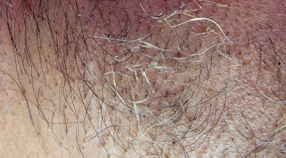
A 38-year-old obese man sought consultation with us for the bad axillary odor and "roughened" texture of his axillary hair on both sides. The condition used to worsen in summer, staining his clothing as he had a tendency of profuse sweating. On examination, creamy yellow concretions along several hair shafts were seen [Fig. 1]. With a clinical diagnosis of trichomycosis axillaris (TA), we treated him with shaving of affected hair followed by topical 3% erythromycin cream and drying clotrimazole powder. His axillary odour entirely disappeared within a couple of weeks. He was advised to continue the medications until the end of summer.
Figure 1.
Trichomycosis (trichobacteriosis) axillaris. Yellow concretions seen along hair shafts of right axilla of an Indian adult male patient.
Trichomycosis axillaris is bacterial colonization of hairs commonly affecting axillae and, sometimes the pubes. It is characterized by concretions along the hair shafts clinically seen commonly as yellow and rarely as red or black nodules. Thus, the term "trichomycosis" is a misnomer and may now better be called as trichobacteriosis.[1]
The concretions contain the bacterial colonies of Corynebacterium tenuis, easily recognized in gram stained preparation of crushed concretions seen under light microscopy as purple rods and coccobacilli. Different taxonomic species of corynebactria are identified. The concretion material is derived from bacterial colonization along the hair shaft containing dried apocrine sweat with a cementing substance generated by the bacteria. Encapsulated corynebacteria entrapped in a biofilm serve as adhering mechanism, which possibly helps to escape immunological attack by the host.[2,3,4]
The condition is easily recognized by the clinicians. Wood's light examination reveals pale-yellow fluorescence. Ultraviolet light enhanced visualization of white prominent nodule represents bacterial colonies and encapsulated biofilm. Rarely, the condition may be confused with pediculosis and Trichosporon aselie infections.[1]
Warm, moist environment, excessive sweating and poor local hygiene are common predisposing factors.[1,3] TA often results into bad odour in axillae and may stain the clothing. Rarely, it may cause hair shaft breakage.[4] Frequent association with other cutaneous diseases caused by corynebacteria forms the so called "corynebacterial triad" with TA, pitted keratolysis (which affects soles) and erythrasma, (which affects inguinal and axillary folds).[5]
Vigorous rubbing of the affected hair while washing, clipping of only the affected hair in early infections or shaving of the entire axilla in severe infection are known physical measures to clear the infection. Topical application of erythromycin or clindamycin or imidazole derivatives and/or benzyl peroxide are generally helpful to contain the infection. Topical ammonium chloride solution and drying powders counter perspiration and their regular use may prevent further recurrences.[1,2,5]
References
- Blaise G, Nikkels AF, Hermanns-Lê T, Nikkels-Tassoudji N, Piérard GE. Corynebacterium-associated skin infections. Int J Dermatol. 2008;47:884–890. doi: 10.1111/j.1365-4632.2008.03773.x. [DOI] [PubMed] [Google Scholar]
- Coyle MB, Lipsky BA. Coryneform bacteria in infectious diseases: clinical and laboratory aspects. Clin Microbiol Rev. 1990;3:227–246. doi: 10.1128/cmr.3.3.227. [DOI] [PMC free article] [PubMed] [Google Scholar]
- Orfanos CE, Schloesser E, Mahrle G. Hair destroying growth of Corynebacterium tenuis in the so-called trichomycosis axillaris. New findings from scanning electron microscopy. Arch Dermatol. 1971;103:632–639. [PubMed] [Google Scholar]
- Shelley WB, Miller MA. Electron microscopy, histochemistry, and microbiology of bacterial adhesion in trichomycosis axillaris. J Am Acad Dermatol. 1984;10:1005–1014. doi: 10.1016/s0190-9622(84)80325-4. [DOI] [PubMed] [Google Scholar]
- Rho NK, Kim BJ. A corynebacterial triad: Prevalence of erythrasma and trichomycosis axillaris in soldiers with pitted keratolysis. J Am Acad Dermatol. 2008;58(2 Suppl):S57–58. doi: 10.1016/j.jaad.2006.05.054. [DOI] [PubMed] [Google Scholar]


