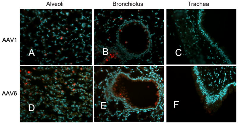
Fig. 2. AAV transduction of mouse airways in vivo.


(a) Balb/c mice were inoculated by the intro-tracheal route with a single dose of 1×10¹¹vg/mouse in a volume of 50ul of AAV1-GFP (panels A, B and C) or AAV6-GFP (D, E, and F). Mouse trachea and lungs were harvested at 1 month p.i.. Paraffin-embedded tissue sections were prepared, and transduced cells were immunolabeled with rabbit anti-GFP antibody (Sigma) followed by Alexafluor594- conjugated goat anti-rabbit antibody (Invitrogen). Nuclei were stained blue with DAPI. Representative images for alveolar (A & D), bronchiolus (B & E), and tracheal regions (C & F) are shown. (b) The numbers of GFP positive cells per image field were quantified and the mean numbers from five random images were shown (± SEM).
