Abstract
Much controversy surrounds the relative role of insulin signaling in the brain in the control of hepatic glucose metabolism. In this issue of the JCI, Ramnanan and colleagues demonstrate that arterial infusion of insulin into the brains of dogs reduces net hepatic glucose output without altering endogenous glucose production. However, this effect was modest and required both prolonged fasting and prolonged exposure of the brain to insulin, raising doubts about the overall physiological relevance of insulin action in the brain on hepatic glucose metabolism. Given the dominant direct role that insulin plays in inhibiting glucose production in the liver, we suggest that the main effect of central insulin on hepatic glucose metabolism may be more chronic and assume greater significance either when portal insulin is deficient, as occurs during exogenous insulin treatment of type 1 diabetes, or when chronic hyperinsulinemia and central insulin resistance develops, as occurs in type 2 diabetes.
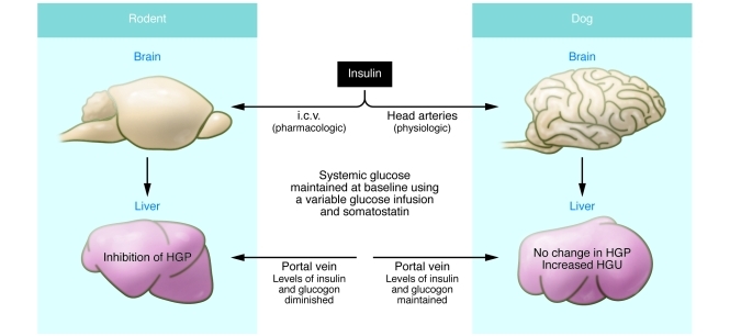
Insulin controls nutrient and metabolic homeostasis via effects on the liver, muscle, and adipose tissue. After a meal, insulin acts on the liver to inhibit net hepatic glucose output (NHGO) — the balance between hepatic glucose production (HGP), which occurs via gluconeogenesis and glycogenolysis, and hepatic glucose uptake, which includes glycogen synthesis. In humans (1) and in large mammals such as the dog (2), the direct action of insulin on the liver plays a dominant role in suppressing NHGO. However, studies in rodents suggest that insulin can also act within the brain to alter hepatic glucose metabolism, primarily by suppressing HGP (Figure 1 and refs. refs. 3, 4). In this issue of the JCI, Ramnanan and colleagues show that raising brain insulin levels in dogs using the physiologically relevant route of carotid and vertebral arterial infusions inhibits NHGO by increasing hepatic uptake of glucose and its incorporation into glycogen (Figure 1 and ref. 5). HGP was not substantially altered, although the expression of gluconeogenic genes in liver was diminished. However, unlike the transient (approximately 1–2 hours) rise in insulin and fall in glucagon levels in the portal circulation that follows most carbohydrate-rich meals, the insulin infusions performed by Ramnanan and colleagues were carried out for 3–4 hours and the dogs were fasted for 42 hours; even under these conditions, only a relatively modest inhibition of NHGO was observed. While the work of Ramnanan and colleagues (5) confirms that insulin can act on the brain to alter hepatic glucose metabolism in dogs, it does not settle the question about the relative importance of central insulin signaling in mediating NHGO under physiological conditions in humans and other mammals.
Figure 1. Effect of insulin delivery to the brain on hepatic glucose metabolism in the rat (3) and dog (5).
In the rat, insulin was administered i.c.v. in supraphysiological doses, whereas in the dog, insulin was administered via direct carotid and vertebral arterial infusion, which raised levels to the upper physiological range. In both cases, plasma glucose was kept at baseline using variable glucose infusion and somatostatin to suppress endogenous insulin and glucagon secretion. However, in the rat, insulin alone was replaced systemically, whereas in the dog, both insulin and glucagon were directly delivered to the liver to maintain portal levels. In both models, changes in hepatic glucose metabolism were observed: in the rat, HGP was inhibited; in the dog, a change in HGP was not seen, but hepatic glucose uptake (HGU) increased, thereby diminishing NHGO.
Insulin and the brain
The brain is an insulin-sensitive organ; various studies have shown that insulin action in the brain affects energy and glucose homeostasis, neuroendocrine function, reward, memory, and learning (3, 6, 7). Neurons in many areas of the brain express insulin receptors (InsRs), and insulin, acting on these receptors, causes activation of PI3K and MAPK signaling pathways (8, 9). Insulin-mediated activation of these pathways can lead to short-term changes in neuronal activity (10) or prolonged changes in gene transcription and neuronal plasticity (11), which occur within the 3- to 4-hour time frame used in the Ramnanan studies (5).
In seminal pancreatic clamp studies by Obici et al. (3), in short-term–fasted rats in which secretion of insulin and glucagon were inhibited using somatostatin and insulin was infused peripherally, i.c.v. delivery of insulin for 4–6 hours inhibited HGP by approximately 30%. The central effects of insulin were thought to signal via the vagus nerve to the liver (4). However, sympathetic innervation to the liver increases hepatic gluconeogenesis and glycogenolysis (12) and might also play a role in mediating the central action of insulin (13). Thus, the collective observations in rodents suggest that insulin signaling in the brain can have an important effect on HGP. But this does not appear to be the case in dogs. Cherrington and colleagues previously found that direct delivery of insulin to the dog liver via the portal vein was the dominant inhibitor of HGP, as opposed to the effects of arterial infusions of insulin into the brain (2). In addition, any significant requirement for a central effect of insulin on neutrally mediated NHGO (4) is not supported by studies conducted in humans with denervated livers (1). Although these differences between rodents on the one hand and dogs and humans on the other might be species specific, they raise important questions regarding the role of insulin signaling in the brain in the control of HGP under short- and long-term physiologic and pathologic conditions.
Are the central effects of insulin physiologically relevant?
Many studies in rodents support a role for insulin acting on the brain as a regulator of peripheral glucose homeostasis. InsR-expressing neurons in the rat are localized in important hypothalamic and hindbrain areas that modulate glucose homeostasis, energy intake and expenditure, and neuroendocrine and autonomic functions (8, 9). In rodent brains, insulin has relatively acute effects on neuronal activity in vitro (14). Furthermore, in rodents, up to 75% of hypothalamic glucose-sensing neurons that alter their activity in response to physiological changes in ambient glucose levels also coexpress InsR and the insulin-dependent GLUT4 glucose transporter, thus suggesting a potential role for insulin-mediated effects on neuronal glucose uptake (15). Although such an effect has not been demonstrated directly, deletion of InsR in GLUT4-expressing neurons of mice promotes the development of diabetes (16). Moreover, administration of InsR antisense or antagonists via the third cerebral ventricle blunts the inhibitory effect of i.c.v. insulin on HGP (3). Finally, deletion of InsR signaling in agouti-related protein (AgRP) neurons in the mouse hypothalamus attenuates the central effect of insulin on HGP (17), whereas restoration of InsR in AgRP neurons in mice lacking InsR elsewhere in the brain and body restores the ability of peripheral insulin infusion to inhibit HGP (18).
On the other hand, there is strong evidence against central actions of insulin having an important role in regulating hepatic glucose metabolism under physiological conditions. First, use of somatostatin and peripheral insulin replacement alone in rodent studies decreases insulin and glucagon levels in the hepatic portal circulation, likely magnifying the effect of centrally delivered insulin on HGP (3). Second, although deletion of InsR from specific hypothalamic neurons can affect hepatic glucose metabolism, the overall effect of decreasing insulin signaling throughout the brain in mice lacking InsR in neurons is not dramatic (19), unless there is concomitant InsR deletion in peripheral insulin-sensitive tissues. Additionally, InsR deletion in the mouse germline likely alters neuronal development and plasticity. Even relatively short-term deletion of InsR from specific neurons is likely associated with changes in neuronal connectivity that could alter the neural circuits involved with glucose homeostasis (11).
There are also several technical issues that cloud interpretation of rodent and dog studies in this field. For example, studies in rats and mice generally use i.c.v. infusion of insulin at supraphysiological doses. This technique has serious shortcomings. Insulin normally enters the brain from plasma by crossing the blood-brain barrier from the arterial circulation. Insulin is also transported into cerebrospinal fluid via the choroid plexus, which contains high levels of InsR (9), from which insulin gains access to the periventricular brain areas but is unlikely to diffuse more than a short distance (20). This limits how much insulin reaches neurons involved in neuroendocrine and autonomic regulation that are distant from the ventricles. While it is often thought that insulin infused into the third cerebral ventricle primarily engages hypothalamic neurons (3, 4), such infusions also expose critical autonomic InsR-expressing neurons in the hindbrain nucleus tractus solitarius and dorsal motor nucleus of the vagus to high levels of insulin (8). It is also noteworthy that the euglycemic hyperinsulinemic or pancreatic clamps used to investigate the central effects of insulin signaling on hepatic glucose metabolism are carried out over long periods of time: 3–6 hours in rodents (3), and, in the case of the Ramnanan studies (5), in dogs that were fasted for 42 hours prior to the clamp. Such experimental conditions in no way mimic the relatively brief (30–180 minutes) rise and fall of circulating insulin that follows a meal or the marked suppression of HGP accompanying physiological increments in insulin, which are near maximal at 1 hour. This difference in insulin kinetics under clamp versus physiological conditions is crucial in the interpretation of the central action of insulin on HGP. In contrast, the direct effect of insulin on the liver is both rapid and dominant in the suppression of HGP (2).
Making sense of the data
To make sense of all the data, we must first ask whether existing studies carried out under a variety of experimental conditions faithfully reproduce physiological conditions. The answer to this question is likely no. Nevertheless, the studies of Ramnanan et al. suggest that, under certain conditions, insulin delivered to the brain from the systemic circulation can alter NHGO (5). However, we believe that current data continue to support the view that insulin normally regulates the liver predominantly via local delivery from the portal circulation. The close proximity of the pancreas to the liver provides high local insulin concentrations, which promote the rapid changes in NHGO observed. In contrast, passage of insulin into the brain is much slower, and its levels in the brain are much lower, than those in the general circulation (21). The contribution of CNS insulin to HGP regulation might, however, be somewhat greater in the setting of insulin-deficient type 1 diabetes, where insulin is delivered systemically.
On the other hand, brain insulin signaling does appear to exert important tonic direct and/or indirect effects that serve to limit excessive HGP. Loss of insulin signaling in neurons expressing Glut4 markedly increases hyperglycemia in mice with peripheral insulin resistance (18). Furthermore, in nondiabetic rats, acute blockade of insulin signaling within the ventromedial hypothalamus, a key glucose-sensing region, immediately stimulates glucagon secretion (7). Impaired insulin secretion also appeared in preliminary studies using virus-induced knockdown of ventromedial hypothalamus InsR (R.S. Sherwin, unpublished observations). Of course, such a reduction in central insulin signaling is similar to what happens in type 2 diabetic individuals exposed to chronic hyperinsulinemia in conjunction with the development of central and peripheral insulin resistance. Thus, central insulin signaling may play its most important role in regulating hepatic glucose metabolism in such individuals when it is attenuated after prolonged hyperinsulinemia. Such a hypothesis does not require that central insulin resistance play a dominant role in the regulation of hepatic glucose metabolism. Rather, it would become an important contributor to the overall disturbance experienced by such individuals and might provide a therapeutic target for remediation of their abnormal glucose metabolism.
In conclusion, the work of Ramnanan et al. (5), together with previous studies from this group (2, 22), demonstrate the utility of the dog as a model in which many experimental variables can be controlled simultaneously. The current study clearly demonstrates that over time, insulin can act centrally to alter hepatic glucose metabolism under such controlled conditions. The daunting challenge going forward is to demonstrate whether such findings apply to any physiologically relevant condition or whether, as we suggest, they might be more relevant to pathological conditions such as those encountered in type 2 diabetes mellitus.
Acknowledgments
This work was supported in part by the Research Service of the Veterans Administration and NIDDK grant DK53181 to B.E. Levin, and by NIH grants DK 20495, P30 DK 45735, and UL1 RR024139 and Juvenile Diabetes Research Foundation grant 4-2004-807 to R.S. Sherwin.
Footnotes
Conflict of interest: Robert S. Sherwin has stock options from Insulet and receives honoraria from Novartis and MannKind for service on data safety monitoring boards.
Citation for this article: J Clin Invest. 2011;121(9):3392–3395. doi:10.1172/JCI59653.
See the related article beginning on page 3713.
References
- 1.Perseghin G, et al. Regulation of glucose homeostasis in humans with denervated livers. J Clin Invest. 1997;100(4):931–941. doi: 10.1172/JCI119609. [DOI] [PMC free article] [PubMed] [Google Scholar]
- 2.Edgerton DS, et al. Insulin’s direct effects on the liver dominate the control of hepatic glucose production. J Clin Invest. 2006;116(2):521–527. doi: 10.1172/JCI27073. [DOI] [PMC free article] [PubMed] [Google Scholar]
- 3.Obici S, Zhang BB, Karkanias G, Rossetti L. Hypothalamic insulin signaling is required for inhibition of glucose production. Nat Med. 2002;8(12):1376–1382. doi: 10.1038/nm1202-798. [DOI] [PubMed] [Google Scholar]
- 4.Pocai A, et al. Hypothalamic K(ATP) channels control hepatic glucose production. Nature. 2005;434(7036):1026–1031. doi: 10.1038/nature03439. [DOI] [PubMed] [Google Scholar]
- 5.Ramnanan CJ, et al. Brain insulin action augments hepatic glycogen synthesis without suppressing glucose production or gluconeogenesis in dogs. . J Clin Invest. 2011;121(9):3713–3723. doi: 10.1172/JCI45472. [DOI] [PMC free article] [PubMed] [Google Scholar]
- 6.Woods SC, Lotter EC, McKay LD, Porte D., Jr Chronic intracerebroventricular infusion of insulin reduces food intake and body weight of baboons. Nature. 1979;282(5738):503–505. doi: 10.1038/282503a0. [DOI] [PubMed] [Google Scholar]
- 7.Paranjape SA, et al. Influence of insulin in the ventromedial hypothalamus on pancreatic glucagon secretion in vivo. Diabetes. 2010;59(6):1521–1527. doi: 10.2337/db10-0014. [DOI] [PMC free article] [PubMed] [Google Scholar]
- 8.Unger JW, Moss AM, Livingston JN. Immunohistochemical localization of insulin receptors and phosphotyrosine in the brainstem of the adult rat. NeuroScience. 1991;42(3):853–861. doi: 10.1016/0306-4522(91)90049-T. [DOI] [PubMed] [Google Scholar]
- 9.Werther GA, et al. Localization and characterization of insulin receptors in rat brain and pituitary gland using in vitro autoradiography and computerized densitometry. Endocrinology. 1987;121(4):1562–1570. doi: 10.1210/endo-121-4-1562. [DOI] [PubMed] [Google Scholar]
- 10.Spanswick D, Smith MA, Mirshamsi S, Routh VH, Ashford ML. Insulin activates ATP-sensitive K+ channels in hypothalamic neurons of lean, but not obese rats. Nat Neurosci. 2000;3(8):757–758. doi: 10.1038/77660. [DOI] [PubMed] [Google Scholar]
- 11.Levin BE, Kang L, Sanders NM, Dunn-Meynell AA. Role of neuronal glucosensing in the regulation of energy homeostasis. Diabetes. 2006;55(suppl 2):S122–S130. [Google Scholar]
- 12.Lautt WW. Autonomic neural control of liver glycogen metabolism. Med Hypotheses. 1979;5(12):1287–1296. doi: 10.1016/0306-9877(79)90096-3. [DOI] [PubMed] [Google Scholar]
- 13.Sakaguchi T, Bray GA. Sympathetic activity following paraventricular injections of glucose and insulin. Brain Res Bull. 1988;21(1):25–29. doi: 10.1016/0361-9230(88)90115-3. [DOI] [PubMed] [Google Scholar]
- 14.Spanswick D, Smith MA, Mirshamsi S, Routh VH, Ashford ML. Insulin activates ATP-sensitive K+ channels in hypothalamic neurons of lean, but not obese rats. Nature Neurosci. 2000;3(8):757–758. doi: 10.1038/77660. [DOI] [PubMed] [Google Scholar]
- 15.Kang L, Routh VH, Kuzhikandathil EV, Gaspers L, Levin BE. Physiological and molecular characteristics of rat hypothalamic ventromedial nucleus glucosensing neurons. Diabetes. 2004;53(3):549–559. doi: 10.2337/diabetes.53.3.549. [DOI] [PubMed] [Google Scholar]
- 16.Lin HV, et al. Diabetes in mice with selective impairment of insulin action in glut4-expressing tissues. Diabetes. 2011;60(3):700–709. doi: 10.2337/db10-1056. [DOI] [PMC free article] [PubMed] [Google Scholar]
- 17.Konner AC, et al. Insulin action in AgRP-expressing neurons is required for suppression of hepatic glucose production. Cell Metab. 2007;5(6):438–449. doi: 10.1016/j.cmet.2007.05.004. [DOI] [PubMed] [Google Scholar]
- 18.Lin HV, et al. Divergent regulation of energy expenditure and hepatic glucose production by insulin receptor in agouti-related protein and POMC neurons. Diabetes. 2010;59(2):337–346. doi: 10.2337/db09-1303. [DOI] [PMC free article] [PubMed] [Google Scholar]
- 19.Bruning JC, et al. Role of brain insulin receptor in control of body weight and reproduction. Science. 2000;289(5487):2122–2125. doi: 10.1126/science.289.5487.2122. [DOI] [PubMed] [Google Scholar]
- 20.Mullier A, Bouret SG, Prevot V, Dehouck B. Differential distribution of tight junction proteins suggests a role for tanycytes in blood-hypothalamus barrier regulation in the adult mouse brain. J Comp Neurol. 2010;518(7):943–962. doi: 10.1002/cne.22273. [DOI] [PMC free article] [PubMed] [Google Scholar]
- 21.Gerozissis K, Rouch C, Lemierre S, Nicolaidis S, Orosco M. A potential role of central insulin in learning and memory related to feeding. Cell Mol Neurobiol. 2001;21(4):389–401. doi: 10.1023/A:1012606206116. [DOI] [PMC free article] [PubMed] [Google Scholar]
- 22.Sindelar DK, Balcom JH, Chu CA, Neal DW, Cherrington AD. A comparison of the effects of selective increases in peripheral or portal insulin on hepatic glucose production in the conscious dog. Diabetes. 1996;45(11):1594–1604. doi: 10.2337/diabetes.45.11.1594. [DOI] [PubMed] [Google Scholar]


