Abstract
Purpose:
To determine if heel height alters sagittal plane knee kinematics when landing from a forward hop or drop landing.
Background:
Knee angles close to extension during landing are theorized to increase ACL injury risk in female athletes.
Methods:
Fifty collegiate females performed two single-limb landing tasks while wearing heel lifts of three different sizes (0, 12 & 24 mm) attached to the bottom of a sneaker. Using an electrogoniometer, sagittal plane kinematics (initial contact [KAIC], peak flexion [KAPeak], and rate of excursion [RE]) were examined. Repeated measures ANOVAs were used to determine the influence of heel height on the dependent measures.
Results:
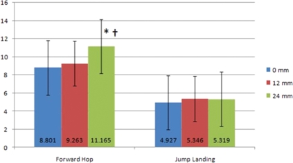
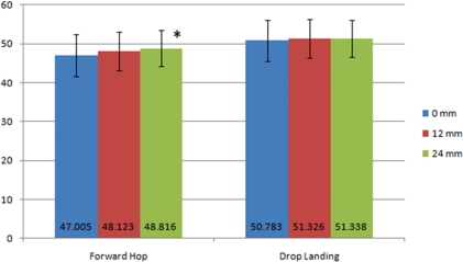
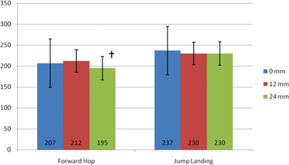
Forward hop task- KAIC with 0 mm, 12 mm, and 24 mm lifts were 8.88±6.5, 9.38±5.8 and 11.28±7.0, respectively. Significant differences were noted between 0 and 24 mm lift (p<.001) and 12 and 24 mm lifts (p=.003), but not between the 0 and 12 mm conditions (p=.423). KAPeak with 0 mm, 12 mm, and 24 mm lifts were 47.08±10.9, 48.18±10.3 and 48.88±9.7, respectively. A significant difference was noted between 0 and 24 mm lift (p=.004), but not between the 0 and 12 mm or 12 and 24 mm conditions (p=.071 and p=.282, respectively). The RE decreased significantly from 2128/sec±52 with the 12 mm lift to 1958/sec±55 with the 24 mm lift (p=.004). RE did not differ from 0 to 12 or 0 to 24 mm lift conditions (p=.351 and p=.086, respectively). Jump-landing task- No significant differences were found in KAIC (p=.531), KAPeak (p=.741), or the RE (p=.190) between any of the heel lift conditions.
Conclusions:
The addition of a 24 mm heel lift to the bottom of a sneaker significantly alters sagittal plane knee kinematics upon landing from a unilateral forward hop but not from a drop jump.
Keywords: ACL, heel lift, kinematics, landing
INTRODUCTION
It is well established that anterior cruciate ligament (ACL) injuries occur more frequently in female compared to male athletes.1–7 However, extensive research into the basis of this gender disparity continues to be inconclusive. Although many theories have been proposed,8 no evidence exists to overwhelmingly support any single cause for the female's propensity towards ACL injury suggesting that the link may be multi-factorial. Furthermore, researchers have struggled to establish consistent suggestions regarding proven methods to limit or prevent these injuries.
Risk factors for sustaining ACL injuries have been categorically classified as those related to environmental, anatomical, hormonal, or biomechanical origins.8 There are obvious limitations in the feasibility of altering some of these risk factors. Given the ability to alter joint kinematics and muscle activity, many researchers have conducted studies that have focused on biomechanical risk factors utilized by female athletes during jumping and landing activities.9–16 Videotape analysis of ACL injuries in both genders has demonstrated that a common characteristic observed at the time of injury is a knee posture close to extension.9–11 Researchers have demonstrated that females repeatedly utilize this kinematic pattern in a variety of sport maneuvers.12–16
The ramifications of a more extended knee posture during landing include increased electrical activity of the quadriceps,13 increased anterior tibio-femoral shear18–21 and increased vertical ground reaction forces.14,22 Likewise, there is a decrease in activity of the hamstrings,13,17–19 and less sagittal plane knee joint excursion occurs throughout the movement resulting in less time to attenuate forces and moments at the joint.23-24 Each of the aforementioned compensations are thought to increase ACL injury risk.5,21,25–27
ACL injury prevention programs have been used to favorably influence kinematic patterns during varied landing tasks in female athletes. Preventative exercise programs designed for female athletes participating in a variety of sports typically consist of plyometric activities and have demonstrated the ability to positively affect lower extremity kinematics and kinetics28–30 in laboratory settings14,31,32 as well as decrease injury rates on the field.37 However, many of these programs require a substantial time investment of 90-minutes or more several times per week,14,30,32,33 often requiring greater than six-weeks of consistent training.28,30–32,34 This time commitment may dissuade coaches and athletes from participating. Among those that do participate, the time component is apt to have negative effects on compliance, which has been demonstrated to be a substantial factor in the overall success of a program.34 Researchers that used training programs requiring only 15 minutes to complete found that only the most elite athletes who were consistently compliant had a decrease in injury rates, while other athletic populations or less compliant subjects saw no changes.34 Furthermore, the duration of the protective effects of these training programs after cessation of participation is unknown.
A prevention strategy that requires minimal time and effort yet can still favorably alter knee biomechanics would be of substantial benefit to female athletes. To this end, evidence exists that increased heel height alters lower extremity biomechanics, including sagittal plane knee postures. Both static-standing and dynamic gait analyses have demonstrated increased knee flexion angles with increased heel height.35–39 It is unknown; however, if these findings will be evident during sports maneuvers, such as jump-landing or single limb hopping. If so, the incorporation of a heel lift may offer an alternative strategy for altering sagittal plane knee kinematics during sporting activities.
Therefore, the purpose of this study was to examine the effect of heel height on knee flexion angles at initial contact, peak flexion and the rate of excursion while performing a drop-landing and a forward hop. The authors hypothesized that increased heel height would significantly increase knee flexion at initial contact, increase peak knee flexion, and decrease the rate of excursion during both tasks.
METHODS
Subjects
Fifty female subjects between the ages of 18 and 25 (age = 20.6 ± 1.5 years, height = 166.2 ± 6.1 cm, weight = 64.3 ± 9.3 kg) were recruited from the university community via consecutive sampling procedures. Pilot data collection using identical procedures described below was performed on 10 female subjects to obtain subject size calculations for the current study. A readily available power analysis software program (G*Power 3.1.10, Dusseldorf, Germany)49 was used to calculate the subject size required to obtain 80% power with p<0.05 based on the pilot data. The sample size needed to achieve this statistical power was 48 subjects. Selection criteria for all subjects included: 1) at least recreationally active; 2) no history of knee surgeries; 3) no acute lower extremity injury within the past six months that required the use of an assistive device for more than one day; 4) no use of foot orthotics; 5) the ability to perform required jumping tasks without limitation. Recreationally active was defined as being engaged in aerobic and/or anaerobic exercise for an average of four to five hours per week. However, this study did not exclude those who participated in more exercise, such as the levels that intercollegiate athletes would engage in. Sixteen of the participants were NCAA athletes participating in volleyball, lacrosse, soccer, or tennis. The 34 non-NCAA athletes participated in activities such as weight lifting, running, exercise classes, and Pilates based exercise for a self-reported average of 5.5 hours per week. Neither group reported having participated in any form of ACL-specific prevention activities in their past. All subjects were asked to read and sign an informed consent form approved by Duquesne University's Institutional Review Board.
Experimental Design & Setting
All data were collected in a laboratory setting. A randomized block design was used to evaluate the effect of heel lift height on sagittal plane knee joint kinematics of subjects participating in two landing activities on their dominant limb only: a drop-landing from a 40 cm high platform and a forward hop over an obstacle. As previously noted, landing is a common injury mechanism and previous authors40,41 have utilized similar tasks to those presented here to analyze lower extremity kinematics in female athletes. Three dependent variables were assessed in the sagittal plane: knee flexion angle at initial contact and peak flexion, and the rate of excursion. Rate of excursion was defined as the change in the knee joint angle (degrees) between initial contact and peak knee flexion divided by the time elapsed during this period (milliseconds). These factors have been shown to differ significantly between genders in landing tasks.40 The independent variable was heel height, with 3 different conditions studied: no lift (0 mm - control), 12 mm, and 24 mm. The lift sizes were chosen based on published heel lift research42–44 and pilot testing. The application of the varying heel lifts was completed in random order. The subjects were not told which heel lift was being utilized in an effort to minimize bias.
Instrumentation
An electrogoniometer (EG) (Biometrics Ltd; Gwent, UK) that sampled at 2000 Hz was used to measure sagittal plane knee joint angles during the drop-landing testing procedure. Recent research has demonstrated intra-tester reliability coefficients for EG measurement of knee angles in static standing with various heel heights at 0.87-0.88.45 Bronner et al46 found that EG reliability correlation and accuracy were high (r≥0.999, SEM≤1.728; respectively) and the concurrent validity correlations to a motion analysis criterion measure were also high (r≥0.991) when analyzing knee angle displacement during dance moves, including jumping. The manufacturer lists the EG's accuracy at ±28 and repeatability at 18 over a 908 range.
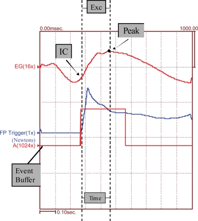
A force plate (Bertec Corporation, Columbus, OH; sampled at 2000 Hz) securely mounted in the sub-floor (such that the top surface of the force plate was level with the laboratory floor) was utilized to mark initial contact with the floor. Specifically, initial contact was determined to be when a minimal amount of force (10 N) applied to the force plate was detected by the acquisition software above a quiet baseline measured 100 ms prior to the event (Figure 1). The acquisition software was programmed to use the force plate as an event marker from which the data were recorded using a trigger sweep mode. The software system continually records and temporarily stores all data. This circular buffering allows the user to have access to data points up to one second prior to the event marker. A total of 1000 ms was recorded: 300 ms pre-contact and 700 ms post-contact for each trial.
Figure 1.
Example of force plate data output.
All hardware was connected to an analog-to-digital conversion board and a desktop computer (Dell; Austin, TX) thereby allowing synchronous collection of data from each of the respective sources. Raw data were acquired and filtered with a software acquisition and analysis system (Run Technologies; Laguna Hills, CA).
Procedures
Identical procedures were followed for each subject. Data collection was completed during one session. Demographic data including height, weight, age, and the level and type of sport or activity was collected. All testing was performed on the dominant lower extremity. Dominance was determined by having the subject jump off a 20 cm wooden box and land on one leg. The lower extremity upon which the subject landed two out of three trials was considered the dominant lower extremity.47 The subjects were asked to demonstrate an ability to perform the landing task to be utilized in this study. Each task was described and the potential participant completed each activity. If an individual was unable to perform both landing tasks without restriction they were excused from participation in the study.
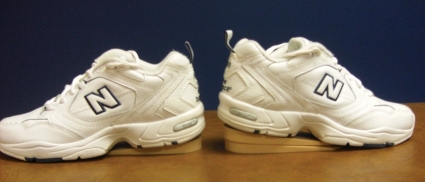
Subjects donned a standardized shoe (NewBalance, Model 600; Boston, MA) that was neutrally balanced (neither a supination or pronation bias) for the testing procedures. Subjects chose their own shoe size. Prior to donning the shoes, the investigator determined the order of the application of the heel lifts by randomly pulling papers labeled with the 3 different height measurements out of a hat. The subject was not told which heel height was being used for each trial. The heel lifts (G&W Heel Lift Incorporated; Cuba, MO) were injection-molded PVC vinyl lifts 12 mm in height. Two heel lifts were combined with double-sided tape provided by the manufacturer to achieve the 24 mm height (Figure 2). The investigator then affixed the appropriate heel lift to the under-surface of the subject's shoe with carpet tape (Ace Hardware Corp.; Oak Brook, IL).
Figure 2.
Heel lifts, placed on the external surface of standard shoe.
Next, the EG was centered over the lateral joint line of the knee with the proximal block of the EG aligned with the greater trochanter and the distal block with the lateral malleolus. The end-blocks of the EG were attached to the subject's lateral leg with double sided adhesive tape and further secured with a thin foam wrap. Extra care was taken to ensure the foam wrap did not interfere with the optical fibers of the device. Once in place, the subject was asked to stand with her knee at 08 as verified by a manual goniometer, at which time the EG was zeroed.
Drop-landing activity.
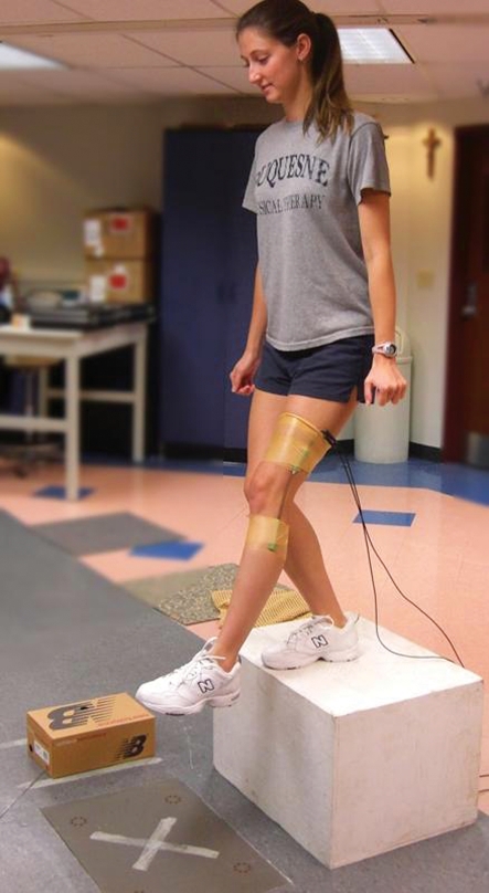
The subject stepped onto the 40 cm high platform and stood facing the force plate with her feet shoulder width apart and toes aligned with the edge of the platform. The platform was located 11 cm from the force plate. The subject then leaned and fell forward off the platform and landed in the middle of the force plate on her dominant lower extremity (Figure 3). The subject was instructed to maintain her balance on the single limb until cued by the investigator to place the other lower extremity on the floor. Failure to land in the middle of the force plate or to maintain single limb balance following the jump resulted in negation of that trial from the data set and the subject was asked to repeat the jump.
Figure 3.
Drop landing activity set up.
Forward hop activity.
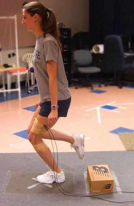
The subject stood with both lower extremities on the floor and feet shoulder width apart 45% of her height away from the force plate. A 12 cm tall box was placed halfway between the subject and the force plate. These procedures are similar to those found in previous studies investigating lower extremity kinematics in landing.40 The subject hopped forward from a static stance over the box and landed in the middle of the platform on the dominant lower extremity (Figure 4). The subject was instructed to maintain her balance on the single limb until cued by the investigator to place the other lower extremity on the floor. Failure to land in the middle of the force plate or to maintain single limb balance following the jump resulted in negation of that trial from the data set and the subject was asked to repeat the hop.
Figure 4.
Forward Hop Activity set up.
Figure 5.
Graphic representation of means and standard deviations for Initial Contact (degrees). Key: *, significantly different between 0 mm and 24 mm; †, significantly different between 12 mm and 24 mm.
Figure 6.
Graphic representation of means and standard deviations for Peak Knee Flexion (degrees). Key: *, significantly different between 0 mm and 24 mm.
Figure 7.
Graphic representation of means and standard deviations for Rate of Excursion (degrees/second). Key: †, significantly different between 12 mm and 24 mm.
Each subject completed five trials of each landing task for all three heel lift conditions for a total of 30 jumps or trials. Most subjects were able to complete successful trials without the need to repeat additional jumps. In subjects who required an additional jump, on average only one negated trial occurred. The order of the landing activities was counterbalanced between each subject with the initial subject's order being determined by flipping a coin. The subject was given a 30-second rest period between each jump to minimize the effect of fatigue. All data were gathered consecutively with a five-minute break between each condition to allow time to affix the heel lift and to give the subject a longer rest period.
Data Reduction
Kinematic, force plate and time data were signal averaged over all five trials of each task using the features available in the acquisition software. The acquisition software capabilities were used to report the values of the dependent measures. Sagittal plane knee joint excursion was calculated by subtracting the averaged knee angle at initial contact (KAIC) from the peak knee flexion (KAPeak) value. Rate of excursion (degrees/second) was calculated by dividing knee excursion by the time difference between maximum and initial contact knee flexion angles:
 |
The knee angle measured at 250 ms was used as the peak knee flexion angle in the instance that peak knee flexion occurred later than 250 ms. Research has suggested that ACL injuries occur within a small time frame,48 and it is unlikely that knee angles occurring after 250 ms impact an athlete's susceptibility to injury. Data were filtered with a linear smoothing function with a 10 ms time constant. All identified dependent measures for each subject were exported from the acquisition software as an ASCII text file and then imported into a spreadsheet program (Microsoft Excel) to be pooled and organized. The data were then transferred to SPSS, a statistical analysis software program, for analysis.
STATISTICAL METHODS
Statistical analysis was completed with a commercially available software package (SPSS; Chicago, IL). Separate repeated measures (randomized block) analysis of variances (ANOVA) were utilized to determine the influence of heel height on knee angle at initial contact, peak flexion and rate of excursion for each landing task. The within subject variable was heel lift height with 3 levels: 0 mm, 12 mm, and 24 mm. The block factor was the individual subjects. When appropriate, post hoc testing using one-tailed paired t-tests with Bonferroni correction were performed to identify any significant differences in the dependent variables between the heel lift conditions. Alpha levels were set a-priori at p < 0.05. The corrected p-value used for each post hoc test was p < 0.017.
RESULTS
Subjects: Fifty females, 16 athletes and 32 recreationally active individuals, between the ages of 18 and 25 (age = 20.6 ± 1.5 years, height = 166.2 ± 6.1 cm, weight = 64.3 ± 9.3 kg) participated in this study. In order to better establish the homogeneity of our sample population Unpaired T-tests were utilized to compare the performance of the athletes and the recreationally active females at each heel lift level for the forward hop tasks. No significant differences were found between the two groups at the 0 mm heel lift level for knee angle at initial contact (p = 0.686), peak knee flexion angle (p = 0.819) or rate of excursion (p = 0.839). No significant differences were found at the 12 mm heel lift level for knee angle at initial contact (p = 0.635), peak knee flexion angle (p = 0.558) or rate of excursion (p = 0.126). No significant differences were found at the 24 mm heel lift level for knee angle at initial contact (p = 0.886), peak knee flexion angle (p = 0.940) or rate of excursion (p = 0.180). (Table 1)
Table 1.
Comparison of the mean, standard deviation and standard error for all dependent measures at each heel lift height for the forward hop between athletes and recreationally active subjects
| Athlete (n=16) | Recreationally Active (n=32) | ||||||
|---|---|---|---|---|---|---|---|
| Mean | SD | SE | Mean | SD | SE | p-value | |
| KA(IC)(°) | |||||||
| 0mm | 8.225 | 7.237 | 1.809 | 9.058 | 6.159 | 1.056 | 0.686 |
| 12mm | 9.840 | 5.276 | 1.319 | 8.992 | 6.097 | 1.046 | 0.635 |
| 24mm | 10.954 | 7.293 | 1.823 | 11.264 | 7.023 | 1.204 | 0.886 |
| KA(PEAK)(°) | |||||||
| 0mm | 47.526 | 10.061 | 2.515 | 46.761 | 11.349 | 1.946 | 0.819 |
| 12mm | 49.389 | 10.061 | 2.515 | 47.528 | 10.557 | 1.810 | 0.558 |
| 24mm | 48.970 | 9.808 | 2.452 | 48.744 | 9.813 | 1.683 | 0.940 |
| RE(°/sec) | |||||||
| 0mm | 204 | 66 | 0.016 | 208 | 46 | 0.008 | 0.839 |
| 12mm | 189 | 43 | 0.011 | 224 | 53 | 0.009 | 0.126 |
| 24mm | 179 | 52 | 0.013 | 202 | 55 | 0.009 | 0.180 |
Abbreviations: KA(IC), knee angle at initial contact; KA(PEAK), peak knee angle; RE, rate of excursion; SD, standard deviation; SE, standard error
Forward Hop Task: Knee angle at initial contact, peak knee flexion angle and rate of excursion (Table 2) were all found to be significantly different (F2,98 = 9.705, p < 0.001, eta2 = 0.165; F2,98 = 4.463, p = 0.014, eta2 = 0.083; F2,98 = 4.108, p = 0.019, eta2 = 0.077; respectively) when testing for heel lift differences. Post-hoc analyses revealed significant differences between the 0 and 24 mm lifts (p < .001) and the 12 and 24 mm lifts (p = .003), but not between the 0 and 12 mm conditions (p = .423). A significant difference was noted between the 0 and 24 mm lift (p = .004), but not between the 0 and 12 mm or the 12 and 24 mm conditions (p = .071 and p = .282, respectively). The mean values for rate of excursion decreased significantly from the 12 mm lift to the 24 mm lift (p = .004). Rate of excursion did not differ from the 0 to 12 mm or the 0 to 24 mm lift conditions (p = .351 and p = .086, respectively).
Table 2.
Mean, standard deviation and standard error for all dependent measures at each heel lift height for the forward hop and drop-landing tasks. (n=50)
| Forward Hop | Drop-Landing | |||||||
|---|---|---|---|---|---|---|---|---|
| 0mm | 12mm | 24mm | p-value | 0mm | 12mm | 24mm | p-value | |
| KA(IC)(°) | *†<0.001 | 0.521 | ||||||
| Mean | 8.801 | 9.263 | 11.165 | 4.927 | 5.346 | 5.319 | ||
| SD | 6.459 | 5.806 | 7.037 | 5.993 | 4.558 | 4.74 | ||
| SE | 0.913 | 0.821 | 0.995 | 0.848 | 0.645 | 0.67 | ||
| KA(PEAK)(°) | *0.014 | 0.741 | ||||||
| Mean | 47.005 | 48.123 | 48.816 | 50.783 | 51.362 | 51.338 | ||
| SD | 10.856 | 10.335 | 9.712 | 10.423 | 9.785 | 9.191 | ||
| SE | 1.535 | 1.462 | 1.373 | 1.474 | 1.384 | 1.3 | ||
| RE(°/sec) | †0.019 | 0.190 | ||||||
| Mean | 207 | 212 | 195 | 237 | 230 | 230 | ||
| SD | 52 | 52 | 55 | 57 | 54 | 56 | ||
| SE | 0.007 | 0.007 | 0.008 | 0.008 | 0.008 | 0.008 | ||
Abbreviations: KA(IC), knee angle at initial contact; KA(PEAK), peak knee angle; RE, rate of excursion; SD, standard deviation; SE, standard error
*significantly different from 0mm condition
†significantly different from 12mm condition
Drop-Landing Task: Data collected during the jump-landing task revealed no significant differences for knee angle at initial contact (F2,98 = 0.657, p = 0.521, eta2 = 0.013), peak flexion (F2,98 = 0.300, p = 0.741, eta2 = 0.006), or rate of excursion (F2,98 = 1.689, p = 0.190, eta2 = 0.033) across the heel lift conditions (Table 2).
DISCUSSION
The purpose of this study was to determine if a heel lift had the ability to alter sagittal plane knee kinematics during landing. These data demonstrated an increase in initial contact and peak flexion angles exhibited by subjects when there was an addition of a 24 mm heel lift compared to the control condition during a forward hop task. Furthermore, the subjects' rate of excursion decreased between the 12 and 24 mm lift conditions during this same task. For the drop-landing task, no significant changes in sagittal plane knee kinematics were observed between conditions. When sub-group comparison data were examined, no differences in the results were found between athletes and non-athletic active individuals in our study. Subjects' sagittal plane knee kinematics responded similarly to added heel height despite any differing level of activity or training.
Forward Hop
Initial Contact and Peak Knee Flexion Measures.
When performing the forward hop task onto a single, dominant limb with participants wearing a neutral, non-wedged athletic shoe the addition of a 12 mm heel lift did not significantly alter initial contact angle. However, a 24 mm heel lift did increase subjects' knee flexion angle at landing. Similar results were seen with peak knee flexion following landing from a forward hop.
The results of this study paralleled research that has previously evaluated the influence of elevated heel height on sagittal plane knee posture in both static and dynamic gait situations.35–39 DeLateur et al35 demonstrated that an increase in heel height was related to an increase in knee flexion angle in static stance. Ebbling et al36 demonstrated that knee flexion angles at initial contact and peak knee flexion angle increased along with the increased heel height during the stance phase of ambulation.36 The data presented in this manuscript also demonstrated that a greater knee flexion angle occurred with increased heel height. The 12 mm lift failed to demonstrate a notable increase in this measure, while the 24 mm lift generated a greater increase in the knee flexion angle.
The discrepancy between the 12 mm and 24 mm lift may suggest that a minimum threshold exists for the effect of heel height on knee angle. At initial contact, the change in knee angle between 12 mm and 24 mm conditions was statistically different. Perhaps a minimum of 24 mm lift height in necessary to impart a change in knee flexion during a forward hop maneuver. Another consideration is that the minimum threshold lies somewhere between 12 mm and 24 mm. Ebbling et al36 did not compare their data to a neutral (0 mm) heel height condition, and no other studies were found that looked at comparable heel heights and knee kinematics to those used in this study. However, Bird et al42 and Traino et al50 demonstrated that a minimum heel height of 20 mm was necessary to generate any significant changes in EMG activity during static stance and gait activities.
Rate of Excursion.
The data is this study demonstrated that an overall lengthening of the time required to achieve peak knee flexion occurred despite the total excursion from initial contact remaining the same. In other words, our participants achieved the same amount of knee flexion range-of-motion over a longer period of time. For example, peak knee flexion was achieved 191.05 ms after initial contact at baseline and 200.99 ms after initial contact when wearing the 24 mm heel lift. No research has been identified that investigated the influence elevated heel height has on excursion or rate of excursion at the knee. It is possible that the source of the slower rate of excursion is related to muscle activity, a variable that we did not explore in this study. Edwards et al51 found that EMG activity in the vastus medialis and vastus lateralis muscles increased with increasing heel height during sit to stand tasks. Kerrigan et al52 demonstrated prolonged knee flexor torque during the stance phase of walking when subjects donned high-heeled shoes. It is possible that an increased level of quadriceps activity could slow the eccentric knee flexion that occurs with landing from a jump thus slowing the rate of flexion. Further research is necessary to investigate this relationship.
The rate of knee excursion may also have been influenced by the kinematic alterations at other joints. Johanson et al44 found that higher heel heights increased ankle dorsiflexion excursion and delayed time to heel off during walking tasks. Ankle joint kinematics were not addressed in this current study. However, the authors speculated that at heel contact the ankle was positioned in more plantarflexion to achieve the increased excursion. If this was true in our study, then it may be possible that the tibia required more time to progress forward over the ankle complex and would alter the mechanical timing at the knee as well. Additional research is required to elucidate this idea.
Drop Landing
Initial Contact and Peak Knee Flexion Measures.
Our data failed to demonstrate any effect of heel height on knee angle at initial contact or peak knee flexion angle while performing a drop-landing task onto a single limb. No past research was identified that explored the influence heel height has on knee posture when landing from a jump, thus it is difficult to explain why the data from the drop-landing maneuver varied so differently from our data from the forward hop. One potential factor is the variance in the specific landing techniques between the two maneuvers. The researchers observed that participants landing from the jump-landing task landed with a more accentuated forefoot landing style compared to landing from the forward hop. While this was only a visual observation, it has been demonstrated in other research that subjects landing from a more vertically oriented jump demonstrate a larger degree of ankle plantarflexion and utilize a forefoot landing strategy.53–55 DiStefano et al55 measured ankle kinematics at initial contact after landing from a jump-landing maneuver and found the average plantarflexion angle to be 37.88. However, studies utilizing a forward hop maneuver describe that the participants rely more on a heel contact style of landing.56–58 While we did not measure ankle kinematics during our study, it is possible that the foot and ankle position of our participants at initial contact when performing the drop-landing task was such that the heel lift would not be in contact with the ground and thus be unable to alter knee kinematics during landing.
Additionally, the rate at which knee flexion occurred may have coincided with the eccentric dorsiflexion movement at the ankle and the participants could have reached peak knee flexion prior to full rearfoot contact, thus not utilizing the heel lift during the knee excursion portion of the maneuver. While DiStefano et al55 did not document measures associated with heel contact, they did demonstrate that the knee achieved peak knee flexion prior to the ankle achieving peak dorsiflexion, suggesting that the rate of excursion of the ankle is slower than that of the knee despite a smaller magnitude of excursion. It seems possible that the joint kinematics at the ankle when landing from a jump-landing maneuver minimize the potential influence of a heel lift on the knee.
Rate of Excursion.
As with the knee angle measurements at initial contact and peak flexion, the rate of excursion results were not significantly changed as a result of the heel lift variables. However, if the heel lift failed to influence the knee position at different points in time throughout the landing maneuver, then it stands to reason that the lift would also have no impact on the rate at which the joint displacement occurred.
Clinical Significance/Relevance
Three dependent measures were examined and were found to be statistically significantly different between conditions. These dependent measures were knee angle at initial contact, maximum knee flexion angle, and rate of excursion during the forward hop task. However, even given the statistically significant differences, one has to question whether these results would be considered clinically important. In order to evaluate clinical importance the effect size related to the interventions can be studied. The representation of effect size utilized by SPSS for an ANOVA is the Eta Squared (Eta2). The value falls in a range from zero (0) to one (1), where a number closer to one represents a more potent independent variable. Portney and Watkins59 described the value 0.5 as representing a “medium” effect size, and constitutes an intervention that causes change that is visible to the eye.59 The effect sizes from our data points found to be statistically significant ranged from 0.077 (rate of excursion) to 0.165 (knee angle at initial contact), representing a small effect size at best. Looking directly at the difference in the means across lift conditions, it is evident that the magnitudes of the changes were quite small. For example, the increase in knee flexion angle at initial contact between the 0 mm and 24 mm lift conditions increased by only 2.368. Though statistically significant, it is unknown whether this change would have a clinically relevant impact. On the surface it appears unlikely that one would predict any significant clinical differences at this level. However, the heel lift intervention strategy is a novel idea and additional research is needed to make any clear determinations.
Landing with the knee in angles closer to extension and achieving peak knee displacement over a shortened period of time have been established as a risk factor for ACL injury.9–11,23,24 Theoretically, increasing knee flexion (both at initial contact and a peak displacement) and slowing the rate of excursion should reduce the impact of these risk factors. This has been the goal of a number of exercise-based intervention programs.14,28–32 A few have been successful in positively altering such kinematics following extensive training.28–30,32 The training; however, typically req-uires a large time and financial commitment.28–30,32 We wanted to examine if an alternative intervention strategy, one requiring less aggressive changes to an individual's typical daily regiment, would have similar results in altering knee kinematics. The current study did not directly examine sagittal plane knee kinematics as related to the strain on the ACL or the risk for injury. However, similar to the exercise-intervention studies,28–30,32 we were able to demonstrate that the addition of a heel lift to athletic shoe wear increased knee flexion at initial contact and at peak displacement while also slowing the rate of excursion following a forward hop maneuver. We are not able to comment on whether the magnitude of this change was enough to alter the level of risk associated with ACL strain in our subjects. Additional research is necessary to elucidate the clinical impact our lift intervention truly has on the female athletic population.
Limitations
Limited data sources: The current study only investigated the influence a heel lift has on sagittal plane knee kinematics during landing tasks in females. This limited the conclusions that could be drawn from the current data to that of knee flexion-extension range-of-motion characteristics. The authors are not able to comment on any potential influence a heel lift might impart on coronal or transverse plane kinematics at the knee. Also, the current study did not examine how alterations in ankle joint kinetics and kinematics may have affected the knee mechanism. Furthermore, EMG data on the musculature around the knee was not collected during the two functional tasks, and thus the authors do not know how this may have been affected by the addition of a heel lift during the landing maneuvers. Researchers have demonstrated that a strong association exists between extended knee posture at initial contact, limited knee excursion, and faster rates of excursion at the knee and the potential risk factors for ACL injury in female athletes.9–11,23,24 Thus, the authors of the current study felt that collecting sagittal plane goniometric measures at the knee was well justified, however, we acknowledge that other data sources should also be explored.
External Validity: The current study was conducted in a controlled environment within a laboratory. The landing activities that were utilized were performed without distraction, beginning in a static position. Therefore, it is impossible to speculate on how a heel lift would influence sagittal plane knee kinematics during a similar task performed during a real-time sporting event. However, as is the nature of clinical research, the authors felt that it was appropriate to conduct a controlled laboratory trial before a field study. Further controlled research is needed to determine if heel lifts are effective enough to warrant research in more “real-life” scenarios.
Another limitation of the current study was that the authors are unable to predict any potential negative effects from utilizing an altered heel lift height in athletic shoe wear. As this intervention has not been investigated for landing tasks previous to the current study, no data exists to suggest whether an elevated heel could have a negative impact on performance, joint integrity, or the overall safety of the individual. Again, future research and longitudinal studies would be necessary to discover if any problems would arise from the use of heel lifts.
Future research
The heel lift intervention investigated in this study is new and unique to ACL injury prevention research. The information gathered offers new insight into the potential for alternative methods to address ACL injury risk factors in female athletes in addition to those previously explored. Additional research questions based on the findings of this study include whether there is a minimum heel lift height needed to impart any biomechanical changes at the knee and what sports maneuvers could be influenced by a lift that size. If future studies do reveal that heel lifts have a substantial role in altering lower extremity biomechanics, and could minimize potential risk factors for injury, it would behoove researchers to then look more directly at the potential influence directly on the ACL.
Conclusions
The addition of a 24 mm heel lift alters sagittal plane knee kinematics, specifically knee angle at initial contact, peak knee flexion angle, and rate of excursion, when performing a forward hop task. However, a 12 mm heel lift failed to impart any significant changes in these measures during the same task. When performing a drop-landing task, the addition of either a 12 mm or 24 mm heel lift did not alter sagittal plane knee kinematics. Further research is needed to understand the potential that various heel lift heights may have on knee biomechanics in all planes of motion, on joints other than the knee, and during sport-related maneuvers other than those explored in this study.
REFERENCES
- 1.Arendt E, Dick R. Knee injury patterns among men and women in collegiate basketball and soccer. Am J Sports Med 1995;23:694–701 [DOI] [PubMed] [Google Scholar]
- 2.Arendt EA, Agel J, Dick R. Anterior Cruciate Ligament Injury Patterns Among Collegiate Men and Women. J Athl Train 1999;34(2):86–92 [PMC free article] [PubMed] [Google Scholar]
- 3.Chandy TA, Grana WA. Secondary school athletic injury in boys and girls: a three-year comparison. Physician Sportmed. 1985; 13:106–111 [Google Scholar]
- 4.De Loes M, Dahlstedt LJ, Thomee R. A 7-year study on risks and costs of knee injuries in male and female youth participants in 12 sports. Scand J Med Sci Sport 2000;10(2):90–97 [DOI] [PubMed] [Google Scholar]
- 5.Ferretti A, Papandrea P, Contenduca F, Mariani PP. Knee ligament injuries in volleyball players. Am J Sports Med.1992;20:203–207 [DOI] [PubMed] [Google Scholar]
- 6.Gwinn DE, Wilckens JH, McDevitt ER, Ross G, Kao T. The relative incidence of anterior cruciate ligament injury in men and women at the United States Naval Academy. Am J Sports Med 2000;28(1):98–102 [DOI] [PubMed] [Google Scholar]
- 7.Malone TR, Hardaker WT, Garrett WE. Relationship of gender to anterior cruciate ligament injuries in intercollegiate basketball players. J. Southern Orthop. Assoc. 1993; 2:36–39 [Google Scholar]
- 8.Griffin LY, Agel J, Albohm MJ, et al. Noncontact anterior cruciate ligament injuries: Risk factors and prevention strategies. J Am Acad Orthop Surg 2000;8(3):141–150 [DOI] [PubMed] [Google Scholar]
- 9.Boden BP, Dean GS, Feagin JA, Garrett WE. Mechanisms of anterior cruciate ligament injury. Orthopedics 2000;23(6):573–578 [DOI] [PubMed] [Google Scholar]
- 10.Ireland ML. Anterior cruciate ligament injury in female athletes: epidemiology. J Athl Train 1999;34(2):150–154 [PMC free article] [PubMed] [Google Scholar]
- 11.Olsen OE, Myklebust G, Engebretsen L, Bahr R. Injury mechanisms for anterior cruciate ligament injuries in team handball: a systematic video analysis. Am J Sports Med 2004;32(4):1002–1012 [DOI] [PubMed] [Google Scholar]
- 12.Chappell JD, Creighton RA, Giuliani C, Yu B, Garrett WE. Kinematics and electromyography of landing preparation in vertical stop-jump: Risks for noncontact anterior cruciate ligament injury. Am J Sports Med 2007;35:235–241 [DOI] [PubMed] [Google Scholar]
- 13.Colby S, Francisco A, Yu B, Kirkendall D, Finch M, Garrett W. Electromyographic and kinematic analysis of cutting maneuvers. Implications for anterior cruciate ligament injury. Am J Sports Med 2000;28(2):234–240 [DOI] [PubMed] [Google Scholar]
- 14.Hewett TE, Stroupe AL, Nance TA, Noyes FR. Plyometric training in female athletes – decreased impact forces and increased hamstring torques. Am J Sports Med 1996;24(6):765–773 [DOI] [PubMed] [Google Scholar]
- 15.Malinzak RA, Colby SM, Kirkendall DT, Yu B, Garrett WE. A comparison of knee joint motion patterns between men and women in selected athletic tasks. Clin Biomech 2001;16:438–445 [DOI] [PubMed] [Google Scholar]
- 16.McLean SG, Lipfert SW, van den Bogert AJ. Effect of gender and defensive opponent on the biomechanics of sidestep cutting. Med Sci Sport Exer 2004;36(6):1008–1016 [DOI] [PubMed] [Google Scholar]
- 17.Baratta R, Solomonow M, Zhou BH, Letson D, Chuinard R, D'Ambrosia R. Muscular coactivation. The role of the antagonist musculature in maintaining knee stability. Am J Sports Med.1988;16:113–22 [DOI] [PubMed] [Google Scholar]
- 18.Li G, Rudy TW, Sakane M, Kanamori A, Ma CB, Woo SL-Y. The importance of quadriceps and hamstring muscle loading on knee kinematics and in-situ forces in the ACL. J Biomech. 1999;32: 395–400 [DOI] [PubMed] [Google Scholar]
- 19.Pandy MG, Shelburne KB. Dependence of cruciate-ligament loading on muscle forces and external load. J Biomech 1997;30(10):1015–1024 [DOI] [PubMed] [Google Scholar]
- 20.More RC, Karras BT, Neiman R, Fritschy D, Woo SL, Daniel DM. Hamstrings – an anterior cruciate ligament protagonist. An in vitro study. Am J Sports Med 1993;21(2):231–237 [DOI] [PubMed] [Google Scholar]
- 21.Renstrom P, Arms SW, Stanwyck TS, Johnson RJ, Pope MH. Strain within the anterior cruciate ligament during hamstring and quadriceps activity. Am J Sports Med 1986;14:83–87 [DOI] [PubMed] [Google Scholar]
- 22.DeVita P, Skelly WA. Effect of landing stiffness on joint kinematics and energetics in the lower extremity. Med Sci Sport Exerc 1992;24(1):108–115 [PubMed] [Google Scholar]
- 23.Kreighbaum E, Barthels KM: Linear momentum and kinetic energy. In: Biomechanics: A Qualitative Approach for Studying Human Movement, Ed 3. New York, NY: Macmillian Publishing Company; 1990 [Google Scholar]
- 24.Lephart SM, Abt JP, Ferris CM. Neuromuscular contributions to anterior cruciate ligament injuries in females. Curr Opin Rheumatol 2002;14:168–173 [DOI] [PubMed] [Google Scholar]
- 25.DeMorat G, Weinhold P, Blackburn T, Chudik S, Garrett W. Aggressive quadriceps loading can induce noncontact anterior cruciate ligament injury. Am J Sports Med 2004;32(2):477–483 [DOI] [PubMed] [Google Scholar]
- 26.Hewett TE, Myer GD, Ford KR, et al. Biomechanical measures of neuromuscular control and valgus laoding of the knee predict anterior cruciate ligament injury risk in female athletes: a prospective study. Am J Sports Med 2005;33(4):492–501 [DOI] [PubMed] [Google Scholar]
- 27.Solomonow M, Baratta R, Zhou BH, et al. The synergistic action of the anterior cruciate ligament and thigh muscles in maintaining joint stability. Am J Sports Med 1987;5(3):207–213 [DOI] [PubMed] [Google Scholar]
- 28.Lephart SM, Abt JP, Ferris CM, et al. Neuromuscular and biomechanical characteristic changes in high school athletes: a plyometric versus basic resistance program. Br J Sports Med 2005;39:932–938 [DOI] [PMC free article] [PubMed] [Google Scholar]
- 29.Myer GD, Ford KR, Palumbo JP, Hewett TE. Neuromuscular training improves performance and lower-extremity biomechanics in female athletes. J Strength Cond Res 2005;19(1):51–60 [DOI] [PubMed] [Google Scholar]
- 30.Myer GD, Ford KR, McLean SG, Hewett TE. The effects of plyometric versus dynamic stabilization and balance training on lower extremity biomechanics. Am J Sports Med. 2006b;34(3):445–455 [DOI] [PubMed] [Google Scholar]
- 31.Irmischer BS, Harris C, Pfeiffer RP, DeBeliso MA, Shea KG. Effects of a knee ligament injury prevention exercise program on impact forces in women. J Strength Cond Res 2004;18(4):703–707 [DOI] [PubMed] [Google Scholar]
- 32.Myer GD, Ford KR, Brent JL, Hewett TE. The effects of plyometric vs dynamic stabilization and balance training on power, balance, and landing force in female athletes. J Strength Cond Res. 2006a;20(2):345–353 [DOI] [PubMed] [Google Scholar]
- 33.Hewett TE, Lindenfeld TN, Riccobene JV, Noyes FR. The effect of neuromuscular training on the incidence of knee injury in female athletes: A prospective study. Am J Sports Med 1999;27(6):699–706 [DOI] [PubMed] [Google Scholar]
- 34.Myklebust G, Engebretsen L, Braekken IH, Skjolberg A, Olsen OE, Bahr R. Prevention of anterior cruciate ligament injuries in female team handball players: a prospective intervention study over three seasons. Clin J Sports Med 2003;13(2):71–78 [DOI] [PubMed] [Google Scholar]
- 35.DeLateur BJ, Giaconi RM, Questad K, Ko M, Lehmann JF. Footwear and posture. Compensatory strategies for heel height. Am J Phys Med Rehabil 1991;70(5):246–254 [PubMed] [Google Scholar]
- 36.Ebbling CJ, Hamill J, Crussemeyer JA. Lower extremity mechanics and energy cost of walking in high-heeled shoes. JOSPT 1994;19(4):190–196 [DOI] [PubMed] [Google Scholar]
- 37.Gollnick PD, Tipton CM, Karpovich PV. Electrogoniometric study of walking in high-heels. Res Q Exerc Sport 1964;35:370–8 [DOI] [PubMed] [Google Scholar]
- 38.Opila-Correia KA. Kinematics of high-heeled gait. Arch Phys Med Rehabil 1990;71(5):304–309 [PubMed] [Google Scholar]
- 39.Opila-Correia KA. Kinematics of high-heeled gait with consideration for age and experience of wearers. Arch Phys Med Rehabil 1990;71(11):905–909 [PubMed] [Google Scholar]
- 40.Lephart SM, Ferris CM, Riemann BL, Myers JB, Fu FH. Gender differences in strength and lower extremity kinematics during landing. Clin Ortho Rel Res 2002;401:163–169 [DOI] [PubMed] [Google Scholar]
- 41.Pappas E, Hagins M, Sheikhzadeh A, Nordin M, Rose D. Biomechanical differences between unilateral and bilateral landings from a jump: gender differences. Clin J Sport Med 2007;17:263–268 [DOI] [PubMed] [Google Scholar]
- 42.Bird AR, Bendrups AP, Payne CB. The effect of foot wedging on electromyographic activity in the erector spinae and gluteus medius muscles during walking. Gait & Posture. 2003;18: 81–91 [DOI] [PubMed] [Google Scholar]
- 43.Johanson MA, Cooksey A, Hillier C, Kobbeman H, Stambaugh A. Heel lifts and the stance phase of gait in subjects with limited ankle dorsiflexion. J Athl Train 2006;41(2):159–165 [PMC free article] [PubMed] [Google Scholar]
- 44.Hong WH, Lee YH, Chen HC, Pei YC, Wu CY. Influence of heel height and shoe insert on comfort perception and biomechanical performance of young female adults during walking. Foot Ankle Intern. 2005; 26(12):1042–1048 [DOI] [PubMed] [Google Scholar]
- 45.Piriyaprasarth P, Morris ME, Winter A, Bialocerkowski AE. The reliability of knee joint position testing using electrogoniometry. BMC Musculoskelet Disord 2008;9(1):6. [DOI] [PMC free article] [PubMed] [Google Scholar]
- 46.Bronner S, Agraharasamakulam S, Ojofeitimi S. Reliability and validity of electrogoniometry measurement of lower extremity movement. J Med Eng Technol 2010;34:232–242 [DOI] [PubMed] [Google Scholar]
- 47.Padua DA, Carcia CR, Wilson SE, Granata KP. Lower extremity stiffness and stiffness recruitment strategies between males and females during hopping. J Motor Behav; 37:111–125, 2005 [DOI] [PMC free article] [PubMed] [Google Scholar]
- 48.Yasuda K, Erickson AR, Beynnon BD, Johnson RJ, Pope MH. Dynamic elongation behavior in the medial collateral and anterior cruciate ligaments during lateral impact loading. J Orthop Res 1993;11(2):190–198 [DOI] [PubMed] [Google Scholar]
- 49.Faul F, Erdfelder E, Lang A-G, Buchner A. G*Power 3: A flexible statistical power analysis program for the social, behavioral, and biomedical sciences. Behavior Research Methods.2007;39:175–191 [DOI] [PubMed] [Google Scholar]
- 50.Triano JJ. Objective electromyographic evidence for use and effects of lift therapy. J Manipulative Physiol Ther 1983;6:13–16 [PubMed] [Google Scholar]
- 51.Edwards L, Dixon J, Kent JR, Hodgson D, Whittaker VJ. Effect of shoe heel height on vastus medialis and vastus lateralis electromyographic activity during sit to stand. J Ortho Surg Res 2008;3:2–8 [DOI] [PMC free article] [PubMed] [Google Scholar]
- 52.Kerrigan DC, Johansson JL, Bryant MG, Boxer JA, Della Croce U, Riley PO. Moderate-heeled shoes and knee joint torques relevant to the development and progression of knee osteoarthritis. Arch Phys Med Rehabil 2005;86:871–875 [DOI] [PubMed] [Google Scholar]
- 53.Brown C, Padua D, Marshall SW, Guskiewicz K. Individuals with mechanical ankle instability exhibit different motion patterns than those with functional ankle instability and ankle sprain copers. Clin Biomech 2008;23:822–831 [DOI] [PubMed] [Google Scholar]
- 54.Cortes N, Onate J, Abrantes J, Gagen L, Dowling E, van Lunen B. Effects of gender and foot-landing techniques on lower extremity kinematics during drop-jump landings. J Applied Biomech 2007;23:289–299 [DOI] [PubMed] [Google Scholar]
- 55.DiStefano LJ, Padua DA, Brown CN, Guskiewicz KM. Lower extremity kinematics and ground reaction forces after prophylactic lace-up ankle bracing. J Athl Train 2008;43:234–241 [DOI] [PMC free article] [PubMed] [Google Scholar]
- 56.Hart JM, Garrison JC, Palmieri-Smith R, Kerrigan DC, Ingersoll CD. Lower extremity joint moments of collegiate soccer players differ between genders during forward jump. J Sport Rehabil 2008;17:137–147 [DOI] [PubMed] [Google Scholar]
- 57.Orishimo KF, Kremenic IJ. Effect of fatigue on single-leg hop landing biomechanics. J Applied Biomech. 2006;22:245–254 [DOI] [PubMed] [Google Scholar]
- 58.van der Harst JJ, Gokeler A, Hof AL. Leg kinematics and kinetics in landing from a single-leg hop for distance. A comparison between dominant and non-dominant leg. Clin Biomech 2007;22:674–680 [DOI] [PubMed] [Google Scholar]
- 59.Portney LG, Watkins MP. Foundations of Clinical Research: Applications to Practice. Norwalk: Appleton & Lange, 1993 [Google Scholar]


