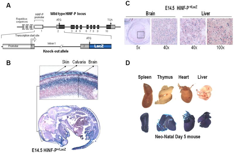
Figure 4. In vivo expression analysis of Hinfp gene during development using HiNFPLacZ/+ mice.

(A) Diagram of the HinfpLacZ allele in which LacZ coding sequences are located immediately downstream of the Hinfp ATG start codon and linked to the native Hinfp promoter (Xie et al., 2009). Mice carrying this allele were examined for expression of β-galactosidase at different embryonic and post-natal stages. (B) Embryos derived from wild type and heterozygous HiNFPLacZ/+ crosses at E14.5 were stained with X-gal in histological sections. One area spanning cranial tissues is enlarged in the inset. (C) Selected tissues as indicated were microscopically examined for β-galactosidase staining in histological sections, (D) Gross anatomical analysis of β-galactosidase staining in tissues from Day 5 neonates derived from wild type and heterozygous HiNFPLacZ/+ mice.
