Abstract
Virulent and moderately virulent strains of Newcastle disease virus (NDV), representing avian paramyxovirus serotype 1 (APMV-1), cause respiratory and neurological disease in chickens and other species of birds. In contrast, APMV-2 is avirulent in chickens. We investigated the role of the fusion (F) and hemagglutinin-neuraminidase (HN) envelope glycoproteins in these contrasting phenotypes by designing chimeric viruses in which the F and HN glycoproteins or their ectodomains were exchanged individually or together between the moderately virulent, neurotropic NDV strain Beaudette C (BC) and the avirulent APMV-2 strain Yucaipa. When we attempted to exchange the complete F and HN glycoproteins individually and together between the two viruses, the only construct that could be recovered was recombinant APMV-2 strain Yucaipa (rAPMV-2), containing the NDV F glycoprotein in place of its own. This substitution of NDV F into APMV-2 was sufficient to confer the neurotropic, neuroinvasive, and neurovirulent phenotypes, in spite of all being at reduced levels compared to what was seen for NDV-BC. When the ectodomains of F and HN were exchanged individually and together, two constructs could be recovered: NDV, containing both the F and HN ectodomains of APMV-2; and APMV-2, containing both ectodomains of NDV. This supported the idea that homologous cytoplasmic tails and matched F and HN ectodomains are important for virus replication. Analysis of these viruses for replication in vitro, syncytium formation, mean embryo death time, intracerebral pathogenicity index, and replication and tropism in 1-day-old chicks and 2-week-old chickens showed that the two contrasting phenotypes of NDV and APMV-2 could largely be transferred between the two backbones by transfer of homotypic F and HN ectodomains. Further analysis provided evidence that the homologous stalk domain of NDV HN is essential for virus replication, while the globular head domain of NDV HN could be replaced with that of APMV-2 with only a minimal attenuating effect. These results demonstrate that the F and HN ectodomains together determine the cell fusion, tropism, and virulence phenotypes of NDV and APMV-2 and that the regions of HN that are critical to replication and the species-specific phenotypes include the cytoplasmic tail and stalk domain but not the globular head domain.
INTRODUCTION
The family Paramyxoviridae consists of enveloped viruses with a nonsegmented, single-stranded, negative-sense RNA genome (23). These viruses have been isolated from a great variety of mammalian and avian species around the world. Many members of the family cause important human and animal diseases, while the disease potential of many other members is not known. The family is divided into two subfamilies, Paramyxovirinae and Pneumovirinae. The subfamily Paramyxovirinae comprises five genera, Rubulavirus, Respirovirus, Morbillivirus, Henipavirus, and Avulavirus. Subfamily Pneumovirinae is divided into two genera, Pneumovirus and Metapneumovirus. All paramyxoviruses isolated from avian species are classified into the genus Avulavirus, except avian metapneumoviruses, which are classified in the genus Metapneumovirus. Avian paramyxoviruses (APMVs) have been divided into nine different serotypes (APMV-1 to -9) based on hemagglutination inhibition (HI) and neuraminidase inhibition (NI) assays (3). APMV-1, which comprises all strains of Newcastle disease virus (NDV), has been extensively characterized because virulent NDV strains cause severe disease in chickens. Complete genome sequences and reverse genetics systems are available for several NDV strains (14, 19, 33). As an initial step toward characterizing other APMV serotypes, complete genome sequences of one or more representative strains of APMV serotypes 2 to 9 have been determined (20, 28, 30, 38, 39, 43, 55, 56). Recently, a reverse genetics system for APMV-2 has also been reported (45). The genome of APMV is similar to those of other paramyxoviruses.
The genomes of NDV and APMV-2 are very similar in organization. Both serotypes encode a nucleoprotein (N), a phosphoprotein (P), a matrix protein (M), a fusion protein (F), a hemagglutinin-neuraminidase protein (HN), and a large polymerase protein (L). Two additional proteins called, V and W, are produced by RNA editing of the P gene. The F and HN proteins form spike-like projections on the outer surface of the viral envelope and are the neutralizing and protective antigens of NDV. Significant sequence divergence in these two proteins exists among APMV serotypes (39). The HN protein possesses receptor-binding and neuraminidase activity, whereas the F protein is directly involved in membrane fusion which is necessary for the entry of the virus. Homotypic interactions between the HN and F proteins are hypothesized to control initiation of the fusion process for most paramyxoviruses (6, 25). The M protein forms the inner layer of the envelope and plays a key role in assembly by interacting with the HN and F proteins as well as ribonucleocapsid (49, 53).
NDV isolates vary greatly in their pathogenicity for chickens, ranging from no apparent disease to severe respiratory and neurological disease causing 100% mortality (32). NDV strains are categorized into three main pathotypes, lentogenic (avirulent), mesogenic (moderately virulent), and velogenic (virulent), based on their pathogenicity in chickens (2). In contrast, strains of APMV-2 appear to be much less virulent and at most have been associated with inapparent to mild respiratory diseases in chickens and turkeys (1). Consistent with their classification as separate serotypes, NDV and APMV-2 have significant sequence divergence. For example, the amino acid sequence identities of the F and HN proteins between the two serotypes are 42% and 36%, respectively (43).
Our understanding of the viral factors responsible for tissue tropism and virulence of APMVs in chickens is incomplete and is based mainly on studies with NDV. In NDV, the amino acid sequence at the F protein cleavage site has been identified as the primary determinant of virulence. Virulent NDV strains have multibasic residues that conform to the preferred cleavage site of the intracellular protease furin (Arg-X-Arg/Lys-Arg↓) present in most cell types. In contrast, avirulent NDV strains typically contain a single basic residue at the F protein cleavage site and are cleaved by extracellular proteases. The ability of the F protein of virulent NDV strains to be cleaved by furin provides the possibility for replication in a wide variety of tissues, whereas the presence of monobasic residues in avirulent NDV strains restricts viral replication to the respiratory and enteric tracts where secretary proteases for cleavage are available. The NDV strain used in the present study, Beaudette C (BC), contains a multibasic cleavage site with a furin motif (Arg-Arg-Gln-Lys-Arg↓) that is readily cleaved in cell culture without the need for exogenous protease. NDV-BC readily forms syncytia in cell culture. The cleavage site of APMV-2 (Lys-Pro-Ala-Ser-Arg↓) contains two basic amino acids of the furin motif, and APMV-2 does not form syncytia in cell culture. Surprisingly, however, APMV-2 replicates efficiently in vitro without added protease, and its replication is not augmented by added protease (43). Recently, the F protein cleavage site sequence of APMV-2 was changed to multibasic residues by reverse genetics, but the change did not increase the pathogenicity of APMV-2 in chickens, indicating that the sequence at the F protein cleavage site is not the major limitation to APMV-2 virulence (45). In addition to the F protein, the HN and L proteins have been shown to contribute to the overall pathogenicity of NDV (5, 8, 15, 37).
In general, the outer surface glycoproteins of enveloped viruses have been shown to play a major roles in the virulence phenotypes of many viruses (7, 10, 12, 18, 24, 27, 29, 52). In the present study, we investigated the roles of the F and HN envelope glycoproteins in APMV pathogenicity by exchanging them between the mesogenic, neurotropic NDV strain BC and the avirulent APMV-2 strain Yucaipa. This took advantage of reverse genetics systems previously established in our laboratory (19, 45). In previous studies, we confirmed that these two viruses differ greatly in virulence and tissue tropism (44). NDV-BC infects neuronal tissue and causes neurological disease, whereas APMV-2 strain Yucaipa does not infect neuronal tissue or cause neurological disease. In cell culture, NDV-BC causes syncytium formation, whereas APMV-2 strain Yucaipa causes a single-cell infection without syncytium formation. Thus, the remarkably contrasting phenotypes of these two APMV serotypes provided the opportunity to investigate phenotypic determinants by exchanging genes.
MATERIALS AND METHODS
Cells and viruses.
The chicken embryo fibroblast cell line (DF1) and human epidermoid carcinoma cell line (HEp-2) were grown in Dulbecco's minimal essential medium (DMEM) with 10% fetal bovine serum (FBS) and maintained in DMEM with 5% FBS. The African green monkey kidney Vero cell line was grown in Eagle's minimum essential medium (EMEM) containing 10% FBS and maintained in EMEM with 5% FBS. The modified vaccinia virus strain Ankara (MVA) expressing T7 RNA polymerase was kindly provided by Bernard Moss (NIAID, NIH) and propagated in primary chicken embryo fibroblast cells in DMEM with 2% FBS. Recombinant NDV strain BC (rNDV) and recombinant APMV-2 strain Yucaipa (rAPMV-2) were generated in our laboratory (19, 45). These viruses were grown in the allantoic cavities of 9-day-old specific-pathogen-free (SPF) embryonated chicken eggs. The ability of the viruses to produce plaque was tested on Vero and DF1 cells under 0.8% methylcellulose overlay. Plaques were visualized by immunoperoxidase staining using virus-specific antiserum. All the infectious NDV and chimeric APMV-2 viruses containing the NDV F and HN experiments were conducted in an enhanced biosafety level 3 (BSL-3) containment facility certified by the USDA following the guidelines of the Institutional Animal Care and Use Committee (IACUC) of the University of Maryland.
Construction of chimeric NDV and APMV-2 antigenomic cDNAs and generation of chimeric viruses.
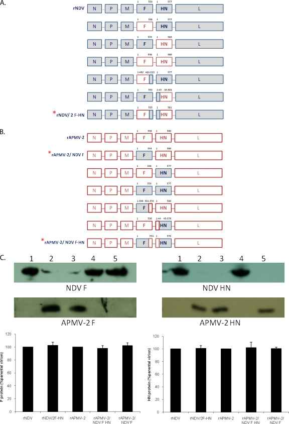
The F and HN open reading frames (ORFs) of APMV-2 strain Yucaipa were placed individually or together into a full-length antigenomic cDNA of NDV strain BC in place of the corresponding NDV F and HN ORF(s) (Fig. 1A). These manipulations were facilitated by the presence of unique restriction enzyme sites (PacI, MluI, and AgeI) located in the untranslated regions (UTRs) flanking the F and HN ORFs in the NDV cDNA. The APMV-2 F and HN ORFs were engineered by overlapping PCR to be flanked by 5′ and 3′ UTRs of the respective NDV F or HN gene along with the compatible restriction enzyme sites. In order to maintain the genome length as a multiple of six, additional nucleotides were introduced at the 3′ UTR (i.e., downstream of the ORF) as necessary. The engineered ORF(s) and flanking sequences were digested with the appropriate restriction enzymes and used to replace the corresponding fragment(s) in the full-length NDV cDNA, resulting in three full-length cDNAs in which the NDV backbone contained replacement of the F ORF, the HN ORF, or both with that of APMV-2 (Fig. 1A). Since specific interaction of the cytoplasmic tails of surface glycoproteins with the M protein has been shown to be critical for the assembly of NDV (17), three additional constructs were made in which only the ectodomains of the APMV-2 F and HN ORFs were exchanged individually and together for those of NDV (Fig. 1A). Briefly, the ectodomain-coding regions of the APMV-2 F and HN ORFs were individually amplified by PCR and joined by overlapping PCR with coding regions for the respective cytoplasmic tail (CT), transmembrane (TM) domain, and UTRs of the NDV F or HN gene. Each fragment was digested with appropriate restriction enzymes and cloned into the digested NDV cDNA as described above, resulting in three full-length NDV cDNAs encoding F and/or HN proteins with ectodomains derived from APMV-2 (Fig. 1A). The same strategy was used to construct a parallel set of six constructs based on a full-length antigenomic cDNA of APMV-2 (Fig. 1B). For three of these constructs, the complete F and HN ORFs of NDV were inserted individually or together placed into the APMV-2 backbone in place of the corresponding APMV-2 sequence(s). An additional three constructs were made in which only the ectodomains of NDV F and HN were exchanged individually or together for those of APMV-2 (Fig. 1B). Insertion of the NDV F and HN sequences into the APMV-2 genome used unique restriction enzyme sites (NotI, PacI, and SnaBI) located in the UTRs of the F and HN genes in APMV-2 cDNA. The nucleotide sequences of exchanged genes in these 12 constructs were confirmed by using primers targeting a downstream region of the M gene and an upstream region of the L gene.
Fig. 1.
Construction of full-length antigenomic cDNAs of NDV (A) or APMV-2 (B) in which the complete F and/or HN glycoprotein or its ectodomain were swapped with that of the other virus, and successful recovery of three of these viruses. NDV and APMV-2 sequences are indicated with shaded and open boxes, respectively. The amino acid lengths of individual proteins or protein domains are indicated. The three chimeric viruses that could be successfully recovered are indicated with asterisks. (A) The coding sequences for the complete ORFs (top three constructs) or ectodomains (bottom three constructs) of F and HN of APMV-2 were inserted individually or together into a full-length antigenomic NDV cDNA in place of the corresponding NDV sequence. Each insert was flanked by 5′ UTR and 3′ UTR and transcription signals of the respective NDV F or HN gene: these flanking sequences are not shown. (B) A parallel set of constructs was made in the APMV-2 backbone. (C) Western blot analysis of virion proteins using polyclonal antibodies raised against the NDV F or HN protein. The parental NDV and APMV-2 viruses and the three chimeric viruses that were successfully recovered were grown in embryonated eggs and subjected to discontinuous sucrose gradients (9) to obtain partially purified stocks. The levels of the F and HN proteins incorporated into virions were quantitated by Western blotting using the sera specific to the individual NDV and APMV-2 glycoproteins, as indicated. Values for all proteins were expressed as percentages of these values for the proteins of respective parental viruses, which were considered to be 100%. Each bar represents the mean and standard error of the mean of three independent experiments. The detected blots by the sera for NDV F protein are F0 protein, corresponding to approximately 50 kDa. Lanes: 1, rNDV; 2, rNDV/2-F-HN; 3, rAPMV-2; 4, rAPMV-2/NDV- F-HN; 5, rAPMV-2/NDV-F.
Infectious chimeric viruses were generated as previously described (19, 45). Briefly, HEp-2 cells were transfected with three plasmids individually encoding the N, P, and L proteins (2.0 μg, 1.0 μg, and 0.5 μg per single well of a six-well dish, respectively) and a fourth plasmid encoding the full-length antigenome (5.0 μg) by using Lipofectamine (Invitrogen, Carlsbad, CA) and simultaneously infected with vaccinia MVA expressing T7 RNA polymerase at a multiplicity of infection (MOI) of 1 PFU/cell. Two days after transfection, the cell culture supernatant was injected into the allantoic cavities of 9-day-old SPF embryonated chicken eggs. Recovery of the virus was confirmed by hemagglutination assay using 1% chicken red blood cells (RBCs). The sequences of the F and HN genes in the recovered chimeric viruses were confirmed by reverse transcription-PCR (RT-PCR) and Western blot analyses. Each chimeric cDNA construct was subjected to at least three independent transfections before being considered negative for virus recovery. In contrast, the frequency of recovery of the parental NDV and APMV-2 viruses by this method has been approximately 80% in studies to date.
Growth characteristics of chimeric viruses.
The growth characteristics of the parental and chimeric viruses were evaluated in DF1 and Vero cells. The ability of the chimeric viruses to produce plaques was tested in DF1 and Vero cells under 0.8% methylcellulose overlay. The plaques were immunostained using polyclonal antibody raised against the N protein of NDV or APMV-2. The multicycle growth kinetics of the F and HN chimeric viruses, along with their respective parental viruses, were evaluated in DF1 cells. Duplicate wells of six-well plates were infected with parental and chimeric viruses at an MOI of 0.01 PFU/cell. After 1 h of adsorption, the cells were washed and then covered with DMEM containing 2% FBS at 37°C in 5% CO2. Supernatants were collected and replaced with an equal volume of fresh medium at 8-h intervals until 72 h after infection. Virus titers in the collected supernatants were quantified in DF1 cells by limiting dilution and immunostaining with N-specific antibodies. Virus titers were expressed as 50% tissue culture infectious dose (TCID50)/ml by the endpoint method of Reed and Muench (36).
Fusion index assay.
The fusogenic abilities of parental and chimeric viruses were evaluated in Vero cells. Briefly, each virus was inoculated into confluent Vero cells in six-well plates at an MOI of 0.1 PFU/cell. After observation of cytopathic effect (CPE) in 48 h, the cells were washed with 0.02% EDTA and then incubated with 1 ml of EDTA for 2 min. The cells were washed and fixed with methanol for 20 min. Cells were stained with hematoxylin-eosin (Hema 3). Fusion was quantitated by expressing the fusion index as the ratio of the total number of nuclei to the number of cells in which these nuclei were observed (i.e., the mean number of nuclei per cell), and the values were expressed relative to rBC as 100%.
HAd assay.
Virus was inoculated into confluent monolayers of Vero cells in six-well plates at an MOI of 0.1 PFU/cell. Twenty-four hours after infection, the washed cells were overlaid with 2% vol/vol suspension of chicken red blood cells (RBCs) (15). The plates were kept at 4°C for 15 min, and then unbound RBCs were removed by two washes with phosphate-buffered saline (PBS). The RBCs bound to the virus-infected cells were lysed with 0.05 M ammonium chloride, and the released hemoglobin was measured at 549 nm. The hemadsorption (HAd) values were expressed relative to the values for rBC as 100%.
NA assay.
The neuraminidase (NA) activity of parental and chimeric viruses was determined by a standard fluorimetric assay (35). In this assay, the substrate [2′-(4-methylumbelliferyl)-α-d-N-acetylneuraminic acid] is cleaved by NA to yield a fluorescent product that can be quantified. The assay mixture contained the substrate (100 μM), serial 2-fold dilutions of supernatants collected from virus-infected cells in 32.5 mM MES (2-N-morpholinoethanesulfonic acid) buffer (pH 6.5), and 4 mM calcium chloride and was incubated at 37°C for 30 min with shaking. The reaction was terminated by adding 0.014 M sodium hydroxide in 83% (vol/vol) ethanol (150 μl). Fluorescence intensity was measured at an excitation of 360 nm and emission of 450 nm, and substrate blanks were subtracted from the sample readings. The NA values were expressed relative to the values for rBC as 100%.
MDT and ICPI.
The pathogenicity of the chimeric viruses was determined by the mean death time (MDT) test in 9-day-old SPF embryonated chicken eggs and by the intracerebral pathogenicity index (ICPI) test in 1-day-old SPF chicks (2). Briefly, for the MDT, a series of 10-fold dilutions of infected allantoic fluid (0.1 ml) was inoculated into the allantoic cavities of five 9-day-old eggs per dilution and incubated at 37°C. The eggs were examined once every 8 h for 7 days, and the time of embryo death was recorded. The MDT was determined as mean time (h) for the minimum lethal dose of virus to kill all the inoculated embryos. The criteria for classifying the virulence of NDV isolates are as follows: <60 h, virulent strains; 60 to 90 h, intermediately virulent strains; and >90 h, avirulent strains. For the ICPI test, 0.05 ml of a 1:10 dilution of fresh infective allantoic fluid for each virus was inoculated into group of 10 1-day-old SPF chicks via the intracerebral route. The birds were observed for clinical symptoms and mortality once every 8 h for a period of 10 days. At each observation, the birds were scored as follows: 0 if normal, 1 if sick, and 2 if dead. The ICPI is the mean of the score per bird per observation over the 10-day period. Highly virulent velogenic viruses give values approaching 2, and the avirulent or lentogenic strains give values close to 0.
Replication in 1-day-old chicks and 2-week-old chickens.
To determine the ability of parental and chimeric viruses to replicate in chicken brains, 10 1-day-old SPF chicks were inoculated with 0.05 ml of a 1:10 dilution of 256 HA units (approximately 1.25 × 10⁶ PFU) of each virus via the intracerebral route. Two birds were sacrificed daily until 5 days postinfection (dpi). Brain tissue samples were collected from the sacrificed birds and then homogenized. Virus titers in the brain tissue samples were determined by limiting dilution and immunoperoxidase assay using polyclonal antibodies against the N proteins in DF1 cells.
To evaluate tropism, six 1-day-old chickens per group were inoculated with each virus (1 × 10⁵ TCID50/bird) via the intracerebral or intranasal route. Three chicks from each group were harvested on days 2 and 3 postinfection, and brain tissue was collected for virus titration by limiting dilution and immunoperoxidase assay as described above. In addition, tissue from day 3 was processed for immunohistochemistry.
To evaluate the tropism of the chimeric viruses in older birds, groups of 10 2-week-old SPF chickens were inoculated with 200 μl of each virus (1 × 10⁵ TCID50/bird) by the intranasal route. Three birds from each group were sacrificed at 3 and 5 dpi, and tissue samples (lung, trachea, spleen, and brain) were collected for virus titration. Virus titers in DF1 cells were determined by limiting dilution as described above. The remaining birds were observed daily for 10 days for any clinical signs.
Immunohistochemistry.
From the experiment described above, brain tissue from 1-day-old chicks infected by the intranasal and intracerebral routes was fixed in phosphate-buffered formalin (10%). Fixed tissues were embedded in paraffin and sectioned (Histoserv, Inc., Germantown, MD). Sections from mock-infected chicks were used as controls. The tissues were deparaffinized, rehydrated, and subsequently immunostained to detect viral N protein by using the following protocol. Briefly, the sections were blocked with 1% bovine serum albumin (BSA) in PBS for 1 h at room temperature, incubated with a polyclonal antibody (1:200 dilution) followed by horseradish peroxidase-conjugated goat anti-rabbit antibodies for 30 min, and then stained with 3-amino-9-ethylcarbazole (AEC) substrate followed by counterstaining with hematoxylin.
Generation of NDV mutants containing the globular head of APMV-2 HN or point mutations in the HN stalk region.
We also attempted to replace only the globular head of the NDV HN protein with that of APMV-2. This would investigate whether the stalk domain had played any role in virus recovery. Briefly, PCR-amplified coding sequence for the cytoplasmic tail, transmembrane, and stalk domains of NDV HN (amino acid sequence positions 1 to 143 of NDV HN) was joined with coding sequence for the HN ectodomain of APMV-2 (amino acid sequence positions 145 to 580 of APMV-2 HN) by overlapping PCR. The amplified fragment was digested and cloned into MluI-AgeI-digested NDV cDNA. The constructed full-length antigenomic NDV cDNA was subjected to transfection, and the encoded virus was successfully recovered (rNDV/HN-2GH) and was passaged as described above. In addition, the role of the stalk domain of NDV HN was investigated by the introduction of point mutations. A previous study has identified four amino acids (alanine, leucine, proline, and leucine at positions 89, 90, 93, and 94, respectively) in the stalk domain of NDV HN as contributors to fusion promotion activity by using a plasmid expression system (25). These four amino acids were individually mutated in the NDV HN into, respectively, glutamine, alanine, alanine, and alanine, following the published study. Each mutated HN fragment was digested and inserted into MluI-AgeI-digested NDV cDNA. The HN mutant cDNAs were used for virus recovery. Of the four HN mutant constructs, only the virus in which alanine-89 was replaced with glutamine-89 (rNDV/HN-A89Q) was recovered. Subsequently, the rNDV/HN-2GH and A89Q mutant viruses were evaluated for in vitro growth, MDT, ICPI, and growth kinetics in the brains of 1-day-old chicks, as described above.
RESULTS
Recovery of recombinant chimeric viruses.
To determine the role of the viral surface glycoproteins in APMV tissue tropism and virulence, the complete ORFs of the F and HN genes were exchanged individually or together between full-length antigenomic cDNAs of NDV and APMV-2. Specifically, the F and HN ORFs in the NDV backbone were replaced individually or together with the corresponding ORFs of APMV-2 (Fig. 1A, first three constructs), and the F and HN ORFs in the APMV-2 backbone were replaced individually or together with the corresponding ORFs of NDV (Fig. 1B, first three constructs). However, of these six chimeric cDNAs, viable virus could be recovered only from the chimeric APMV-2 cDNA that contained the ORF of NDV F gene (Fig. 1B). Our inability to recover the other five chimeric viruses may have been due to the absence of the homologous interactions between the glycoprotein cytoplasmic tails and internal proteins, as was previously shown to contribute to the specificity of virion assembly in several viruses (46). Thus, the recovery of APMV-2 containing the F protein of NDV in place of its own was an unexpected result.
We next sought to recover chimeric viruses in which only the ectodomains of the F and HN proteins were exchanged individually or together between NDV and APMV-2. These viruses would retain the homologous cytoplasmic tails and transmembrane domains. Thus, we constructed a set of three NDV antigenomic cDNAs in which the coding sequences for the F and HN ectodomains were replaced individually or together with the corresponding ectodomain(s) of APMV-2 (Fig. 1A, bottom three constructs), and a set of three APMV-2 antigenomic cDNAs in which the coding sequences for the F and HN ectodomains were replaced individually or together with the corresponding ones from NDV (Fig. 1B, bottom three constructs). We were successful in recovering two of these viruses, namely, NDV, in which both the F and HN ectodomains were replaced by those of APMV-2 (rNDV/2-F-HN), and the corresponding APMV-2 virus bearing the ectodomains of NDV F and HN (rAPMV-2/NDV-F-HN). This supported the idea that the presence of the homologous cytoplasmic tails and transmembrane regions is important for NDV replication and implied that it was necessary to have a matched pair of the F and HN ectodomains.
The viral proteins in partially purified parental and chimeric viruses were analyzed by Western blotting using sera raised against the NDV F or HN protein (Fig. 1C). This confirmed that the correct NDV surface protein(s) was present where expected. The protein composition of the parental and chimeric viruses was verified by Coomassie blue staining of the purified viral proteins separated by SDS-PAGE (data not shown) and Western blotting (Fig. 1C). This indicated that all of the chimeric viruses contained major structural proteins of the expected sizes and amounts, suggesting that the chimeric viruses do not have significant selective disadvantages in virion assembly.
Cytopathic effect of recombinant chimeric viruses in vitro.
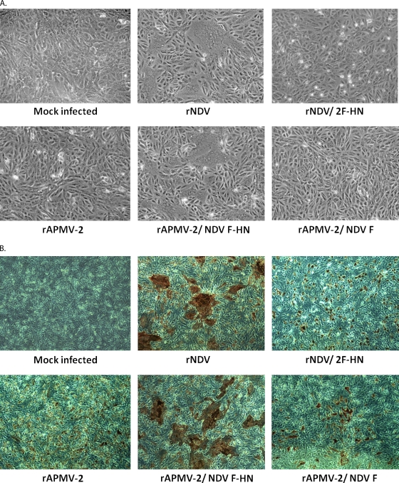
To investigate the role of the envelope glycoproteins in the viral CPE in cell culture, Vero cells or DF1 cells were infected with the chimeric and parental viruses. The two parental viruses produce distinctive CPEs. NDV is a typical syncytium-forming paramyxovirus, whereas APMV-2 produces single-cell infection leading to cell rounding and detachment of infected cells (43). We compared the CPEs of rNDV/2-F-HN, rAPMV-2/NDV-F-HN, and rAPMV-2/NDV-F on the basis of these properties in Vero cells, visualizing CPE by photomicroscopy directly (Fig. 2A) and following immunostaining with antibodies to the respective N protein (Fig. 2B). This showed that rAPMV-2/NDV-F-HN produced syncytia that were similar in number and size to those of the rNDV parent. In contrast, rNDV/2-F-HN produced CPE that was characterized by cell rounding and detachment of virus-infected cells, similar to that of the rAPMV-2 parent. Interestingly, rAPMV-2/NDV-F produced CPE similar to that of parental rAPMV-2. The chimeric and parental viruses also showed similar trends in CPE production in DF1 cells, as observed with Vero cells (data not shown). These results demonstrated that the CPEs of NDV and APMV-2 were determined by the origin of the two envelope glycoprotein ectodomains, irrespective of the origin of the viral backbone. In addition, the formation of the large syncytium characteristic of NDV depended on the presence of both NDV F and HN proteins.
Fig. 2.
Cytopathic effect in Vero cells infected with chimeric NDV and APMV-2 viruses. (A) Vero cells in six-well plates were infected with each of the parental and chimeric viruses at a multiplicity of infection (MOI) of 0.01 PFU/cell and incubated for 48 h. (B) The viral plaques in the infected cells were visualized by immunoperoxidase staining using polyclonal antibody raised against the N protein of NDV (top row) or APMV-2 (bottom row).
Biological activities of chimeric viruses.
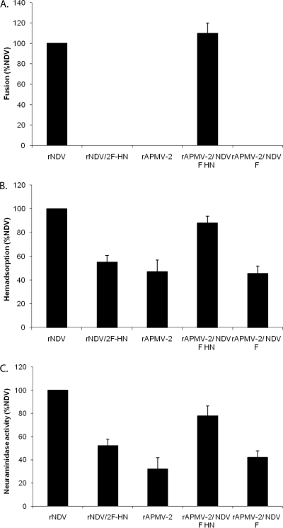
Next, we investigated whether the exchange of the F and HN proteins affected the NA, HAd, and fusion activities of the parental and chimeric viruses (Fig. 3). The fusion activities of rNDV and rAPMV-2/NDV-F-HN did not differ significantly, indicating that the NDV F and HN ectodomains are sufficient to induce efficient cell fusion irrespective of the origin of the viral backbone. With regard to NA and HAd, parental rNDV was more active than parental rAPVM-2, and the activities of the chimeric viruses were intermediate and determined in large part by the origin of the F and HN ectodomains (Fig. 3). For example, the HAd activity of rAPMV-2 was 47% of that of NDV. Exchange of the F and HN ectodomains of NDV with those of APMV-2 (rNDV/2-F-HN) resulted in a value of 55%, similar to that of parental APMV-2. Conversely, modification of APMV-2 to contain the ectodomains of NDV F and HN (rAPMV-2/NDV-F-HN) resulted in a value of 88%. Expression of NDV F alone by APMV-2 (rAPMV-2/NDV-F) had little effect on the phenotype of APMV-2. Similar findings were made for NA activity (Fig. 3). This altered activity could affect viral infectivity and growth kinetics in vitro and in vivo.
Fig. 3.
Biological activities of parental and chimeric viruses. The fusion index (A), HAd (B), and NA (C) were determined in virus-infected Vero cells at an MOI of 0.1 PFU/cell. All values are expressed relative to rNDV as 100%. Each bar represents the mean and standard error of the mean of duplicate samples.
Replication of the chimeric and parental viruses in DF1 cells.
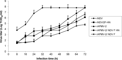
The multicycle replication of chimeric and parental viruses was evaluated in DF1 cells (Fig. 4). There was a great difference in the kinetics and magnitude of replication between the two parental viruses. rNDV reached the highest titer at 32 h postinfection (hpi), and its titers remained consistent up to the end of the experiment at 72 hpi, whereas rAPMV-2 grew slowly, and its highest titer was at 72 hpi and was 3.0 log10 lower than that of rNDV. As shown in Fig. 4, the kinetics and magnitude of replication of the chimeric viruses depended in large part on the origin of the F and HN proteins. rAPMV-2/NDV-F-HN replicated more efficiently than the other chimeric viruses and eventually reached the same maximum titer as rNDV at 72 hpi. Thus, inclusion of the NDV F and HN ectodomains in the APMV-2 backbone resulted in a 3.0-log10 increase in final titer compared to rAPMV-2. rNDV/2-F-HN and rAPMV-2/NDV-F replicated to titers that were intermediate between the extremes of rNDV and rAPMV-2. This observation for rAPMV-2/NDV-F indicated that NDV F alone substantially increased the replication efficiency of APMV-2, although the effect was substantially less than that achieved with the ectodomains of both F and HN. This observation for rAPMV-2/NDV-F-HN indicated that the inclusion of the NDV F and HN ectodomains in the APMV-2 backbone substantially increased its efficiency of replication.
Fig. 4.
In vitro growth characterization of chimeric viruses in DF1 cells. The growth characteristics of parental and chimeric viruses were determined by multicycle growth curve in DF1 cells infected with an MOI of 0.01 PFU/cell. The viral titers were determined by limiting dilution on DF1 cells and immunostaining with a polyclonal antibody raised against the respective N protein. Each bar represents the mean and standard error of the mean of duplicate samples.
Evaluation of the parental and chimeric viruses by MDT and ICPI assays.
The pathogenicity of the parental and chimeric viruses was evaluated with the standard pathogenicity tests used for NDV, namely, MDT in embryonated eggs and ICPI in 1-day-old chicks (see Materials and Methods) (Table 1). The pathogenicities of NDV and APMV-2 varied greatly by these standard tests: NDV strain BC had MDT and ICPI values of 57 h and 1.60, respectively, whereas the respective values for APMV-2 were >120 h and 0.00, indicating that NDV-BC had moderate virulence and APMV-2 was avirulent. The MDT and ICPI values of rNDV/2-F-HN were >120 h and 0.00, respectively, which are very similar to the values for rAPMV-2 and indicated that the F and HN ectodomains of APMV-2 strongly attenuated NDV. In contrast, the MDT and ICPI values of rAPMV-2/NDV-F-HN were 64 h and 1.23, respectively, indicating that the presence of the NDV F and HN ectodomains substantially increased the virulence of APMV-2. Although the level of pathogenicity of rAPMV-2/NDV-F-HN was greatly enhanced, it was slightly lower than that of parental rNDV, indicating that other viral proteins probably contribute to the overall virulence of NDV (37). Interestingly, the pathogenicity of rAPMV-2/NDV-F was slightly increased by the ICPI and MDT tests. This suggests that the NDV F protein alone can increase the pathogenicity to a small extent but that the homotypic HN protein is required in addition for a significant increase in pathogenicity.
Table 1.
Pathogenicities of parental and chimeric viruses in embryonated eggs and chicks
| Virus | MDTa | ICPIb |
|---|---|---|
| rNDV | 57 h | 1.60 |
| rAPMV-2 | >120 h | 0.00 |
| rNDV/2 F-HN | >120 h | 0.00 |
| rAPMV-2/NDV F-HN | 64 h | 1.23 |
| rAPMV-2/NDV F | 120 h | 0.28 |
MDT, mean time (h) for the minimum lethal dose of virus to kill all of the inoculated embryos. Pathotype definition: virulent strains, <60 h; intermediately virulent strains, 60 to 90 h; avirulent strains, >90 h.
Pathogenicity of NDV in 1-day-old SPF chicks was evaluated by the ICPI assay. Pathotype definition: virulent strains, 1.5 to 2.0; intermediate virulent strains, 0.7 to 1.5; avirulent strains, 0.0 to 0.7.
During the ICPI tests, it was observed that chicks infected with rNDV began to show clinical signs at 2 dpi, and 100% mortality was observed at 3 dpi. Due to the rapid death, distinguishable neurological signs were not observed in these infected chicks. However, the group of chicks infected with rAPMV-2/NDV-F-HN began to develop clinical signs at 3 dpi, and at 4 dpi, all chicks were paralyzed with distinctive neurological signs, such as twisting of the head and neck. Since these disease signs are not seen with parental APMV-2, this indicates that the inclusion of the ectodomains of NDV F and HN was sufficient to confer neurovirulence. The group of chicks infected with rAPMV-2/NDV-F also showed mild neurological signs, including paralysis, at 6 dpi, but most of them recovered thereafter. Conversely, chicks infected with rNDV/2-F-HN had no apparent clinical signs during the 8-day postinfection period, as also was observed for the chickens infected with rAPMV-2 parent. These results showed that the NDV F alone conferred neurovirulence, although the additional inclusion of NDV HN protein substantially increased this activity. These results also indicated that neurovirulence was conferred to APMV-2 by transfer of the ectodomains of NDV F and HN and that the phenotype was ablated in NDV by transfer of the ectodomains of APMV-2 F and HN.
Replication of chimeric and parental viruses in the brains of chicks and evaluation of tissue tropism.
To further evaluate the role of the F and HN proteins in neurotropism, the growth kinetics of the parental and chimeric viruses were determined by inoculating 1-day-old chicks by the intracerebral route and sacrificing chicks and harvesting brain tissue at daily intervals for quantitative virology (Fig. 5). Neurovirulent rNDV reached the highest titer—more than 6.0 log10 TCID50/g—in the brain at 2 dpi, resulting in the death of all of the infected chicks at 3 dpi. In contrast, replication of rAPMV-2 was not detectable on any day, and the virus was completely avirulent as already noted. rAPMV-2/NDV-F-HN replicated to a maximum titer of more than 4.0 log10 TCID50/g, at 3 dpi, whereas rAPMV-2/NDV-F had a maximum titer of nearly 3.0 log10 TCID50/g. Conversely, replication of rNDV/2-F-HN was not detected on any day, resembling rAPMV-2. These results showed that the NDV F protein alone conferred neurotropism to APMV but that this was substantially increased by the additional inclusion of the homotypic HN protein. The observation that rAPMV-2/NDV-F-HN did not replicate as efficiently as rNDV and was less virulent is another indication that other NDV proteins may contribute to these phenotypes. Conversely, transfer of the APMV-2 F and HN ectodomains to NDV completely ablated its neurotropism.
Fig. 5.
Growth kinetics of parental and chimeric viruses in the brains of 1-day-old chicks. Ten 1-day-old SPF chicks were inoculated with 256 HA units (approximately 1.28 × 10⁶ PFU) of parental or chimeric virus via the intracerebral route. Two birds in each group were sacrificed daily until 5 dpi. The virus titers in the collected samples were determined by limiting dilution in DF1 cells and immunostaining with a polyclonal antibody raised against the respective N protein.
Next, we evaluated the ability of the parental and chimeric viruses to spread to the brain following intranasal inoculation, a route of inoculation that mimics natural infection. Groups of 1-day-old chicks were inoculated by the intranasal route with the parental and chimeric viruses (Table 2). For comparison, additional groups were inoculated intracerebrally as positive controls (Table 2). Virus titration of brain tissue harvested at 2 and 3 dpi demonstrated that inoculation with rNDV by either route resulted in a high level of replication in the brain, whereas inoculation with APMV-2 by either route resulted in a lack of detectable replication. This showed that rNDV is neuroinvasive. The replication of rAPMV-2/NDV-F-HN and rAPMV-2/-NDV-F was somewhat less efficient than that of rNDV, but for each virus there was no difference between the two routes of infection. Thus, each of these viruses was equally able to spread to the brain following intranasal inoculation. These results indicated that the presence of the NDV F protein alone conferred neuroinvasiveness but that homotypic HN was required in addition for efficient replication at that site. Conversely, inclusion of the APMV-2 F and HN ectodomains into the rNDV backbone completely attenuated the virus for replication in the brain following inoculation by either route.
Table 2.
Replication of parental and chimeric viruses in the brains of 1-day-old chicks
| Virus | Virus titer (log10 TCID50/g of brain)a |
|||
|---|---|---|---|---|
| Intracerebral infection |
Intranasal infection |
|||
| 2 dpi | 3 dpi | 3 dpi | 4 dpi | |
| rNDV | 6.12 | 5.83 | 5.21 | 5.8 |
| rAPMV-2 | N.D.b | N.D. | N.D. | N.D. |
| rNDV/2-F-HN | N.D. | N.D. | N.D. | N.D. |
| rAPMV-2/NDV-F-HN | 2.65 | 3.86 | 3.30 | 3.61 |
| rAPMV-2/NDV-F | N.D. | 1.81 | 1.86 | 1.92 |
One-day-old chicks in groups of 10 were infected with the indicated parental or chimeric virus (1 × 10⁵ TCID50/bird) via the intracerebal or intranasal route, and two chicks per group were harvested on days 1 to 5 for virus titration. Brain tissue was collected and homogenized, and virus titers were determined by limiting dilution on DF1 cells with immunostaining using antibodies against the respective N protein.
N.D., not detected.
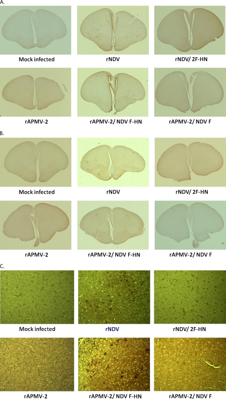
The replication of the chimeric and parental viruses in brain tissue was further evaluated by immunohistochemical analysis of brain tissue harvested on day 3 from this same experiment (Fig. 6). Viral antigens were detected in the brain tissues that had been infected with rNDV and rAPMV-2/NDV-F-HN by either the intracerebral (Fig. 6A and C) or the intranasal (Fig. 6B) route. These results confirmed the replication of the chimeric and parental viruses in chicken brain. However, there was a more extensive distribution of viral antigens in brain tissue with parental rNDV compared to rAPMV-2/NDV-F-HN following either route of inoculation. This was another indication that, although F is sufficient for neuroinvasiveness and the F and HN ectodomains together are required for substantial replication in the brain, maximum replication may depend on other NDV proteins.
Fig. 6.
Detection of viral antigens of parental and chimeric viruses in the brains of 1-day-old chicks infected by the intracerebral or intranasal route. As described in Table 2, footnote a, chicks were inoculated with each virus (1 × 10⁵ TCID50/bird) by the intracerebral (A and C) or intranasal (B) route, and brain tissue was harvested for immunohistopathology at 3 dpi. The tissues were fixed in phosphate-buffered formalin and sections were prepared and stained using an antibody against the respective N protein.
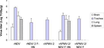
We next sought to determine the replication and tissue tropism of the chimeric and parental viruses in 2-week-old chickens. Chickens were inoculated intransally with the chimeric and parental viruses, and birds were sacrificed on days 3 and 5 and the following tissues were harvested for quantitative virology: trachea, lungs, spleen, and brain (Fig. 7). Of the two parental viruses, rNDV replicated to moderate or high titers in each of the sampled tissues in each of the birds, whereas replication of rAPMV-2 was detected only in the trachea. rAPMV-2/NDV-F-HN resembled rNDV in its ability to replicate in the brain, trachea, lungs, and spleen, whereas rNDV/2-F-HN and rAPMV-2/NDV-F resembled rAPVM-2 in being restricted to the upper respiratory tract (Fig. 7). Thus, while rAPMV-2/NDV-F was able to spread to and replicate in the brains of 1-day-old chicks following intranasal inoculation, it was unable to spread detectably beyond the upper respiratory tract in older birds. These results showed that the F and HN ectodomains together contribute to the virulence and tissue tropism of APMVs in 2-week-old chickens.
Fig. 7.
Replication of parental and chimeric viruses in 2-week-old chickens. Groups of 10 2-week-old chickens were inoculated with 1 × 10⁵ TCID50 of parental or chimeric virus by the intranasal route. Three birds from each group were sacrificed at 3 (shown) and 5 (not shown) dpi, and brain, trachea, lung, and spleen tissue was collected and virus titers determined by limiting dilution and immunoperoxidase staining. The day 3 results are shown because the titers were higher; the day 5 results were of lower titer but otherwise similar.
Role of the HN stalk domain in promoting fusion activity.
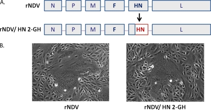
The preceding results strongly suggested that homotypic interaction between the F and HN proteins is a prerequisite for NDV replication, syncytium formation, and pathogenicity. In particular, differences in CPE and pathogenicity between rAPMV-2/NDV-F-HN and rAPMV-2/NDV-F were clearly distinguishable, indicating a strong effect due to the presence of homotypic HN in addition to F. To investigate the specific region of the homotypic HN protein involved in fusion and pathogenicity of NDV, the globular head domain of NDV HN protein was replaced with that of APMV-2 (Fig. 8A). The recovered chimeric virus (rNDV/HN-2-GH) was strongly fusogenic in infected Vero cells, resembling the rNDV parent (Fig. 8B). This indicated that the stalk domain of NDV HN protein, but not the globular head domain, plays a major role in promoting fusion activity (Fig. 8B).
Fig. 8.
Replacement of the globular head (GH) domain of the NDV HN protein to create the chimeric virus rNDV/HN-2GH. The globular head domain of NDV was replaced with that of APMV-2 (A). Fusion activity of the parental rNDV or chimeric rNDV/HN-2GH virus was determined in Vero cells, visualized at 48 hpi (B).
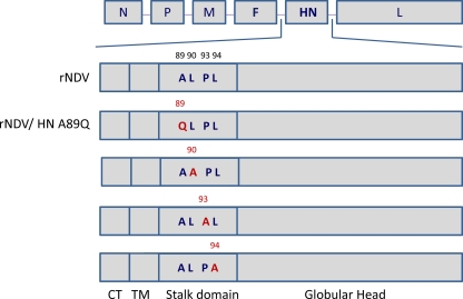
To identify critical amino acid residues involved in F and HN interaction, we modified the NDV HN protein by introducing single amino acid substitutions at positions in the stalk domain that were previously suggested to be important based on plasmid cotransfection experiments (25). Substitutions were attempted at four different positions (Fig. 9). Among the four constructs, we were able to recover only the one with an alanine-to-glutamate substitution at position 89 (rNDV/HN-A89Q). This suggests that the substitutions at the other three positions could not be tolerated, implying that those amino acid residues are crucial for NDV replication (Fig. 9).
Fig. 9.
Construction of HN stalk domain mutant NDVs. Four amino acids (alanine, leucine, proline, and leucine at amino acid positions 89, 90, 93, and 94, respectively) in the HN stalk domain were individually mutated into glutamine or alanine. Among the four constructs, one rNDV with substitution of glutamine for alanine (rNDV/HN-A89Q) was recovered.
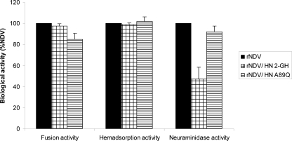
To evaluate the effect of the HN globular head and A89Q substitutions on the biological activities of NDV, the fusion, HAd, and NA activities were determined (Fig. 10). The rNDV/HN-2-GH virus had similar levels of fusion and HAd activities compared to the parental virus but had reduced NA activity (50%). This suggests that the globular head domains of the NDV and APMV-2 HN proteins have functionally similar sialic acid binding activities but differ in NA activity. In contrast, rNDV/HN-A89Q exhibited only very small reductions in fusion and NA activities, and no difference in HAd activity, compared to the parental virus.
Fig. 10.
Biological activities of HN mutant NDVs. The NA, HAd, and fusion index of the mutant viruses were determined in virus-infected Vero cells at an MOI of 0.1. Values for all viruses were expressed as percentages of these values for rNDV, which were considered to be 100%. Each bar represents the mean and standard error of the mean of duplicate samples.
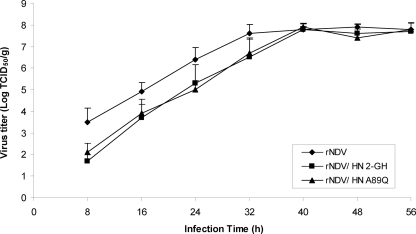
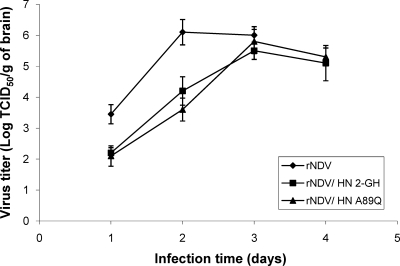
Multicycle growth kinetics of rNDV and the HN globular head and A89Q substitution viruses were compared in DF1 cells (Fig. 11). This showed that these two mutant viruses replicated approximately 10-fold less efficiently than the parental virus up to 32 hpi, but thereafter the titers were the same as for rNDV. Evaluation of pathogenicity by MDT and ICPI assays also demonstrated attenuation of these mutant viruses. The MDT and ICPI values were 56 h and 1.61 for rNDV, 73 h and 1.35 for rNDV/HN-2-GH, and 75 h and 1.37 for rNDV/HN-A89Q, respectively. These viruses also were evaluated for replication in the brains of 1-day-old chicks following intracerebral inoculation (Fig. 12). This showed that the rNDV/HN-2GH and A89Q viruses replicated at 1 to 2 log10 lower than the parental virus at 1 and 2 dpi but were comparable on days 3 and 4 (Fig. 12). Thus, substitution of the globular head domain, or a single amino acid substitution in the stalk domain, was mildly attenuating.
Fig. 11.
In vitro growth characterization of HN mutant NDVs in DF1 cells. The growth characteristics of rNDV and HN mutant viruses were examined by multicycle growth curve in DF1 cells (MOI of 0.01 PFU/cells). The viral titers were determined by immunoperoxidase assay. Each bar represents the mean and standard error of the mean of duplicate samples.
Fig. 12.
Growth kinetics of the HN mutant NDVs in the brains of 1-day-old chicks. One-day-old SPF chicks were inoculated with each virus via the intracerebral route. Virus titers in the brain samples were determined by immunoperoxidase assay in DF1 cells.
DISCUSSION
The characteristics of in vitro CPE and pathogenicity vary greatly among the APMV serotypes. The BC strain of NDV causes extensive cell fusion, spreads systemically in vivo, replicates in most tissues including the brain, and causes a fatal neurological disease in chickens. In contrast, APMV-2 causes single-cell infections in vitro, remains largely restricted to the upper respiratory tract in vivo, does not replicate in brain tissue even when inoculated directly, and does not cause any apparent disease in chickens. The availability of reverse genetics systems for NDV and APMV-2 allowed us to use a chimeric approach to identify the APMV genes responsible for pathogenicity and virulence in chickens. In this study, we evaluated the F and HN envelope glycoproteins because the envelope glycoproteins of numerous viruses have been shown to be important determinants of pathogenesis (10, 12, 15, 16, 27, 29, 52). NDV and APMV-2 represent two distinct serotypes, and their F and HN glycoproteins have 41% and 35% amino acid sequence identity, respectively (43). It has been shown for a number of paramyxoviruses, including NDV, that homotypic interaction between the F and HN proteins is required for fusion and syncytium formation (11, 22, 51). It also is generally thought that the glycoproteins interact with internal virion proteins through their cytoplasmic tails (CTs). There is no significant amino acid sequence identity between the CT domains of the F and HN proteins of the two serotypes, raising the possibility that the CT domains of one serotype might not interact with the internal proteins of the other.
As a first step, we attempted to exchange the entire ORF of the F and HN genes individually and together between NDV and APMV-2. Despite several attempts, in which the parental viruses evaluated in parallel as positive controls were successfully rescued in each case, we were unable to rescue any of the chimeric viruses except for APMV-2/NDV-F, in which the entire F gene ORF of APMV-2 was replaced with that of NDV. The inability to recover all of the other viruses in which complete glycoproteins were swapped individually or together reinforced the idea that homologous interaction between the CT domains of the glycoproteins and internal proteins is required for virus replication. Thus, the rescue of rAPMV-2/NDV-F was an unexpected exception. Analysis of the proteins of purified virions showed that there was no apparent difference in the amounts of F protein between rAPMV-2 and rAPMV-2/NDV-F, indicating that incorporation of the NDV F protein into rAPMV-2 was not impaired. Also, rAPMV-2/NDV-F was more efficient than rAPMV-2 in multicycle growth in cell culture (as well as in vivo), suggesting that there was no obvious impairment. Although the exact internal viral protein(s) with which the APMV F protein interacts during virion formation is not known, it has been suggested that the NDV F protein interacts with N and/or HN proteins but not with the M protein (31). Sequence comparison of the N protein between the two serotypes showed that they have 41% identity. Therefore, it is interesting that, in spite of the low degree of sequence identity in cognate proteins between the two serotypes, there was successful recovery of a chimeric virus with apparently unimpaired capacity for growth. It might be that a conserved structure, such as a constellation of charged amino acids, is important for interactions with N, HN, or other viral protein rather than a conserved sequence. Alternatively, it might be that interaction of the CT domain of APMV F protein with internal proteins is not as critical as has been previously reported.
In light of the low level of sequence relatedness, particularly for the CT domains, we also constructed chimeric viruses in which only the ectodomains of F and/or HN were swapped, thus leaving the homologous CT and transmembrane domains intact. We were successful in recovering the two recombinant chimeric viruses in which the F and HN ectodomains were exchanged together. Despite many attempts, we were unable to rescue viruses in which the ectodomain of only one of the two glycoproteins was exchanged between NDV and APMV-2. These results supported the idea that homologous CT domains are important for virus replication and that type-specific interactions between F and HN proteins also are important for these APMVs. Similar requirements of homotypic F and HN has been observed with other pairs of chimeric paramyxoviruses, such as human parainfluenza virus type 1 (HPIV-1) and human parainfluenza virus type 3 (HPIV-3) (48), HPIV-1 and bovine parainfluenza virus type 3 (BPIV-3) (40), the morbilliviruses rinderpest virus and peste des petits ruminants virus (4), and bovine respiratory syncytial virus and bovine parainfluenza virus (42). The present study also illustrated the importance of homologous F and HN interactions for fusion, since the presence of NDV F paired with APMV-2 HN in the virus rAPMV-2/NDV-F was not sufficiently fusogenic to form syncytia, although presumably it was sufficiently active to initiate infection. This extends previous studies in which plasmid-based expression of cleaved F protein alone did not result in cell-to-cell fusion in cell culture (21). The observation that rAPMV-2/NDV-F replicated efficiently (and thus presumably mediated efficient virus-cell fusion) despite its inability to produce syncytia (cell-to-cell fusion) indicates that the latter activity is not a reliable surrogate marker for the former activity. In most paramyxoviruses, the homotypic F and HN glycoproteins have been known to be physically associated with each other on the cell surface, indicating the attachment protein-dependent mode of fusion (13, 57).
We found that the substitution of NDV F protein alone into rAPMV-2 conferred the ability to replicate in the brains of 1-day-old chicks when infected via the intracerebral route, indicative of neurotropism, or via the intranasal route, indicative of neuroinvasiveness. For rAPMV-2/NDV-F, the titers in the brain following intranasal inoculation were the same as those following intracerebral inoculation, as also was observed for rNDV and rAPMV-2/NDV-F-HN. This implies that NDV F alone mediated efficient spread to the brain (neuroinvasiveness) and that neuroinvasion was not augmented by the presence of the homologous HN protein. On the other hand, the magnitude of replication in the brain (neurotropism) was substantially increased by the presence of the homologous HN protein. The level of neurological disease signs was consistent with the level of replication in the brain and thus was greatest for rNDV, followed by rAPMV-2/NDV-F-HN, followed by rAPMV-2/NDV-F, with no replication or disease signs observed for rAPMV-2 or rNDV/2-F-HN. Thus, replacement of the APMV-2 F protein with that of NDV conferred neurotropism, neuroinvasiveness, and neurovirulence in 1-day-old chicks, but replication in the brain and disease signs were greatly increased by the additional inclusion of homotypic HN. The situation for rAPMV-2/NDV-F in 2-week-old birds inoculated intranasally was somewhat different. In this case, the presence of NDV F alone was insufficient to allow the virus to spread beyond the upper respiratory tract, indicating that the requirement for homotypic HN for viral spread and replication beyond the respiratory tract was greater in these older birds. Although swapping the F and HN proteins of APMV-2 with those of NDV resulted in the acquisition of neurotropism, neuroinvasiveness, and neurovirulence, the level of replication and virulence of rAPMV-2/NDV-F-HN remained lower than those of parental rNDV. This suggested that internal proteins also contribute to viral pathogenicity (8, 37). Conversely, replacing the F and HN ectodomains of NDV with those of APMV-2 was sufficient to completely eliminate syncytium formation, neurotropism, neuroinvasiveness, and neurovirulence, with the result that the phenotypes of rNDV/2-F-HN and rAPMV-2 were very similar. Thus, these reciprocal exchanges showed that the F and HN proteins of NDV and APMV-2 are the major determinants of the virulent and avirulent phenotypes, respectively.
We also constructed an additional chimeric virus (rNDV/2-GH) in which the globular head of the NDV HN protein was substituted with that of APMV-2, thus leaving the homologous CT, TM, and stalk domains intact. This construct was readily recovered, implying that these domains are sufficient to provide most of the necessary homologous interaction, whereas the globular head is exchangeable, as observed with the generation of recombinant NDV vaccine containing the globular head of the APMV serotype 4 HN protein in place of its own (34). This substitution resulted in a 50% reduction in NA activity and a slight attenuation of the virus in vitro but did not affect cell-to-cell fusion. In previous studies, in vitro expression of chimeric HN proteins with segments derived from heterologous paramyxoviruses suggested that specificity for the homologous F protein is determined by the stalk domain of HN, although certain regions in the transmembrane anchor and globular head have also been reported to contribute to F protein specificity (11, 26, 41, 47, 50, 51, 54). The present results, which involve complete virus rather than cDNA-expressed proteins, are consistent with the stalk domain playing a major role in homologous interactions. When evaluated for pathogenicity in embryonated eggs and 1-day-old chicks, the rNDV/2-GH virus was slightly attenuated compared to the rNDV parent with regard to MDT, ICPI, and virus titers in the brain. Thus, while the homologous CT, TM, and stalk domains of HN are crucial for virus replication in vitro and in vivo, the globular head can be exchanged with only a modest effect.
Previously, mutational analysis of the NDV HN stalk domain using a plasmid-based expression system demonstrated that amino acid residues 89, 90, 93, and 94 can affect fusion activity and the ability of HN to interact with F (25). These mutations did not have detectable negative effects on the structure or other functions of the HN protein, implying that they were specific to the fusion-promoting activity (25). In the present study, only the substitution at position 89 could be recovered into infectious virus (rNDV/HN-A89Q). This implies that the other positions, namely, 90, 93, and 94, are essential for virus replication, perhaps due to a role in interaction with F. The A89Q mutation had marginal effects on fusion, HAd, or NA activities and had only a modest effect on virus growth in vitro, MDT, ICPI, and replication in the brains of 1-day-old chicks; thus, it is unlikely to be a critical residue.
In summary, our study highlighted the importance of the F and HN surface proteins in the replication and phenotypes of NDV and APMV-2. The two contrasting phenotypes of NDV and APMV-2 could largely be transferred between the two viral backbones by exchange of the ectodomains of F and HN together. In one case, NDV F alone could be swapped into APMV-2. This showed that NDV F alone conferred neurotropism, neuroinvasiveness, and neurovirulence (all at reduced levels) in 1-day-old chicks but not in older birds. This probably was not simply due to the presence of the multibasic F cleavage site of the NDV F protein, since our previous study indicated that increasing the cleavability of the APMV-2 F protein did not increase the virulence of the virus (45). Homotypic interaction with NDV HN was necessary for syncytium formation in vitro, efficient replication in vitro and in the brains of 1-day-old chicks, spread beyond the upper respiratory tract in older birds, and substantial neurovirulence. The essential domains in the NDV HN protein were dissected further, demonstrating the need for the homologous CT and stalk domains but not the globular head domain. This suggests that the construction of additional chimeras will give further insights into the basis for neurovirulence. In addition, APMV-2 bearing the surface glycoproteins of a more attenuated strain of NDV may provide a live, genetically marked vaccine that is more attenuated than current live NDV vaccines and less able to mutate into a more virulent form.
ACKNOWLEDGMENTS
We thank Daniel Rockemann, Yonas Araya, and our laboratory members for excellent technical assistance and LaShae Green for proofreading of the manuscript. We thank Bernard Moss (NIAID, NIH) for providing the vaccinia T7 recombinant virus.
This research was supported by National Institute of Allergy and Infectious Diseases (NIAID) contract no. N01A060009 (85% support) and NIAID, National Institutes of Health (NIH) Intramural Research Program (15% support).
The views expressed herein do not necessarily reflect the official policies of the Department of Health and Human Services, nor does mention of trade names, commercial practices, or organizations imply endorsement by the U.S. Government.
Footnotes
Published ahead of print on 15 June 2011.
REFERENCES
- 1. Alexander D. J. 1982. Avian paramyxoviruses-other than Newcastle disease virus. Worlds Poult. Sci. J. 38:97–104 [Google Scholar]
- 2. Alexander D. J. 1989. Newcastle disease, p. 114–120 In Purchase H. G., Arp L. H., Domermuth C. H., Pearson J. E. (ed.), A laboratory manual for the isolation and identification of avian pathogens, 3rd ed The American Association of Avian Pathologists, Kendall/Hunt Publishing Company, Dubuque, IA [Google Scholar]
- 3. Alexander D. J. 2003. Avian paramyxoviruses 2–9, p. 88–92 In Saif Y. M. (ed.), Diseases of poultry, 11th ed Iowa State University Press, Ames, IA [Google Scholar]
- 4. Das S. C., Baron M. D., Barrett T. 2000. Recovery and characterization of a chimeric rinderpest virus with the glycoproteins of peste-des-petits-ruminants virus: homologous F and H proteins are required for virus viability. J. Virol. 74:9039–9047 [DOI] [PMC free article] [PubMed] [Google Scholar]
- 5. de Leeuw O. S., Koch G., Hartog L., Ravenshorst N., Peeters B. P. 2005. Virulence of Newcastle disease virus is determined by the cleavage site of the fusion protein and by both the stem region and globular head of the haemagglutinin-neuraminidase protein. J. Gen. Virol. 86:1759–1769 [DOI] [PubMed] [Google Scholar]
- 6. Deng R., Wang Z., Mirza A. M., Iorio R. M. 1995. Localization of a domain on the paramyxovirus attachment protein required for the promotion of cellular fusion by its homologous fusion protein spike. Virology 209:457–469 [DOI] [PubMed] [Google Scholar]
- 7. Dietzschold B., et al. 1985. Differences in cell-to-cell spread of pathogenic and apathogenic rabies virus in vivo and in vitro. J. Virol. 56:12–18 [DOI] [PMC free article] [PubMed] [Google Scholar]
- 8. Dortmans J. C. F. M., Rottier P. J. M., Koch G., Peeters B. P. H. 2010. The viral replication complex is associated with the virulence of Newcastle disease virus. J. Virol. 84:10113–10120 [DOI] [PMC free article] [PubMed] [Google Scholar]
- 9. Duesberg P. H., Robinson W. S. 1965. Isolation of the Newcastle disease virus. Proc. Natl. Acad. Sci. U. S. A. 54:794–800 [DOI] [PMC free article] [PubMed] [Google Scholar]
- 10. Duprex W. P., et al. 1999. The H gene of rodent brain-adapted measles virus confers neurovirulence to the Edmonston vaccine strain. J. Virol. 73:6916–6922 [DOI] [PMC free article] [PubMed] [Google Scholar]
- 11. Gravel K., Morrison T. G. 2003. Interacting domains of the HN and F proteins of Newcastle disease virus. J. Virol. 77:11040–11049 [DOI] [PMC free article] [PubMed] [Google Scholar]
- 12. Hiramatsu K., Tadano M., Men R., Lai C. J. 1996. Mutational analysis of a neutralization epitope on the dengue type 2 virus (DEN2) envelope protein: monoclonal antibody resistant DEN2/DEN4 chimeras exhibit reduced mouse neurovirulence. Virology 224:437–445 [DOI] [PubMed] [Google Scholar]
- 13. Hu X., Ray R., Compans R. W. 1992. Functional interactions between the fusion protein and hemagglutinin–neuraminidase of human parainfluenza viruses. J. Virol. 66:1528–1534 [DOI] [PMC free article] [PubMed] [Google Scholar]
- 14. Huang Z., Krisnamurthy S., Panda A., Samal S. K. 2001. High-level expression of a foreign gene from the 3′ proximal first locus of a recombinant Newcastle disease virus. J. Gen. Virol. 82:1729–1736 [DOI] [PubMed] [Google Scholar]
- 15. Huang Z., et al. 2004. The hemagglutinin-neuraminidase protein of Newcastle disease virus determines tropism and virulence. J. Virol. 78:4176–4184 [DOI] [PMC free article] [PubMed] [Google Scholar]
- 16. Iacono K. T., Kazi L., Weiss S. R. 2006. Both spike and background genes contribute to murine coronavirus neurovirulence. J. Virol. 80:6834–6843 [DOI] [PMC free article] [PubMed] [Google Scholar]
- 17. Kim S. H., Yan Y., Samal S. K. 2009. Role of the cytoplasmic tail amino acid sequences of Newcastle disease virus HN protein in incorporation of virion, cell fusion, and pathogenicity. J. Virol. 83:10250–10255 [DOI] [PMC free article] [PubMed] [Google Scholar]
- 18. Kovamees J., Rydbeck R., Orvell C., Norrby E. 1983. Hemagglutinin-neuraminidase (HN) amino acid alterations in neutralization escape mutants of Kilham mumps virus. Virus Res. 17:117–129 [DOI] [PubMed] [Google Scholar]
- 19. Krishnamurthy S., Huang Z., Samal S. K. 2000. Recovery of a virulent strain of Newcastle disease virus from cloned cDNA: expression of a foreign gene results in growth retardation and attenuation. Virology 278:168–182 [DOI] [PubMed] [Google Scholar]
- 20. Kumar S., Nayak B., Collins P. L., Samal S. K. 2008. Complete genome sequence of avian paramyxovirus type 3 reveals an unusually long trailer region. Virus Res. 137:189–197 [DOI] [PMC free article] [PubMed] [Google Scholar]
- 21. Lamb R. A. 1993. Paramyxovirus fusion: a hypothesis for changes. Virology 197:1–11 [DOI] [PubMed] [Google Scholar]
- 22. Lamb R. A., Paterson R. G., Jardetzky T. S. 2006. Paramyxovirus membrane fusion: lessons from the F and HN atomic structures. Virology 344:30–37 [DOI] [PMC free article] [PubMed] [Google Scholar]
- 23. Lamb R. A., Parks G. D. 2007. Paramyxoviridae: the viruses and their replication, p. 1449–1496 In Knipe D. M., Howley P. M. (ed.), Fields virology, 5th ed Lippincott Williams & Wilkins, Philadelphia, PA [Google Scholar]
- 24. Matloubian M., Somasundaram T., Kolhekar S. R., Selvakumar R., Ahmed R. 1990. Genetic basis of viral persistence: single amino acid change in the viral glycoprotein affects ability of lymphocytic choriomeningitis virus to persist in adult mice. J. Exp. Med. 172:1043–1048 [DOI] [PMC free article] [PubMed] [Google Scholar]
- 25. Melanson V. R., Iorio R. M. 2004. Amino acid substitutions in the F-specific domain in the stalk of the Newcastle disease virus HN protein modulate fusion and interfere with its interaction with the F protein. J. Virol. 78:13053–13061 [DOI] [PMC free article] [PubMed] [Google Scholar]
- 26. Melanson V. R., Iorio R. M. 2006. Addition of N-glycans in the stalk of the Newcastle disease virus HN protein blocks its interaction with the F protein and prevents fusion. J. Virol. 80:623–633 [DOI] [PMC free article] [PubMed] [Google Scholar]
- 27. Morimoto K., Foley H. D., McGettigan J. P., Schnell M. J., Schold B. D. 2000. Reinvestigation of the role of the rabies virus glycoprotein in viral pathogenesis using a reverse genetics approach. J. Neurovirol. 6:373–381 [DOI] [PubMed] [Google Scholar]
- 28. Nayak B., Kumar S., Collins P. L., Samal S. K. 2008. Molecular characterization and complete genome sequence of avian paramyxovirus type 4 prototype strain duck/Hong Kong/D3/75. Virol. J. 5:124. [DOI] [PMC free article] [PubMed] [Google Scholar]
- 29. Ni H., Barrett A. D. T. 1998. Attenuation of Japanese encephalitis virus by selection of its mouse brain membrane receptor preparation escape variants. Virology 241:30–36 [DOI] [PubMed] [Google Scholar]
- 30. Paldurai A., Subbiah M., Kumar S., Collins P. L., Samal S. K. 2009. Complete genome sequences of avian paramyxovirus type 8 strains goose/Delaware/1053/76 and pintail/Wakuya/20/78. Virus Res. 142:144–153 [DOI] [PMC free article] [PubMed] [Google Scholar]
- 31. Pantua H. D., McGinnes L. W., Peeples M. E., Morrison T. G. 2006. Requirements for the assembly and release of Newcastle disease virus-like particles. J. Virol. 80:11062–11073 [DOI] [PMC free article] [PubMed] [Google Scholar]
- 32. Pedersen J. C., et al. 2004. Phylogenetic relationships among virulent Newcastle disease virus isolates from the 2002–2003 outbreak in California and other recent outbreaks in North America. J. Clin. Microbiol. 42:2329–2334 [DOI] [PMC free article] [PubMed] [Google Scholar]
- 33. Peeters B. P. H., de Leeuw O. S., Koch G., Gielkens A. L. J. 1999. Rescue of Newcastle disease virus from cloned cDNA: evidence that cleavability of the fusion protein is a major determinant for virulence. J. Virol. 73:5001–5009 [DOI] [PMC free article] [PubMed] [Google Scholar]
- 34. Peeters B. P., de Leeuw O. S., Verstegen I., Koch G., Gielkens A. L. 2001. Generation of a recombinant chimeric Newcastle disease virus vaccine that allows serological differentiation between vaccinated and infected animals. Vaccine 19:1616–1627 [DOI] [PubMed] [Google Scholar]
- 35. Potier M., Mameli L., Belisle M., Dallaire L., Melancon S. B. 1979. Fluorimetric assay of neuraminidase with a sodium (4-methylumbelliferyl-α-d-N-acetylneuraminate) substrate. Anal. Biochem. 94:287–296 [DOI] [PubMed] [Google Scholar]
- 36. Reed L. J., Muench H. A. 1938. A simple method of estimating fifty percent endpoints. Am. J. Hyg. (Lond.) 27:493–497 [Google Scholar]
- 37. Rout S. N., Samal S. K. 2008. The large polymerase protein is associated with the virulence of Newcastle disease virus. J. Virol. 82:7828–7836 [DOI] [PMC free article] [PubMed] [Google Scholar]
- 38. Samuel A. S., Kumar S., Madhuri S., Collins P. L., Samal S. K. 2009. Complete sequence of the genome of avian paramyxovirus type 9 and comparison with other paramyxoviruses. Virus Res. 142:10–18 [DOI] [PMC free article] [PubMed] [Google Scholar]
- 39. Samuel A. S., Paldurai A., Kumar S., Collins P. L., Samal S. K. 2010. Complete genome sequence of avian paramyxovirus (APMV) serotype 5 completes the analysis of nine APMV serotypes and reveals the longest APMV genome. PLoS One 5:e9269. [DOI] [PMC free article] [PubMed] [Google Scholar]
- 40. Schmidt A. C., et al. 2000. Bovine parainfluenza virus type 3 (BPIV3) fusion and hemagglutinin-neuraminidase glycoproteins make an important contribution to the restricted replication of BPIV3 in primates. J. Virol. 74:8922–8929 [DOI] [PMC free article] [PubMed] [Google Scholar]
- 41. Stone-Hulslander J., Morrison T. G. 1999. Mutational analysis of heptad repeats in the membrane-proximal region of Newcastle disease virus HN protein. J. Virol. 73:3630–3637 [DOI] [PMC free article] [PubMed] [Google Scholar]
- 42. Stope M. B., Karger A., Schmidt U., Buchholz U. J. 2001. Chimeric bovine respiratory syncytial virus with attachment and fusion glycoproteins replaced by bovine parainfluenza virus type 3 hemagglutinin-neuraminidase and fusion proteins. J. Virol. 75:9367–9377 [DOI] [PMC free article] [PubMed] [Google Scholar]
- 43. Subbiah M., Xiao S., Collins P. L., Samal S. K. 2008. Complete sequence of the genome of avian paramyxovirus type 2 (strain Yucaipa) and comparison with other paramyxoviruses. Virus Res. 137:40–48 [DOI] [PMC free article] [PubMed] [Google Scholar]
- 44. Subbiah M., et al. 2010. Pathogenesis of two strains of avian paramyxovirus serotype 2, Yucaipa and Bangor, in chickens and turkeys. Avian Dis. 54:1050–1057 [DOI] [PMC free article] [PubMed] [Google Scholar]
- 45. Subbiah M., Khattar S. K., Collins P. L., Samal S. K. 2011. Mutations in the fusion protein cleavage site of avian paramyxovirus serotype 2 that increase cleavability and syncytia formation but do not increase viral virulence in chickens. J. Virol. 85:5394–5405 [DOI] [PMC free article] [PubMed] [Google Scholar]
- 46. Takimoto T., Bousse T., Coronel E. C., Scroggs R. A., Portner A. 1998. Cytoplasmic domain of Sendai virus HN protein contains a specific sequence required for its incorporation into virions. J. Virol. 72:9747–9754 [DOI] [PMC free article] [PubMed] [Google Scholar]
- 47. Tanabayashi K., Compans R. W. 1996. Functional interaction of paramyxovirus glycoproteins: identification of a domain in Sendai virus HN which promotes cell fusion. J. Virol. 70:6112–6118 [DOI] [PMC free article] [PubMed] [Google Scholar]
- 48. Tao T., et al. 2000. Replacement of the ectodomains of the hemagglutinin-neuraminidase and fusion glycoproteins of recombinant parainfluenza virus type 3 (PIV3) with their counterparts from PIV2 yields attenuated PIV2 vaccine candidates. J. Virol. 74:6448–6458 [DOI] [PMC free article] [PubMed] [Google Scholar]
- 49. Teng M. N., Whitehead S. S., Collins P. L. 2001. Contribution of the respiratory syncytical virus G glycoprotein and its secreted and membrane-bound forms to virus replication in vitro and in vivo. Virology 289:283–296 [DOI] [PubMed] [Google Scholar]
- 50. Tsurudome M., et al. 1995. Identification of regions on the hemagglutinin-neuraminidase protein of human parainfluenza virus type 2 important for promoting cell fusion. Virology 213:190–203 [DOI] [PubMed] [Google Scholar]
- 51. Tsurudome M., et al. 1998. Identification of regions on the fusion protein of human parainfluenza virus type 2 which are required for haemmagglutinin-neuraminidase proteins to promote cell fusion. J. Gen. Virol. 79:279–289 [DOI] [PubMed] [Google Scholar]
- 52. Tucker P. C., Lee S. H., Bui N., Martinie D., Griffin D. E. 1997. Amino acid changes in the Sindbis virus E2 glycoprotein that increase neurovirulence improve entry into neuroblastoma cells. J. Virol. 71:6106–6112 [DOI] [PMC free article] [PubMed] [Google Scholar]
- 53. Waning D. L., Schmitt A. P., Leser G. P., Lamb R. A. 2002. Roles for the cytoplasmic tails of the fusion and hemagglutinin-neuraminidase proteins in budding of the paramyxovirus simian virus 5. J. Virol. 76:9284–9297 [DOI] [PMC free article] [PubMed] [Google Scholar]
- 54. Wild T. F., Fayolle J., Beauverger P., Buckland R. 1994. MV fusion: role of the cysteine-rich region of the fusion glycoprotein. J. Virol. 68:7546–7548 [DOI] [PMC free article] [PubMed] [Google Scholar]
- 55. Xiao S., et al. 2009. Complete genome sequence of avian paramyxovirus type 7 (strain Tennessee) and comparison with other paramyxoviruses. Virus Res. 145:80–91 [DOI] [PMC free article] [PubMed] [Google Scholar]
- 56. Xiao S., et al. 2010. Complete genome sequences of avian paramyxovirus serotype 6 prototype strain Hong Kong and a recent novel strain from Italy: evidence for the existence of subgroups within the serotype. Virus Res. 150:61–72 [DOI] [PMC free article] [PubMed] [Google Scholar]
- 57. Yao Q., Hu X., Compans R. W. 1997. Association of the parainfluenza virus fusion and hemagglutinin–neuraminidase glycoproteins on cell surfaces. J. Virol. 71:650–656 [DOI] [PMC free article] [PubMed] [Google Scholar]


