Description
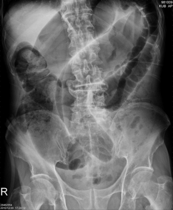
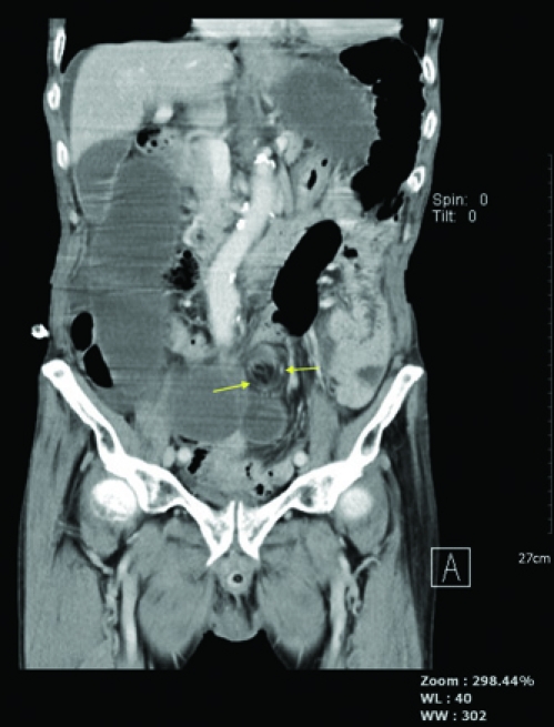
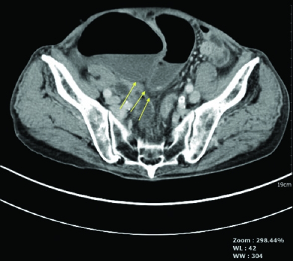
The patient was an 80-year-old man, who had suffered from lower abdominal pain for 3 days and previously had intermittent abdominal discomfort for approximately 1 month. He had not passed any stool for more than 10 days, and had not visited the emergency department prior to the current admission. Upon arrival, his vital signs were stable, and his haemogram demonstrated leukocytosis, with a white blood cell count of 18.0 × 10³/ul (normal range at our institution is 3.4–9.1 × 10³/ul). Physical examination revealed diffuse abdominal tenderness with guarding, and rebound tenderness, which was greater in the lower abdomen. Supine plain film of the abdomen revealed a distended sigmoid loop with an inverted U configuration and a coffee bean sign (figure 1).1 2 The sigmoid colon overlapped with the liver, and extended cephalad to the transverse colon (i.e. northern exposure sign) (figure 1).2 We strongly suspected sigmoid volvulus. CT revealed a whirl sign (figure 2 arrows)2 and transition points (figure 3 arrows).2 Laparotomy confirmed the diagnosis of sigmoid volvulus. The plain film coffee bean sign, northern exposure sign, as well as the CT whirl sign and/or transition point, are indicative of bowel obstruction, including mural thickening and dilatation of the bowel loops. Consequently, these imaging markers aid in the diagnosis of sigmoid volvulus. Thus, physicians need to be aware of the coffee bean, northern exposure sign, whirl sign and transition point, as they are indicative of acute abdominal obstruction, which may be life-threatening and require emergency surgical intervention.
Figure 1.

Plain film of abdomen showed distended sigmoid loop with inverted U configuration and coffee bean sign. Sigmoid overlaps liver and extends cephalad to transverse colon (northern exposure sign).
Figure 2.

CT of the abdomen showed twisting whirl sign.
Figure 3.

CT of the abdomen showed beak-shaped transition point.
Footnotes
Competing interests None.
Patient consent Obtained.
References
- 1.Feldman D. The coffee bean sign. Radiology 2000;216:178–9 [DOI] [PubMed] [Google Scholar]
- 2.Levsky JM, Den EI, DuBrow RA, et al. CT findings of sigmoid volvulus. AJR Am J Roentgenol 2010;194:136–43 [DOI] [PubMed] [Google Scholar]


