Abstract
Aim:
The present study was done to evaluate the role of scrape cytology in the diagnosis of ovarian neoplasm and its utilization for teaching pathology residents.
Materials and Methods:
This was a prospective study on 50 solid/solid-cystic ovarian neoplasms sent in 10% buffered formalin. Scrapings obtained from the fresh cut surface of tumors were smeared uniformly on to glass slides, immediately fixed in 95% ethyl alcohol and stained with hematoxylin and eosin stain.
Results:
The overall diagnostic accuracy of scrape cytology has been satisfactory with 92% of cases correlating with the final diagnosis. Characteristic cytological pattern was noted in various types of surface epithelial, sex cord stromal and germ cell tumors. The technique had limited value in mucinous tumors to distinguish borderline cases from invasive carcinoma. Two mucinous carcinomas were diagnosed as borderline mucinous tumor and two endometrioid carcinomas were misinterpreted as cystadenocarcinoma on scrape cytology. Formalin did not interfere or produce any remarkable changes in cytomorphology.
Conclusions:
Scrape cytology is a simple, rapid, accurate, inexpensive adjunctive cytodiagnostic technique and its routine utilization in ovarian lesions could aid in expanding the cytological knowledge of ovarian neoplasms.
Keywords: Ovarian neoplasm, scrape cytology, utility
Introduction
Fine-needle aspiration (FNAC), Fine-needle cytology (FNC), image-guided aspiration cytology, imprint cytology and squash preparation are common techniques practiced routinely to study cytology of tissues. However, the diagnosis of ovarian neoplasms depends mainly on histopathological examination due to the simple reason that ovaries are inaccessible for cytological techniques except when approached through imaging techniques. Even though ovarian masses could be approached by laparoscopy and ultrasound-guided aspiration, there are controversial views regarding their safety.[1,2] Ascitic fluid is submitted for cytology to stage ovarian neoplasms. A positive fluid cytology suggests an advanced stage, though the categorization of tumor may still be difficult for the cytopathologist. The present study has been done to evaluate the role of scrape cytology in diagnosis of ovarian neoplasm and its utilization for teaching pathology residents.
Materials and Methods
This was a prospective study done on 50 ovarian neoplasms [Table 1]. Grossly solid tumors/solid -cystic ovarian neoplasms were included in the study. Scrapings obtained from the fresh cut surface of tumors sent in 10% buffered formalin were smeared uniformly on to glass slide [Figure 1]. Smears were then immediately fixed in 95% ethyl alcohol and stained with hematoxylin and eosin (H and E) stain.
Table 1.
Final histopathological diagnosis with cytological correlation by scrape cytology
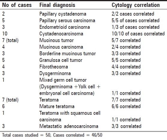
Figure 1.
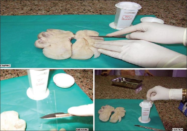
Procedure of scrape cytology includes scraping from tumor mass, smearing and fixation in 95% ethyl alcohol
Cytodiagnosis on smears were made based on cell preservation, cellularity, pattern and cell morphology which was then compared with histopathology to determine the overall correlation. The interpretation of cytology slides was assisted by available clinical data and gross pathologic findings.
Results
The overall diagnostic accuracy of scrape cytology was satisfactory with 92% of cases correlating with final histopathological diagnosis [Table 1]. Characteristic cytological pattern was noted in various types of surface epithelial, sex cord stromal and germ cell tumors [Table 2] [Figures 2–6]. Four of the study cases that did not correlate with final diagnosis included two mucinous carcinomas misdiagnosed as borderline mucinous tumor and two endometrioid carcinomas misinterpreted as cystadenocarcinoma on scrape cytology. One case was a mixed germ cell tumor with dysgerminoma, embryonal carcinoma and yolk sac components. However, only features of dysgerminoma and embryonal carcinoma were identified with certainty on cytosmears of this case. Formalin did not interfere or produce any remarkable changes in cytomorphology. All cases showed good cell preservation.
Table 2.
Cytomorphological features of ovarian neoplasms as seen on scrape cytology
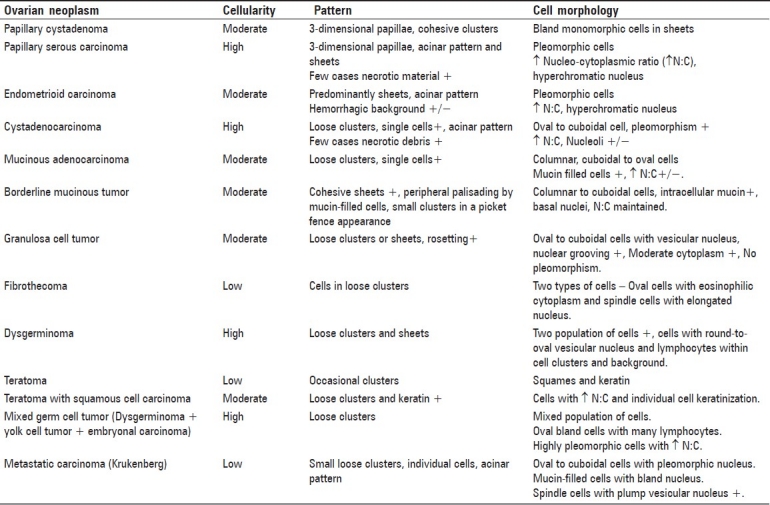
Figure 2.
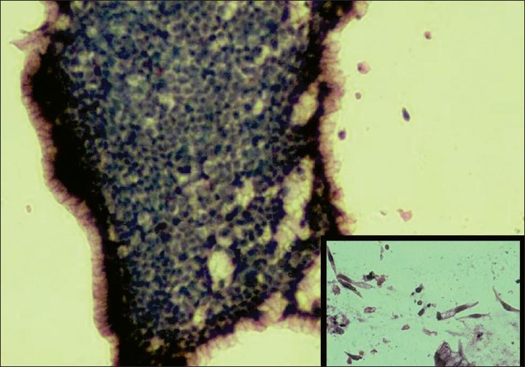
Borderline mucinous tumor with cohesive sheets of bland cells and peripheral palisading by mucin-filled cells (H and E, ×200); inset shows picket fence arrangement of mucin secreting columnar cells (H and E, ×200)
Figure 6.
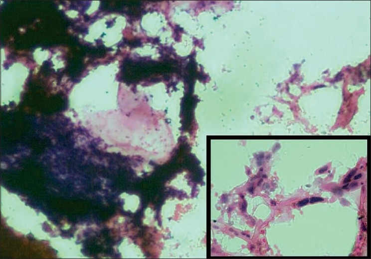
Teratoma with squamous cell carcinoma show flakes of keratin, cellular tissue fragments (H and E, ×200); inset highlights atypical cells with individual cell keratinization (H and E, ×1000)
Figure 3.
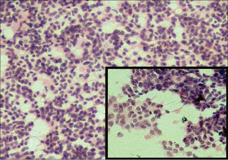
Granulosa cell tumor with loose clusters of uniform round cells and bland nuclei arranged in microacinar pattern; inset shows nuclear grooving in tumor cells (H and E, ×400)
Figure 4.
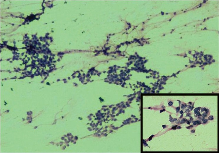
Embryonal carcinoma component composed of highly pleomorphic tumor cells and bizarre nuclei with few clusters arranged in vague glandular pattern (H and E, ×200); inset show higher magnification of the same with visible mitotic figures (H and E, ×400)
Figure 5.
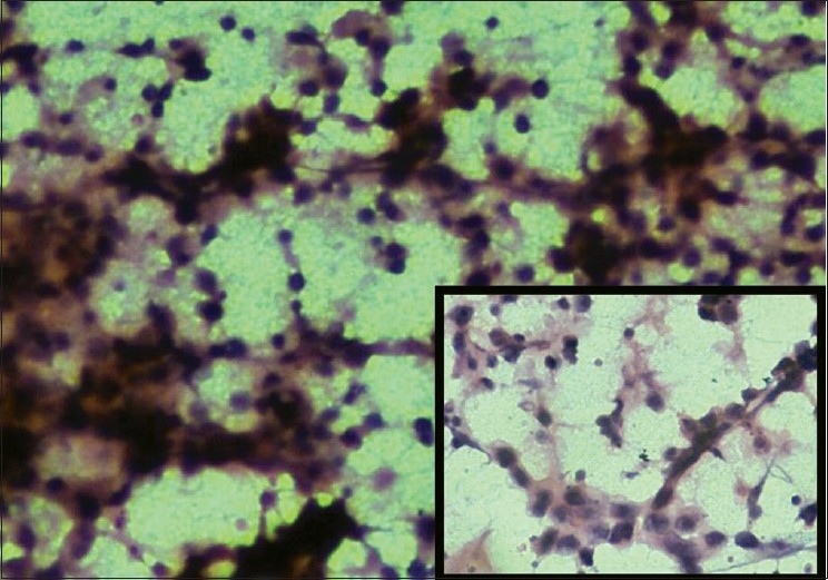
Dysgerminoma with loose clusters of cells having indistinct cytoplasmic margins with scattered lymphocytes (H and E, ×400); inset highlights the same on higher magnification (H and E, ×1000)
Discussion
Dudgeon and Patrick were first to describe the use of imprint smears of fresh tissues in rapid microscopic diagnosis of tumors.[3] Following this, several studies done in the past have discussed the use of imprint and touch preparation especially as a tool for intraoperative diagnosis.[4–6] Scrape cytology is a modification of imprint cytology and its diagnostic accuracy is better than imprint cytology.[5] Scraping of the cut surface prior to smearing facilitates the harvesting of cells. Hence, scrape cytology could be preferred over touch preparation/imprint cytology as the former technique would yield much more material than the latter.[3]
In our study, we analysed the scrape cytology of ovarian neoplasms sent in formalin. In spite of utilizing formalin-fixed specimens, architecture and cell morphology remained well preserved causing no interference in cytological interpretation. This highlights the utility of scrape cytology technique for studying cytomorphology of formalin-fixed cases, provided the gross specimens are available.
Specific ovarian neoplasms show characteristic cytological pattern and morphology suggesting an almost accurate diagnosis. The knowledge of pattern and cell morphology would help in identification of ovarian neoplasms on cytology. Khunamornpong et al.[7] studied role of scrape cytology of ovaries in intraoperative consultation of ovarian lesions and found it to be a useful rapid cytodiagnostic tool.Similar to our findings, their study also revealed that it was the borderline group which showed inconsistent results especially mucinous tumors. A more accurate diagnosis would require histologic architecture evaluation and extensive tissue sampling.[7] Although, cytomorphology of endometrioid carcinoma is quite distinctive, its overlapping cytological features with cystadenocarcinoma might result in misdiagnosis of one for the other. However, both represent malignant surface epithelial tumors (adenocarcinoma) in general.
Cytology of ovarian neoplasms at times may be difficult to interpr et alone without any accompanying data. Relevant clinical data and gross morphological features are valuable information that would be necessary while evaluating cytological smears. The availability of these details while studying cytosmears would be one of the reasons for a high accuracy rate in our study.
Michael et al.[8] compared cytology and frozen section in a study on intraoperative consultation of ovarian lesions and found that intraoperative fine-needle aspirations (FNAs) and scrapes were superior to imprints.In addition, scrape is an easier method yielding greater cellularity.[8] The ability to render immediate diagnosis on scrape cytology highlights its role and its potential usage in intraoperative consultation in institutions unequipped with frozen section facility.[5,7] With respect to ovarian neoplasms this would require the cytologist to be familiar with ovarian lesions and experience in the interpretation of ovarian cytosmears. Pathologist must also be aware of potential diagnostic pitfalls such as evaluation based on few cells.[2] The material obtained would also prove useful for teaching residents the cytomorphology of ovarian lesions. This could be kept as an archival material for reference and future learning. An immediate feedback at gross conference could be an additional benefit with such rapid and easy technique.[9]
To conclude, scrape cytology is a simple, quick, accurate, inexpensive adjunctive cytodiagnostic technique and its routine utilization in ovarian lesions could aid in expanding the knowledge of cytology of ovarian neoplasms.
Footnotes
Source of Support: Nil
Conflict of Interest: None declared.
References
- 1.Uguz A, Ersoz C, Bolat F, Gokdemia A, Vardar MA. Fine needle aspiration cytology of ovarian lesions. Acta Cytol. 2005;49:144–8. doi: 10.1159/000326122. [DOI] [PubMed] [Google Scholar]
- 2.Ganjei P. Fine-needle aspiration cytology of the ovary. Clin Lab Med. 1995;15:705–26. [PubMed] [Google Scholar]
- 3.Shirley SE, Escoffery CT. Usefulness of touch preparation cytology in postmortem diagnosis: A study from the University Hospital of the West Indies. Int J Pathol. 2005;3:2. [Google Scholar]
- 4.Lee TK. The value of imprint cytology in tumour diagnosis: a retrospective study of 522 cases in northern China. Acta Cytol. 1982;26:169–71. [PubMed] [Google Scholar]
- 5.Shidham VB, Dravid NV, Grover S, Kher AV. Role of scrape cytology in rapid intraoperative diagnosis: Value and limitations. Acta Cytol. 1984;28:477–82. [PubMed] [Google Scholar]
- 6.Suen KC, Wood WS, Syed AA, Quenville NF, Clement PB. Role of imprint cytology in intraoperative diagnosis: value and limitations. J Clin Pathol. 1978;31:328–37. doi: 10.1136/jcp.31.4.328. [DOI] [PMC free article] [PubMed] [Google Scholar]
- 7.Khunamornpong S, Siriaunkgul S. Scrape cytology of the ovaries: potential role in intraoperative consultation of ovarian lesions. Diagn Cytopathol. 2003;28:250–7. doi: 10.1002/dc.10273. [DOI] [PubMed] [Google Scholar]
- 8.Michael CW, Lawrence WD, Bedrossian CW. Intraoperative consultation in ovarian lesions: A comparison between cytology and frozen section. Diagn Cytopathol. 1996;15:387–94. doi: 10.1002/(SICI)1097-0339(199612)15:5<387::AID-DC6>3.0.CO;2-9. [DOI] [PubMed] [Google Scholar]
- 9.Schnadig VJ, Molina CP, Aronson JF. Cytodiagnosis in the autopsy site: A tool for improving autopsy quality and resident education. Arch Pathol Lab Med. 2007;131:1056–62. doi: 10.5858/2007-131-1056-CITASA. [DOI] [PubMed] [Google Scholar]


