Abstract
Over the decades, causes of genital elephantiasis have changed only to become elusive to etiological diagnosis. This is a case of 20 year old male who presented with genital elephantiasis occurring due to lymphatic obstruction caused by chromoblastomycosis and super added erysipelas. The diagnosis of chromoblastomycosis was clenched by biopsy. We describe this case for the rarity of its occurrence.
Keywords: Genital elephantiasis, biopsy, chromoblastomycosis
INTRODUCTION
Genital elephantiasis means massive enlargement of genitals. It commonly affects the young and productive age group. Early diagnosis is important as genital elephantiasis is not completely reversible with medical therapy; late intervention often needs surgical reduction. Furthermore, it leads to mental anguish and physical disability as it interferes with walking and sexual intercourse and indirectly interferes with economic livelihood.
Over the decades, causes of genital elephantiasis have changed to become more elusive to etiological diagnosis. In the pre penicillin era, syphilis was the frequent cause of genital elephantiasis. Then was the age of bacterial STI's – donovanosis, LGV; the incidence of which declined with the advent of effective antibiotics. This was succeeded by increase in reporting of non STI cases like filariasis, tuberculosis, leishmaniasis, HIV associated and very rarely chromoblastomycosis. We report a case of genital elephantiasis due to chromoblastomycosis for its rarity of occurrence.
CASE REPORT
A 20-year-old male, truck driver, presented with a primary complaint of genital swelling developed over the last six months. He also had concomitant bizarre and protean skin lesions over lower abdomen, face, neck and thighs along with discharging lesion over right axilla. He gave a history of lesions over face appearing first and other lesions presenting slowly over two years, all with centrifugal expansion. The genital swelling was associated with recent onset pain and itching. The patient had a history of multiple sexual exposures. There was no history of discharge per urethra, genital ulcer or trauma. He also had history of antituberculous and antifilarial treatment with no alleviation of symptoms.
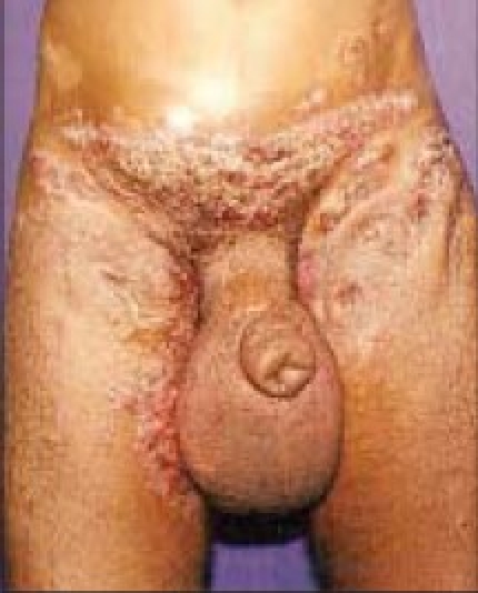
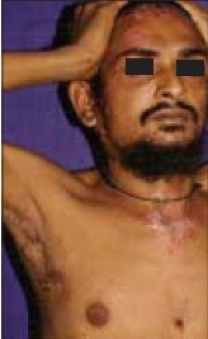
Examination revealed pallor and generalized lymphadenpathy. The patient had grotesque swelling over scrotum and penis [Figure 1a]. There were multiple, well defined large plaques with warty surface over lower abdomen, encircling the upper medial and posterior thighs, superimposed with crusting and discharge [Figure 1a]. Similar warty papules were seen over forehead and neck with satellite lesions peripherally and a discharging sinus over left axillary region [Figure 2a].
Figure 1a.

Swelling over scrotum and penis. Warty crusted lesions over lower abdomen, groin and thighs
Figure 2a.

Protean lesions over forehead and neck. Discharging sinus over right axilla
The differential diagnosis of cutaneous tuberculosis, bacterial STI with complications, filariasis and immunosuppression associated symptoms were kept. Hematological investigations, FNAC, chest X-ray, and USG abdomen were done- all of which were normal. Serum HIV, VDRL with titre were non reactive. The case was reanalyzed and subjected to surgical biopsy to evaluate for less considered diagnosis of chromoblastomycosis in case of genital elephantiasis. Biopsy was suggestive of foreign body granuloma showing muriform body; pseudo epitheliomatous hyperplasias of the epidermis and on staining, fungal hyphae were seen favoring the diagnosis of chromoblastomycosis. Fungal culture was not feasible.
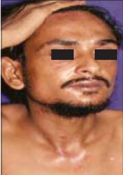
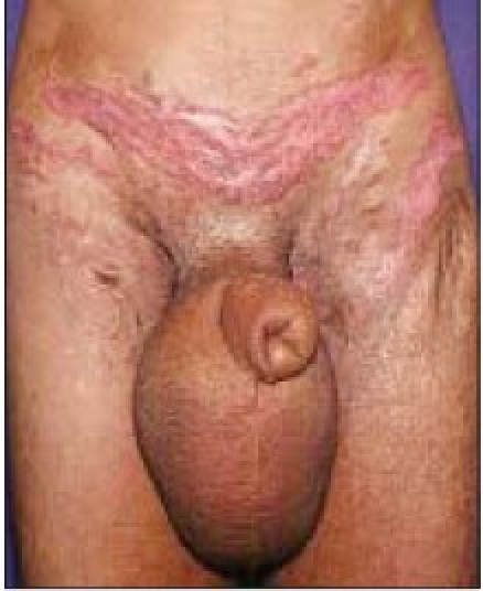
The patient was then put on oral fluconazole 200mg twice a day and cycles of injection amikacin 750mg/day with three weeks on and two weeks off dosing pattern for three months along with cryotherapy, to which he responded well and the lesions healed with mild scarring over a period of one month [Figures 1b, 2b]. The scrotal swelling did not resolve much. He was then advised surgery for reduction of secondary lymphedema induced genital elephantiasis.
Figure 2b.

Healed lesions over forehead and neck
Figure 1b.

Posttreatment healing of lesions with scarring. No change in genital swelling
DISCUSSION
Chromoblastomycosis is a chronic fungal infection of the skin and subcutaneous tissue caused by traumatic (often unnoticed or unremembered) inoculation of pigmented fungi which produce muriform bodies and clinically producing slow growing exophytic lesions. The diagnosis is clenched by biopsy. Usually disease is localized but lymphatic and hematogenous dissemination may occur. It is complicated by secondary bacterial infection and later lymphatic stasis occurs as a sequel to infection.
As per history, our patient developed initial lesion over forehead which later disseminated to axilla and lower abdomen. When he presented to us, he had developed complications of chromoblastomycosis such as erysipelas (over abdominal and groin lesion), lymph stasis and consequently regional elephantiasis.
Guptaet al. reported erysipelas as a frequent complication in patients with genital elephantiasis, as the defense mechanism of the skin is impaired due to chronic lymphatic obstruction and compromised blood supply. Repeated episodes of erysipelas may further increase the lymphatic obstruction and elephantiasis. Such patients benefit from long term penicillin therapy and vaccination against streptococci.[1]
Genital elephantiasis occurs due to diverse causes like LGV, Donovanosis, tuberculosis, filariasis in endemic areas. The causative organism of LGV, C. trachomatis is a lymphotropic organism which causes thrombo-lymphangitis, peri-lymphangitis and involvement of the draining lymph nodes. Genital elephantiasis in LGV is seen approximately 1-20 yrs after infection, occurring as a combination of chronic edema, sclerosing fibrosis, and active diffuse lympho-granulomatous infiltration in the subcutaneous tissues. Polymerase chain reaction (PCR) has been useful in identification of L1 to L3 serovars of C. trachomatis. According to a study conducted by Younget al, genital PCR gives better results than routine culture.[2]
In donovanosis, genital elephantiasis occurs as a result of constriction of lymphatics (pseudo elephantiasis). Nucleic acid amplification test offers the highest sensitivity for diagnosis of Donovanosis, but is not commercially available. Demonstration of donovan bodies with bipolar staining in large foamy mononuclear cells from the genital ulcers is the mainstay of diagnosis.
Primary elephantiasis can be differentiated from secondary elephantiasis by early onset in prior and lymphangiography. Nelsonet al. reported penile and scrotal elephantiasis caused by indolent Chlamydia trachomatis, which responded slowly to treatment with doxycycline.[3]
Murphyet al. reported a case of granulomatous lymphangitis in an 11-year-old boy, and proposed of it as important differential diagnosis of chronic idiopathic swelling of the genitalia, in younger individuals. Such cases should be followed closely for co existing or subsequent development of Crohn's disease. The overlap between granulomatous lymphangitis of the genitalia, Crohn's disease and orofacial granulomatosis suggests that granulomatous lymphangitis of the genitalia may represent a forme fruste of Crohn's disease.[4]
Pandhiet al. reported a case of lupus vulgaris presenting with genital elephantiasis and mimicking LGV. The skin biopsy of the genital lesion and chest X-ray confirmed the diagnosis of tuberculosis.[5]
The main treatment modality for chromoblastomycosis is antifungal chemotherapy like itraconazole, terbinafine, flucytosine. However, we treated our patient with fluconazole because of easy availability of drug, and the patient responded well. For genital elephantiasis surgical reduction is needed. Our patient was not willing for surgery. Mc Dougal reported no recurrence after a follow up of six months to 10 years in male patients with non STI related lymphedema treated with reduction procedures.[6]
Footnotes
Source of Support: Nil,
Conflict of Interest: None declared.
REFERENCES
- 1.Gupta S, Ajith C, Kanwar AJ, Sehgal VN, Kumar B, Mete U. Genital elephantiasis and sexually transmitted infections-revisited. Int J STD AIDS. 2006;17:157–66. doi: 10.1258/095646206775809150. [DOI] [PubMed] [Google Scholar]
- 2.Young H, Scott GR, Patrizio C, Moyes A, Horn K, Sutherland S. PCR testing of genital and urine specimens compared with culture for the diagnosis of chlamydial infection in men and women. Int J STD AIDS. 1998;9:661–5. doi: 10.1258/0956462981921314. [DOI] [PubMed] [Google Scholar]
- 3.Nelson RA, Albert GL, King RL., Jr Penile and scrotal elephantiasis caused by indolent Chlamydia trachomatis infection. Urology. 2003;61:224. doi: 10.1016/s0090-4295(02)02078-2. [DOI] [PubMed] [Google Scholar]
- 4.Murphy MJ, Kogan B, Carlson AJ. Granulomatous lymphangitis of the scrotum and penis: Report of a case and review of the literature of genital swelling with sarcoidal granulomatous inflammation. J Cutan Pathol. 2001;28:419–24. doi: 10.1034/j.1600-0560.2001.028008419.x. [DOI] [PubMed] [Google Scholar]
- 5.Pandhi D, Reddy BS, Rajpal M, Chowdhry S, Rajpal S. Elephantiasic lupus vulgaris mimicking lymphogranuloma venereum: A case report. Ind J Tub. 2002;49:107. [Google Scholar]
- 6.Mc Dougal WS. Lymphedema of the external genitilia. J Urol. 2003;170:711–6. doi: 10.1097/01.ju.0000067625.45000.9e. [DOI] [PubMed] [Google Scholar]


