Fig. 1.

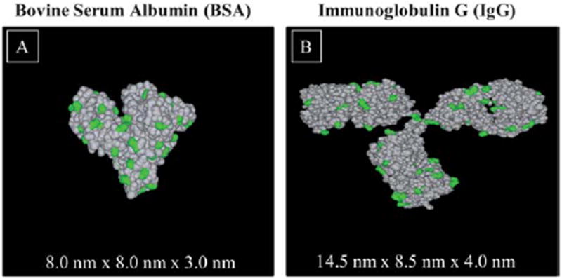
3D space filling models of protein structures revealing lysine residues. (A) N-form conformation of bovine serum albumin (BSA), and (B) immunoglobulin G (IgG). The lysine (Lys) residues are highlighted in green.

3D space filling models of protein structures revealing lysine residues. (A) N-form conformation of bovine serum albumin (BSA), and (B) immunoglobulin G (IgG). The lysine (Lys) residues are highlighted in green.