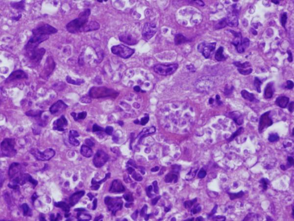
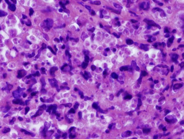
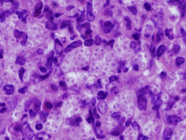
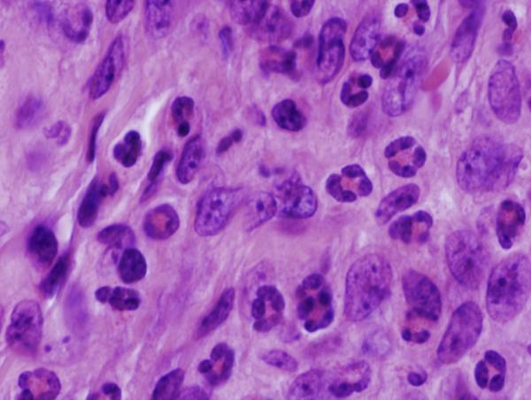
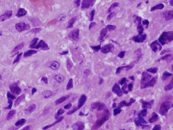
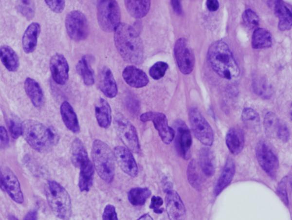
Figure 8. Footpad histology.
Histological sections (H&E stain, × 1,000 magnification) showing the L. major footpad lesion in an infected and untreated BALB/c mouse footpad biopsy as compared to an infected and AmB-PMA treated (400 μg of AmB/mouse) BALB/c mouse footpad biopsy. (a) Infected and untreated at day 14. L. major amastigotes are seen in macrophages. (b) Infected and untreated at day 28. Numerous amastigotes are seen in the phagolysosomes of macrophages. (c) Infected and untreated at day 50. Numerous amastigotes persist in macrophage phagolysosomes. (d) Infected and treated at day 28. Neutrophils are present. There are no amastigotes. (e) Infected and treated at day 50. There are no neutrophils or amastigotes present. The lesion shows an infiltrate of histiocytes, lymphoid cells and plasma cells. (f) Infected and treated at day 80. The presence of myofibroblasts indicates that lesion healing is in its final stages. There are no amastigotes.

