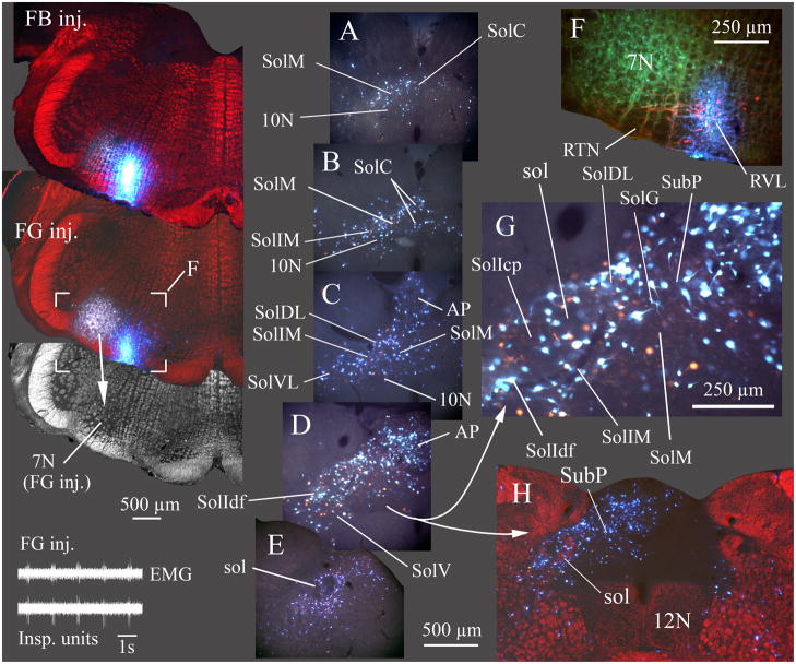
Fig. 4. Most caudal NTS nuclei and area postrema project to the RTN/RVL region.
In this example (case 8045), two retrograde tracers, FG and Fast Blue (FB) were injected at the caudal end of the facial nucleus. FG was injected where inspiratory unit activity was recorded (inset lower left) and FB was injected slightly caudal and ventral to the FG injection (100 μm caudal, 300 μm medial) but where no respiratory unit activity was evident. The FG injection overlapped the caudal facial nucleus (7N; in grayscale darkfield image from section with FG injection site). In addition to overlap of 7N, some dye extended to the superficial layers of the ventral medulla containing the RTN (F, detail of area marked on section with FG injection). FG and FB injections both overlapped TH immunolabeled neurons in RVL (red neurons in F). Sections A–E progress from caudal to rostral with A ~ 300 μm caudal and E ~ 900 μm rostral to obex. G is an enlarged view of the area in D.
A–E and G are true color images taken with a liquid crystal RGB filter and uv excitation. Retrograde labeling from both the FG and FB injections was widespread, preferentially in ipsilateral caudal NTS and ipsilateral area postrema. H is at about the same level as D and illustrates the preferential ipsilateral retrograde labeling in NTS and AP. Labeled cells were found in every nucleus of the ipsilateral caudal NTS (A–E, and G) except SolCe. In most NTS nuclei, FG and FB labeled cells were coextensive, although not always double labeled (see detail in G). FB labeled cells generally appeared brighter than FG labeled cells in the aqueous media used to coverslip this material; double-labeled cells appeared as an intense blue-white color. EMG in inset is inspiratory motor activity recorded from the chest wall.
Injection sites for FB and FG and the higher power image in H are depicted as pseudo-colored images composed by placing a monochrome darkfield image of the relevant section in the red channel of a blank RGB image, then combining this “red darkfield image” with a true color image of both injected dyes (blue FB and gold FG). The true color image was placed in a layer superficial to the darkfield image and combined with the latter using the “lighten” command in Photoshop CS3® which resulted in replacement of only the darker red areas by the brighter colors of the injection sites or, in H, by the retrograde labeled neurons. EMG in inset shows inspiratory motor activity. ECG activity was manually reduced on the EMG trace to facilitate visualization of the respiratory cycle.

