Summary
Topical capsaicin formulations are used for pain management. Safety and modest efficacy of low-concentration capsaicin formulations, which require repeated daily self-administration, are supported by meta-analyses of numerous studies. A high-concentration capsaicin 8% patch (Qutenza™) was recently approved in the EU and USA. A single 60-min application in patients with neuropathic pain produced effective pain relief for up to 12 weeks. Advantages of the high-concentration capsaicin patch include longer duration of effect, patient compliance, and low risk for systemic effects or drug–drug interactions. The mechanism of action of topical capsaicin has been ascribed to depletion of substance P. However, experimental and clinical studies show that depletion of substance P from nociceptors is only a correlate of capsaicin treatment and has little, if any, causative role in pain relief. Rather, topical capsaicin acts in the skin to attenuate cutaneous hypersensitivity and reduce pain by a process best described as ‘defunctionalization’ of nociceptor fibres. Defunctionalization is due to a number of effects that include temporary loss of membrane potential, inability to transport neurotrophic factors leading to altered phenotype, and reversible retraction of epidermal and dermal nerve fibre terminals. Peripheral neuropathic hypersensitivity is mediated by diverse mechanisms, including altered expression of the capsaicin receptor TRPV1 or other key ion channels in affected or intact adjacent peripheral nociceptive nerve fibres, aberrant re-innervation, and collateral sprouting, all of which are defunctionalized by topical capsaicin. Evidence suggests that the utility of topical capsaicin may extend beyond painful peripheral neuropathies.
Keywords: capsaicin, nerve growth factor, neuropathic pain, nociceptor, TRPV1
Editor's key points.
Topical capsaicin is used in pain management.
The mechanism of action (MoA) was thought to be by depletion of substance P.
A more likely MoA is described as ‘defunctionalization’, and involves alteration of several mechanisms involved in pain.
A new higher concentration (8%) patch shows promise in pain management.
Topical capsaicin formulations are widely used to manage pain. Low-concentration creams, lotions, and patches intended for daily skin application have been available in most countries since the early 1980s. Prescriptions are usually not needed for these self-administered medicines, which often have not been reviewed formally by drug regulatory authorities. The recent approval in the EU and USA of a prescription-strength high-concentration single-administration capsaicin 8% patch (Qutenza™) with a duration of action over many weeks invites an examination of recent advances in the understanding of capsaicin's mechanism and site of action.
In this review, which does not cover other naturally occurring or synthetic TRPV1 agonists, we discuss the potential utility of topically administered capsaicin for the management of pain in classical peripheral neuropathies and other hypersensitivity disorders, some of which are currently considered as idiopathic. Furthermore, we seek to elucidate the molecular and cellular basis of capsaicin treatment, and clarify misunderstandings, particularly with respect to the involvement of substance P depletion.
Pain management with topical capsaicin
Capsaicin has played an important role in folk medicine, often on the basis of using like to treat like, for example, treating burning pain with a substance which causes burning pain.1 The first formal report of the pain-reducing properties of topical capsaicin in the West appeared in 1850 as a recommendation to use an alcoholic hot pepper extract on burning or itching extremities.2 Creams, lotions, and patches containing capsaicin, generally in the range of 0.025–0.1% by weight, are now sold in many countries, often without the requirement of a prescription, for the management of neuropathic and musculoskeletal pain. Clinical studies of these medications, usually involving three to five topical skin applications per day for periods of 2–6 weeks, have generally suggested modest beneficial effects against various pain syndromes, including post-herpetic neuralgia (PHN), diabetic neuropathy, and chronic musculoskeletal pain.3,4 Since low-concentration, capsaicin-based products often result in contamination of the patient's environment (clothing, bedding, contact lenses, etc.) and each application may be associated with a burning sensation, poor patient compliance with these products is often cited as a likely contributor to limited efficacy.5
In an attempt to evaluate whether pain relief could be achieved by a single exposure to a much higher concentration of topical capsaicin, 10 patients with intractable pain syndromes were treated with a compounded high-concentration 5–10% w/w cream.6 Patients were provided regional anaesthesia for tolerability and airborne contamination of treatment rooms occurred. Based on encouraging results, a high-concentration capsaicin-containing (8%) patch designated NGX-4010 and then given the trade name Qutenza™ was developed and evaluated.7
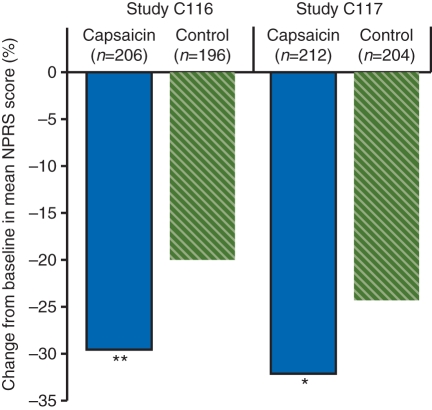
The capsaicin 8% patch is designed to rapidly deliver capsaicin into the skin while minimizing unwanted systemic or environmental exposure of capsaicin to patients and health-care providers. Phase 1 data suggested that a single 60-min patch application was adequate to induce nociceptor defunctionalization, as measured by reversible reduction in intra-epidermal nerve fibres (ENFs), marked by the structural nerve marker protein gene product (PGP) 9.5 immunostaining, and small, reversible alterations in cutaneous nociceptor function.8,9 Phase 3 studies demonstrated efficacy against PHN10,11 (Fig. 1) and painful HIV-AN (associated neuropathy).12 For both neuropathic pain syndromes, efficacy was observed to last for 12 weeks. Blinding was provided by a control patch which contained sufficient capsaicin to induce pain and erythema in a substantial number of subjects.
Fig 1.
Efficacy of capsaicin 8% patch in post-herpetic neuralgia patients. Per cent change from baseline in mean numeric pain rating scale (NPRS) score during weeks 2–8 (the primary endpoint) in two similarly designed randomized, double-blind, multicentre trials (C11610 and C11711). Capsaicin 8% w/w or control (capsaicin 0.04% w/w) patches were applied once for 60 min to the painful areas and patients were followed for 12 weeks. Mean baseline NPRS scores per group ranged from 5.7 to 6.0. *P=0.011, **P=0.001 vs control. Taken from McCormack.7
In 2009, Qutenza™ was approved for the treatment of peripheral neuropathic pain in non-diabetic adults in the EU, and in the USA to manage neuropathic pain associated with PHN.7 One important aspect of this formulation relative to low-concentration capsaicin formulations is removal of the potential for variability in administration and a lack of patient compliance, as its use occurs under the supervision of a health-care professional, and it requires a single application for 30 or 60 min. Furthermore, the environmental contamination issues associated with home use are avoided.
Capsaicin pharmacology
Capsaicin is a highly selective and potent (low nanomolar affinity) exogenous agonist for the TRPV1 receptor, a trans-membrane receptor-ion channel complex which provides integrated responses to temperature, pH, and endogenous lipids.13 Temperatures of 43°C or higher or acidity of pH of <6.0 can directly activate the channel, but combinations of these two stimuli can activate the channel at substantially lower temperatures or pH values. Numerous putative endogenous agonists for TRPV1 have been identified; these include anandamide, N-acyldopamines, other long-chain unsaturated fatty acids, and lipoxygenase compounds such as leukotriene B4 and 12-(S) and 15-(S)-hydroperoxyeicosatetraenoic acid.13 Recently, oxidized metabolites of linoleic acid have been added to the list of potential endogenous agonists.14 Responsiveness of TRPV1 receptors to these activators is also highly regulated by the phosphorylation state of the channel complex, the presence of ancillary proteins, and an ever-growing array of putative allosteric modulators.15
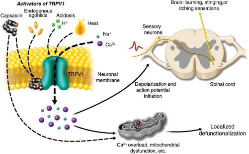
When activated by a combination of heat, acidosis, or endogenous/exogenous agonists, TRPV1 may open transiently and initiate depolarization mediated by the influx of sodium and calcium ions. In the nociceptive sensory nerves which selectively express TRPV1 (mostly C- and some Aδ-fibres), depolarization results in action potentials, which propagate into the spinal cord and brain, and may be experienced as warming, burning, stinging, or itching sensations (Fig. 2).
Fig 2.
Activation of TRPV1 by capsaicin results in sensory neuronal depolarization, and can induce local sensitization to activation by heat, acidosis, and endogenous agonists. Topical exposure to capsaicin leads to the sensations of heat, burning, stinging, or itching. High concentrations of capsaicin or repeated applications can produce a persistent local effect on cutaneous nociceptors, which is best described as defunctionalization and constituted by reduced spontaneous activity and a loss of responsiveness to a wide range of sensory stimuli.
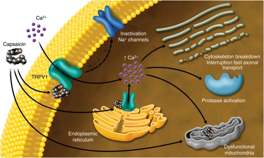
In contrast to transient activation which follows normal environmental stimuli or inflammatory responses to tissue injury, activation of TRPV1-expressing nerve fibres by exposure to a chemically stable exogenous agonist, such as capsaicin, can generate a biochemical signal with a persistent effect. The TRPV1 channel is highly calcium permeable (with a calcium:sodium permeability ratio that starts at about 8:1 and increases to about 25:1 during prolonged capsaicin exposures),16 which allows significant amounts of calcium to flow down its steep electrochemical gradient into nerve fibres. Furthermore, as TRPV1 is also expressed on intracellular organelles, external capsaicin application can cause release of calcium from the endoplasmic reticulum17 and induce additional intracellular calcium release from internal stores via calcium-dependent calcium release.18 Taken together, these multiple sources of calcium provide a robust intracellular signal which can overwhelm local calcium sequestration mechanisms. Consequently, sustained high levels of intracellular calcium can activate calcium-dependent enzymes such as proteases,19 and can induce the depolymerization of cytoskeletal components such as microtubules.20,21 Moreover, osmotic swelling due to the chloride accumulation that must accompany influxes of positively charged ions also occurs.1 An additional effect of high concentrations of capsaicin, which does not involve TRPV1, is a direct inhibition of mitochondrial respiration. Numerous mitochondria are present in the peripheral terminals of nociceptors and may congregate there in response to nerve growth factor (NGF).22 At concentrations much higher than required to activate TRPV1, capsaicin can compete with ubiquinone to inhibit directly electron chain transport.23 Consequently, capsaicin can dissipate mitochondrial transmembrane potential,24 and does so with an EC50 of 6.9 μM in sensory neurones.25 In accord with these widely recognized effects, if TRPV1-expressing sensory nerve fibres are exposed to high concentrations of capsaicin or to lower concentrations in a continuous fashion, high levels of intracellular calcium and the associated enzymatic, cytoskeletal, and osmotic changes, and the disruption of mitochondrial respiration lead to impaired local nociceptor function for extended periods26 (Fig. 3).
Fig 3.
Multiple mechanisms underlie capsaicin-induced defunctionalization. Inactivation of voltage-gated Na+ channels and direct pharmacological desensitization of plasma membrane TRPV1 receptors may contribute to an immediate reduction on neuronal excitability and responsiveness. More persistent effects may be due to the overwhelming of intracellular Ca2+ buffering capacity by extracellular Ca2+ entering through TRPV1 and being released from intracellular stores, with subsequent activation of calcium-dependent proteases and cytoskeleton breakdown. Microtubule depolymerization may interrupt fast axonal transport. At concentrations far in excess of those required to activate TRPV1, capsaicin can also render mitochondria dysfunctional by directly inhibiting electron chain transport. Thus mitochondria are a key convergence point for defunctionalization.
The term ‘desensitization’ is often used to describe these local effects of capsaicin on sensory nerve function, but is unsatisfactory in several respects. The use of this nomenclature in capsaicin literature arose many years ago from psychophysical studies of human subjects who displayed reduced reactions to painful stimuli applied to skin areas pretreated with capsaicin.1 Unfortunately, once the capsaicin receptor (TRPV1) was recognized as a unique molecule entity, the psychophysical use of ‘desensitization’ evolved into pharmacological use, which denotes the reduction of responsiveness of receptors, ion channels, or intracellular signalling pathways after prolonged or repeated agonist exposures. In the continued presence of exogenous agonists such as capsaicin, pharmacological desensitization of TRPV1 itself may indeed contribute acutely to analgesic efficacy. However, transient effects on TRPV1 are quite unlikely to account for the persistent pain relief seen clinically after either single treatments with high-concentration capsaicin or repetitive administration of low-concentration capsaicin. Hence, the emerging preferred term for the persistent local effects of capsaicin is ‘defunctionalization’,26,27 which avoids conceptual confusion with the intrinsic desensitisation of the TRPV1 receptor.
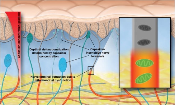
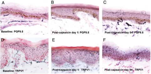
Loss of mitochondrial function due to calcium overload and inhibition of metabolism may render affected nerve processes unable to maintain plasma membrane integrity and thus cause collapse of nerve endings to the depth where the capsaicin exposure was insufficient to irreversibly overwhelm mitochondrial function (Fig. 4). If nerve fibres in skin retract or ‘degenerate’ to the depth at which mitochondrial function was preserved, it is expected that markers for any constituent of those fibres will show reductions. Indeed, many immune-histochemical studies using antibodies to PGP 9.5 or other nerve fibre proteins provide evidence that capsaicin can produce highly localized loss of nociceptive nerve fibre terminals in the epidermis and dermis28 (Fig. 5). However, it is important to recognize that defunctionalization and nerve terminal degeneration are distinct phenomena, each with potentially different time-courses. In the simplest case, a loss of electrical excitability (the loss of function) may occur via depolarization block or sodium channel inactivation, independently of a loss of axonal integrity. Other important physiological roles such as fast axonal transport of growth factors can also be compromised by capsaicin without axonal collapse.29 Even though a general parallel between capsaicin-induced functional and structural changes is expected, there may be differences in the time-courses. For instance, in a recent study of the time-course of recovery of cerebral-evoked potential amplitudes to cutaneous heat-pain laser stimuli after topical capsaicin treatment, functional recovery occurred before the conventional cutaneous nerve markers PGP 9.5 and TRPV1 showed significant recovery30 (Fig. 5). In contrast, fibres marked by GAP-43, which is expressed by regenerating nerve fibres, did show good correlation with the functional responses.
Fig 4.
The site of action of topical capsaicin is in the skin, and pain relief is not mediated by transdermal systemic delivery. Owing to near insolubility in water, capsaicin is not readily absorbed into the microvasculature. When cutaneous nociceptors are hypersensitive and sometimes spontaneously active, localized defunctionalization of capsaicin-responsive nerve fibre terminals in the epidermis and dermis can reduce the afferent barrage which may drive pain syndromes. Inset shows how mitochondrial dysfunction leads to nerve terminal retraction.
Fig 5.
Topical capsaicin treatment leads to a reversible loss of ENFs. Human leg (calf) skin biopsies pre-capsaicin treatment (baseline; a, PGP 9.5; d, TRPV1), 1 day post (b, PGP 9.5; e, TRPV1), and 54 days post (c, PGP 9.5; f, TRPV1) capsaicin treatment. Biopsies were immunostained with antibodies to structural nerve marker PGP 9.5, and heat and capsaicin receptor TRPV1. There was a marked loss of ENFs and sub-ENFs after capsaicin treatment for 3 days (day 1 biopsy), with regeneration of a majority of ENFs by day 54. Magnification ×40.
A persistent confusion which continues to appear in the medical literature involves the role of ‘substance P depletion’ in capsaicin-induced pain relief. The neurogenic inflammation which follows application of topical capsaicin is due to the vascular actions of substance P and calcitonin gene-related peptide (CGRP) released from C-fibres. Mast cell degranulation is contributory but not necessary.31 There is no evidence that the neurogenic inflammation which accompanies topical capsaicin administration is related to prolonged pain relief, even though it has long been appreciated that systemic capsaicin can cause substance P release by nociceptors.32 In the early and mid-1980s, researchers observed that skin substance P levels were also significantly reduced after topical treatment with capsaicin.33 At that time, substance P was thought to be a fundamentally important signal for pain neurotransmission (hence the substantial efforts to develop substance P receptor antagonists), and the coincidental reduction of substance P content was inferred to play a causal relationship in capsaicin-induced pain relief. Since then, substance P receptor antagonists have failed as analgesics in a number of clinical trials,34 and it is now widely recognized that of all the neuropeptides released by C-fibres, CGRP is a more likely potential contributor to pain pathophysiology, particularly in migraine.35 If nociceptive nerve fibres retract from the epidermis and dermis then all markers they contain will be lost, and substance P is just one of many. The reduction of substance P content in skin after topical capsaicin administration is thus consequent to this process of nerve fibre defunctionalization and retraction. The ‘substance P depletion’ hypothesis was used to describe the mechanism of action (MoA) of the low-concentration capsaicin formulations which became available in the 1980s, and, unfortunately over the years, this hypothesis continues to be repeated even in recent review articles and textbooks.
Site of action and pharmacokinetics
There is no evidence that topical capsaicin works through a transdermal systemic delivery into tissues other than the skin. Indeed, capsaicin is a very lipophilic, non-water-soluble compound and resists diffusion into aqueous solutions such as blood, and shows limited potential for transdermal delivery across human skin. Even when capsaicin is absorbed systemically, the duration of exposure is very short. The oral bioavailability of capsaicin was recently reported in humans: after ingestion of 26.6 mg of capsaicin, the pharmacokinetic parameters were a Cmax of 2.5 (0.1) ng ml−1, Tmax of 47.1 (2.0) min, and T1/2 of 24.9 (5.0) min.36 There are no published data from low-concentration formulations, but after 60 or 90 min capsaicin 8% patch treatments for painful peripheral neuropathy, plasma concentrations were also very low (with a population Cmax of 1.86 ng ml−1) and transient (mean elimination half-life of 1.64 h).37 The longer elimination half-life of topical capsaicin relative to oral exposure is likely to reflect its slow release from the skin at the patch application site. Capsaicin is metabolized rapidly by several cytochrome (CYP) enzymes present in the human liver, but in vitro studies show that its metabolism in human skin is quite slow.38 The implication for topical capsaicin-containing analgesics is that capsaicin can reside at the site of action (i.e. skin) relatively unchanged, whereas any capsaicin which is transdermally absorbed is rapidly eliminated.
Rapid delivery of capsaicin may promote, rather than reduce, the tolerability of topical capsaicin. Some of the defunctionalization mechanisms discussed above can occur very rapidly and in vitro loss of capsaicin responsiveness may develop within 20 s.39 By driving cutaneous nociceptors to a defunctionalized state quickly, the inevitable pungency may be greatly mitigated. Indeed, in clinical studies with capsaicin 8% patch, <2% of patients asked for early removal of the patch due to intolerance.7
With respect to the site(s) of capsaicin action within the skin, most studies point to the highest level of TRPV1 expression in nociceptive sensory nerve fibres, although there have been several reports of TRPV1 expression in skin cells other than neurones, particularly in cultured cells. For instance, one report40 provided indirect evidence that TRPV1 activation mediated functional responses in a human keratinocyte cell line (HaCaT). However, when freshly dispersed human keratinocytes were exposed to capsaicin by another group, no functional response was observed at pharmacologically relevant concentrations and 300 μM capsaicin was cytotoxic independent of TRPV1.41 Such studies emphasize that the presence of mRNA or even measurable protein does not necessarily ensure a physiological role for that protein in a cell type, and that the interpretation of data from immortal cell lines can be problematic because their phenotypes may diverge from the source tissue. The specificity of TRPV1 immuno-detection in neural elements, primarily in human dorsal root ganglion sensory neurones and subsequently in peripheral nerves, has been established repeatedly for high-titre, region-specific antibodies42–44 (Fig. 5). Thus, much of the evidence supports neuronal rather than non-neuronal TRPV1 as the primary mediator of nociceptive processes in the skin.
Hyperactive cutaneous nociceptors
Consistent with the MoA described above, and the selective expression of TRPV1, the preferential target for topically administered capsaicin appears to be cutaneous nociceptors. Defunctionalization of these nociceptors would be expected to produce pain relief if they are spontaneously active or hypersensitive, or help maintain pain syndromes by retrograde transport of excitatory trophic factors which contribute to neuronal hyperexcitability. Indeed, evidence exists of a role for both of these mechanisms in painful peripheral neuropathies and some other chronic pain syndromes.
Direct correlations between aberrant activity of peripheral nociceptors and pain reported by patients have not often been observed due to the technical complexity of measuring electrical activity in small-diameter nerve fibres in patients. A technique known as microneurography, which measures action potentials extracellularly, can be used, but this diagnostic procedure is somewhat invasive and may cause discomfort.45 In patients with painful small-fibre polyneuropathy, both polymodal and mechanically insensitive C-fibres were hyperexcitable, as indicated by reduced receptor thresholds, spontaneous discharges, and exaggerated responses to stimulation.46 Interestingly, it was suggested that the clinical and electrophysiological profiles of these patients resembled the effects of experimental capsaicin application to the skin. Studies of hyperactive nociceptors in patients with erythromelalgia (burning pain of the feet, in some patients due to mutations in NaV 1.7 voltage-dependent sodium channels) or diabetic neuropathy showed spontaneous activity in nociceptive fibres, sensitization of mechano-insensitive C-fibres, and a reversal of the proportion of the two main subtypes of C-fibres (which indicates a loss of function of polymodal nociceptors).47 Microneurography studies also suggest that hypersensitivity of C-fibres—particularly those classified as ‘silent’ or mechanically insensitive under normal conditions—can contribute to tactile allodynia.48 Therefore, tactile allodynia may not arise only from inappropriate spinal integration of electrical signals arising from A-fibres.
Complementing human clinical data, spontaneous activity of distal nociceptive fibres after nerve injury has been recorded extensively in animal models, and correlated directly with pain behaviour. Animal studies have increasingly focused on aberrant electrical activity in injured nerve fibres to the study of intact nociceptors after mechanical or metabolic injuries to adjacent nerve fibres.49 Thus, consequent to peripheral nerve lesions in primate and rodent models, spontaneous activity (with an incidence up to ∼50%) develops in uninjured nociceptors that share the same innervation territory of the transected fibres.50–52
Nociceptor density and hyperactivity
The projections of nociceptors into target organs can be visualized and quantified by immunostaining of antigens selectively expressed in neurones. PGP 9.5 is the most commonly studied structural marker because it stains most nerve fibres; substance P, CGRP, GAP-43, TRPV1, and others have also been used.53 For the assessment of small-fibre neuropathy without relying upon punch biopsies, rapid stimulation of cutaneous nerve fibres using a contact heat-evoked potential stimulator (CHEPS) and measurement of evoked potential has proven to be a useful non-invasive measure, which correlates with TRPV1 nerve fibres in skin biopsies.54
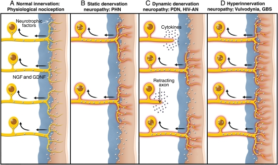
Immunohistochemical analyses have indicated that the density of ENFs in the epidermis is decreased in a wide range of neuropathic pain syndromes, including PHN,55–57 painful diabetic neuropathy (PDN),58,59 painful HIV-associated neuropathy (HIV-AN),60 complex regional pain syndrome,61 small-fibre neuropathy,62–64 metabolic syndrome,65 and Fabry disease.66 Moreover, data suggest a positive correlation between the extent of ENF loss and the severity of pain in PHN,57 PDN,58,59 and HIV-AN.60 Thus, in most pain syndromes considered to be neuropathic, sensory neurone axon density in the target tissue (commonly the skin) is decreased. In contrast, in some other neuropathic pain conditions, such as in proximal inflammatory or compressive disorders affecting the spinal nerve root or dorsal root ganglion (e.g. Sjogren's syndrome), which do not involve significant loss of cell bodies or axons distal to the dorsal root ganglion, there may be changes which are not length-dependent, with regionally preserved nociceptor innervation of target organs. Spared nerve fibres may also sprout in the skin in neuropathic conditions such as PHN, although overall density of ENFs is generally reduced. Why density changes of cutaneous nerve fibres can lead to chronic pain and particularly hypersensitivity will be discussed below (Fig. 6).
Fig 6.
Alterations in skin innervation can be used to categorize neuropathic pain syndromes of diverse aetiologies. (a) Innervation of the skin serves to protect organisms through normal nociception. Growth factors (e.g. NGF and GDNF) are constantly produced in the skin and transported retrogradely to sensory neurone cell bodies. (b) When cell bodies are lost or nerves cut and cannot regrow (‘static’ denervation neuropathies), reduced cutaneous innervation results in intact sensory fibres being exposed to abnormally high levels of neurotrophins; this hypertrophic microenvironment is known to enhance excitability and promote sprouting. (c) In ‘dynamic’ denervation neuropathies, cyclic metabolic or other types of stress render cell bodies unable to maintain their longest axons. During cycles of retraction and regrowth, pro-inflammatory cytokines or other mediators may produce axonal excitation. In addition, intact nerve terminals are subject to a hypertrophic environment. (d) In a class of neuropathies or ‘dynias’ best exemplified by vulvodynia or Gullian–Barré syndrome (GBS), immune system activation (or perhaps other factors) have caused local regions of hyperinnervation by cutaneous nociceptors and these nerve terminals display hyperexcitability.
It is now widely accepted that nociceptors may develop hyperexcitable electrophysiological properties, due to exposure to relatively abnormal concentrations of neurotrophins such as NGF or glial cell line-derived neurotrophic factor (GDNF), or pro-inflammatory cytokines, as hypothesized and discussed previously.67–69 In chronic pain syndromes associated with denervation (Fig. 6b), pain intensity may correlate with reduced nociceptor immunostaining because when there are only a small number of intact nociceptive endings, it is more likely those endings will have access to an abnormally high supply of the neurotrophins produced by the skin and ensheathing Schwann cells. Although NGF has an important role in controlling the survival and development of small-diameter neurones—both sensory and sympathetic—it has become clear that NGF also serves as an important signal for neuroimmune and inflammatory processes in mature organisms.69 In normal human skin, NGF-immunoreactivity is predominantly in basal keratinocytes.70 Production of NGF may be up-regulated by inflammation or denervation of skin; rats show a rapid and prolonged increase (5- to 10-fold) of NGF mRNA in denervated skin, distal nerves, and basal keratinocytes.71 In response to enhanced NGF supply, intact nociceptors may respond by becoming hyperactive, sprout, or both. As a direct excitatory stimulant, NGF causes immediate excitation of nociceptors,72 resulting in prolonged hyperalgesia and allodynia.73,74 In addition to this direct and rapid effect, retrograde transport of NGF to sensory neurone cell bodies may lead to the up-regulation of pro-excitatory proteins such as TRPV1 and voltage-activated sodium channels,75 and down-regulation of anti-excitatory proteins such as voltage-activated potassium channels.76 The recent clinical successes of the anti-NGF neutralizing antibody tanezumab provide direct evidence for the role of NGF in chronic pain syndromes.77 Similar success has been achieved by relocating painful injured nerves from NGF-rich (subcutaneous) to NGF-poor (muscle) regions, with appropriate changes in NGF levels.78
Although not as extensively studied as NGF, oversupply of other neurotrophins produced in the skin such as GDNF79 or artemin80 can also hypersensitize cutaneous nociceptors. We postulate that much of the hyperactivity of nociceptors in peripheral neuropathic pain syndromes of ‘static denervation’ (Fig. 6b), ‘dynamic denervation’ (Fig. 6c), and ‘hyperinnervation’ (Fig. 6d) types is due to the relatively hyperstimulating hypertrophic environment to which intact or regenerating cutaneous nociceptors are exposed.
Pro-inflammatory cytokines can also directly activate and modify gene expression in sensory neurones, and there are several sources of these molecules in close proximity to peripheral nerves. Schwann cells, which have often been thought of as having only a passive support role for peripheral nerves, are able to secrete pro-inflammatory cytokines,81 including via a purinergic P2X7-mediated mechanism.82 Wallerian degeneration is a post-traumatic process of the peripheral nervous system whereby damaged axons and their surrounding myelin sheaths are phagocytosed by infiltrating macrophages or leucocytes. During the process of infiltration of inflamed or damaged peripheral nerves, these immune system cells are known to secrete pro-inflammatory cytokines.49 Pain initiation by pro-inflammatory cytokines may be particularly important in ‘dynamic’ denervation neuropathies such as PDN or HIV-AN (Fig. 6c).
According to the concept depicted in Figure 6b, diabetic and some types of traumatic neuropathy would fall under the rubric of ‘classical denervation neuropathy’, because either there is long-term ‘dying back’ of nerve fibres such as in established PDN or severed large nerve trunks. This would also apply to severe PHN with loss of cell bodies in the DRG, or in chronic PHN when any surviving nerve fibres in skin will have sprouted to their maximum capacity. For early PHN, early PDN, HIV-AN, cancer-chemotherapy-induced neuropathy, partial nerve injury, and post-nerve repair, and others, denervation and nerve sprouting is a dynamic process which can fluctuate depending on the metabolic health of the sensory neurones and the degree to which they are exposed to toxins, etc. (Fig. 6c). Sensory neuronal cell bodies which survive, and their axons which sprout, may lead to hypersensitivity disorders. Within this group, there are chronic painful syndromes associated with overall increased nociceptor density; examples include vulvodynia,83,84 burning mouth syndrome,85 interstitial cystitis,86,87 notalgia paresthetica,88 rectal hypersensitivity,43 gastro-oesophageal reflux disease,89 inflammatory and irritable bowel diseases,90–92 post-surgical breast pain,93 and allergic rhinitis.94 These conditions do not reflect nerve injury or disease per se, but may fall under the definition of ‘neuropathic’ pain as pain arising as direct consequence of a lesion or disease affecting the somatosensory system.95 Denervation may be followed by aberrant re-innervation, proliferation with or sprouting of distal axons associated with these pain syndromes (Fig. 6d).
Vulvodynia constitutes a very interesting exemplar of chronic ‘dynias’ or idiopathic pain syndromes with regional changes in skin innervation. It is characterized by painful burning sensations, allodynia, hyperalgesia, and itching, usually localized in the region of the vulvar vestibules.96 Vulvar tissue arises from the same urogenital progenitors as bladder; hence it might not be surprising to find parallels involving hyperproliferation of nociceptors in bladder tissue.97 In vulvodynia patients, the hypersensitivity of vulvar C-fibres is well documented,98,99 and immunohistological evaluation of small-diameter nociceptive nerve fibres shows increased densities relative to normal subjects.83 Moreover, TRPV1 expression appears to be significantly increased in these proliferated nociceptors.87 Patterns of enhanced TRPV1 expression similar to vulvodynia occur in rectal hypersensitivity syndrome, which includes faecal urgency and incontinence as symptoms.43 Increases in TRPV1 expression appear to correlate with decreases in heat and distension sensory thresholds.
As previously noted, regional innervation of the skin is heterogeneous in many types of peripheral neuropathy. During sensory examination of chronic pain patients with denervation neuropathies, clinicians commonly observe areas of reduced or absent thermal or tactile sensitivity immediately adjacent to areas of hypersensitivity, allodynia, or spontaneous pain. To illustrate, in PHN, patients have been classified as displaying either ‘irritable nociceptors’ or ‘de-afferentation’,55 and later it was appreciated that both of these phenomena could appear in the same patient.100 Given the high level of skin innervation heterogeneity, including collateral or border zone sprouting in many neuropathies, diagnostic approaches based on single punch biopsies and sensory examinations of small areas could be misleading, and might inappropriately lead to patients with a diagnosis of de-afferentation to not be treated locally with potentially effective pain medicines. As previously discussed, the lower the density of cutaneous nociceptors, the more likely those nociceptors are to be hyperactive and the more readily they may respond to topical capsaicin.
Additional potential uses for topical capsaicin
Other chronic pain syndromes may also be responsive to topical capsaicin. For example, low-concentration topical capsaicin has been evaluated in four small studies as a treatment for vulvar vestibulitis (VVS), a highly localized form of vulvodynia, described above.101–104 Daily administration of low-concentration capsaicin patches placed directly on the lower back have been evaluated in two controlled clinical studies. Pain reductions were observed both back pain studies, without notable or systemic side-effects.105,106 Topical, low-concentration capsaicin has been evaluated as a treatment for osteoarthritis (OA) in multiple double-blind vehicle-controlled clinical trials. From these data, albeit limited by the potential for inadequate blinding in some studies due to the lack of vehicle pungency, it can be inferred that topical capsaicin appears modestly effective, either as a monotherapy or as an adjunctive therapy.107
If the primary cause of chronic musculoskeletal pain lies deep within joints and topical capsaicin does not provide substantial transdermal delivery, then the apparent efficacy of topical capsaicin in lower back pain and OA could be postulated to be by CNS modulatory mechanisms. Another possibility is that alterations in, or sensitization of, cutaneous nociceptors could be a contributing factor. Such alterations might be driven by unusually high concentrations of cytokines or growth factors in joints which could either diffuse some distance away from the primary site of inflammation or promote excitatory phenotypes in nearby collateral axons. To assess sensory function in the skin overlying the joints of patients with rheumatoid arthritis, capsaicin was injected intradermally over those joints.108 Capsaicin-induced axon reflex vasodilatation was significantly greater over affected joints when compared with age-matched normal controls and there was also a correlation between axon reflex vasodilatation and visual analogue pain score apparent in the rheumatoid group.
Non-pain indications for topical capsaicin for which there is some evidence include itch, psoriasis, and allergic rhinitis.109,110 Based on the premise that intranasally delivered drugs might exert direct or selective effects on the trigeminal sensory nerves thought to be hyperactive in headache,111 controlled studies of intranasal capsaicin have suggested efficacy in short-term prophylaxis in episodic cluster headache112,113 and migraine.114
Safety and adverse effects
Capsaicin has been widely consumed orally by humans throughout the world over centuries and comprehensive reviews of its safety have not identified serious toxicity,115,116 even though some have conjectured that one or more hepatic metabolites of capsaicin may be mutagenic at very high concentrations.117 Although the presumed lack of toxicity of capsaicin in food does not preclude adverse effects related to its actions on the skin, topical capsaicin is also generally regarded as safe, either for medical3 or for cosmetic116 uses. The primary adverse effects seem to be local, transient, application site reactions, mainly pain and erythema. Aside from acute, local application site reactions, published non-clinical118 and clinical7 data pertaining to the capsaicin 8% patch do not suggest any special safety concerns regarding skin exposure to capsaicin or capsaicin metabolites. Transient increases in arterial pressure associated with the pain experienced during the application procedure were observed during clinical trials.7
The theoretical concern most relevant to chronic pain management is that in peripheral neuropathies associated with cutaneous denervation, defunctionalization of cutaneous nociceptors could leave areas of skin without sufficient protective sensation to prevent or avoid injury. In addition, cutaneous C-fibres may play a role in blood flow regulation and wound repair, as the substance P and neurokinin A released by C-fibres can stimulate growth of fibroblasts and epithelial cells, while CGRP can stimulate growth of keratinocytes and epithelial cells.119 However, these theoretical effects have not been observed in clinical practice. The safety and efficacy of multi-week applications of low-concentration capsaicin creams (0.025 to 0.075% w/w) have been evaluated in six large double-blind trials with comparators and in other smaller studies with no reports relating to either loss of protective sensations or impaired wound healing.110 When the effects of topical capsaicin on sensory nerve fibre function have been examined specifically in diabetic neuropathy patients, there have also been neither detectable changes in sensory function nor adverse effects relating to cutaneous perfusion.120,121 Similarly, in an experimental model which uses a 48 h application of 0.1% w/w capsaicin cream under occlusion to induce nearly complete loss of immunostaining for ENFs and a very robust effect on dermal nociceptors, no effect on wound healing has been reported in PDN and HIV-AN patients after repeated 3 mm punch biopsies in capsaicin-treated areas on the leg.28,122
Limited safety data are currently available for capsaicin 8% patch for diabetic neuropathy, which may have led the EMA to restrict use of this treatment to non-diabetic neuropathy patients.7 Treatment with capsaicin 8% patch did not result in changes suggestive of detrimental effects on sensory function in patients with painful diabetic neuropathy123 or painful HIV-AN12 at 12 weeks after treatment. Similarly, neurosensory testing in HIV-AN and PHN patients treated with up to four treatments over a period of 48 weeks revealed no evidence of impairment.124 The reversibility of both sensory function and ENF innervation after capsaicin 8% patch treatment has been documented in healthy subjects, with small reductions in tactile and sharp pain sensations returning to normal within 12 weeks and ENF density to within a few per cent of normal by 24 weeks.9 Because of the high selectivity of capsaicin for the TRPV1 receptor and the selective expression of TRPV1 in nociceptive sensory nerves, other skin sensory nerve endings may remain intact and functional even with pronounced defunctionalisation and reduction of cutaneous nociceptors.26 Capsaicin-insensitive nerve endings include those which arise from Aβ-fibres which transduce tactile and proprioceptive stimuli, as well as a subpopulation of C-fibres and the majority of Aδ-fibres, which are primarily responsible for mediating pin-prick.125 Non-TRPV1-expressing nerve fibres are also capable of transducing thermal stimuli, as they express TRP receptors such as TRPV2 (which is activated at 52°C) and TRPV3 (which is activated at 39°C).126 It should be remembered that ENF density measurements are a diagnostic tool, one of the several used to evaluate symptomatic small-fibre neuropathies. Age- and length-related reductions of ENF density occur in healthy individuals who display no symptoms of sensory loss.127 Therefore, perhaps it should not be surprising that the complex cutaneous innervation allows for selective defunctionalization of a subpopulation of cutaneous nociceptors (i.e. those capsaicin-sensitive) without significant loss of protective sensation. A recent study using topical capsaicin in an occlusive 48 h application reported elimination of immunostaining for both sensory and autonomic nerve markers.128 However, such cutaneous nerve fibre losses have not been observed with either the high-concentration patch8,9 or low-concentration creams.129
In conclusion, directed TRPV1 agonist therapies, in which nociceptor defunctionalization is restricted to discrete target organs such as the skin, may be an attractive treatment to control localized pain or hypersensitivity. There is good evidence for hyperactivity and proliferation of cutaneous nociceptive C-fibres in numerous pain syndromes, and thus topical TRPV1 receptor agonist-mediated defunctionalisation warrants evaluation as an approach for their management. Although the ‘substance P depletion’ hypothesis was attractive in the 1980s, subsequent advances in our understanding of cutaneous innervation and the responses to topical capsaicin have rendered this hypothesis irrelevant. Reduction of substance P content in the skin is just one of many consequences of defunctionalisation, and there is no evidence that this process is related causally to pain relief.
An important advantage of the topical capsaicin approach is that this drug is poorly absorbed transdermally in humans and there appear to be few systemic adverse effects or even local effects other than transient application-site reactions such as pain and erythema. The recent approval of the capsaicin 8% patch, which is designed to be used episodically, thus provides an alternative to low-concentration capsaicin-based medicines by providing for longer term pain relief in some patients, while avoiding the requirement for repeated daily self-administration, lack of patient compliance, and possible home environmental contamination. Given the common use of topical capsaicin in a wide variety of chronic pain syndromes and other conditions, we look forward to further clinical evaluations of capsaicin 8% patch and other innovative topical capsaicin formulations.
Conflict of interest
K.B. is an employee of NeurogesX, Inc., which is the developer of Qutenza.
Funding
NeurogesX contributed to the payment of the Open Access charge.
Acknowledgement
We thank Prof. L. Plaghki, Faculty of Medicine, Université Catholique de Louvain, Brussels, Belgium, for the capsaicin treatment skin biopsies shown in Figure 5 (similar data are presented in our joint publication).30
References
- 1.Szallasi A, Blumberg PM. Vanilloid (capsaicin) receptors and mechanisms. Pharmacol Rev. 1999;51:159–212. [PubMed] [Google Scholar]
- 2.Turnbull A. Tincture of capsaicin as a remedy for chilblains and toothache. Dublin Free Press. 1850;1:95–6. [Google Scholar]
- 3.Derry S, Lloyd R, Moore RA, McQuay HJ. Topical capsaicin for chronic neuropathic pain in adults. Cochrane Database Syst Rev. 2009:CD007393. doi: 10.1002/14651858.CD007393.pub2. [DOI] [PMC free article] [PubMed] [Google Scholar]
- 4.Hempenstall K, Nurmikko TJ, Johnson RW, A'Hern RP, Rice AS. Analgesic therapy in postherpetic neuralgia: a quantitative systematic review. PLoS Med. 2005;2:e164. doi: 10.1371/journal.pmed.0020164. doi:10.1371/journal.pmed.0020164. [DOI] [PMC free article] [PubMed] [Google Scholar]
- 5.Altman R, Barkin RL. Topical therapy for osteoarthritis: clinical and pharmacologic perspectives. Postgrad Med. 2009;121:139–47. doi: 10.3810/pgm.2009.03.1986. doi:10.3810/pgm.2009.03.1986. [DOI] [PubMed] [Google Scholar]
- 6.Robbins WR, Staats PS, Levine J, et al. Treatment of intractable pain with topical large-dose capsaicin: preliminary report. Anesth Analg. 1998;86:579–83. doi: 10.1097/00000539-199803000-00027. [DOI] [PubMed] [Google Scholar]
- 7.McCormack PL. Capsaicin dermal patch: in non-diabetic peripheral neuropathic pain. Drugs. 2010;70:1831–42. doi: 10.2165/11206050-000000000-00000. doi:10.2165/11206050-000000000-00000. [DOI] [PubMed] [Google Scholar]
- 8.Malmberg AB, Mizisin AP, Calcutt NA, Von Stein T, Robbins WR, Bley KR. Reduced heat sensitivity and epidermal nerve fiber immunostaining following single applications of a high-concentration capsaicin patch. Pain. 2004;111:360–7. doi: 10.1016/j.pain.2004.07.017. doi:10.1016/j.pain.2004.07.017. [DOI] [PubMed] [Google Scholar]
- 9.Kennedy WR, Vanhove GF, Lu SP, et al. A randomized, controlled, open-label study of the long-term effects of NGX-4010, a high-concentration capsaicin patch, on epidermal nerve fiber density and sensory function in healthy volunteers. J Pain. 2010;11:579–87. doi: 10.1016/j.jpain.2009.09.019. doi:10.1016/j.jpain.2009.09.019. [DOI] [PubMed] [Google Scholar]
- 10.Backonja M, Wallace MS, Blonsky ER, et al. NGX-4010 C116 Study Group. NGX-4010, a high-concentration capsaicin patch, for the treatment of postherpetic neuralgia: a randomised, double-blind study. Lancet Neurol. 2008;7:1106–12. doi: 10.1016/S1474-4422(08)70228-X. doi:10.1016/S1474-4422(08)70228-X. [DOI] [PubMed] [Google Scholar]
- 11.Irving G, Irving GA, Backonja M, et al. The NGX-4010 C117 Study Group. A multicenter, randomized, double-blind, controlled study of NGX-4010, a high-concentration capsaicin patch, for the treatment of postherpetic neuralgia. Pain Med. 2011;12:99–109. doi: 10.1111/j.1526-4637.2010.01004.x. doi:10.1111/j.1526-4637.2010.01004.x. [DOI] [PubMed] [Google Scholar]
- 12.Simpson DM, Brown S, Tobias J NGX-4010 C107 Study Group. Controlled trial of high-concentration capsaicin patch for treatment of painful HIV neuropathy. Neurology. 2008;70:2305–13. doi: 10.1212/01.wnl.0000314647.35825.9c. doi:10.1212/01.wnl.0000314647.35825.9c. [DOI] [PubMed] [Google Scholar]
- 13.Alawi K, Keeble J. The paradoxical role of the transient receptor potential vanilloid 1 receptor in inflammation. Pharmacol Ther. 2010;125:181–95. doi: 10.1016/j.pharmthera.2009.10.005. doi:10.1016/j.pharmthera.2009.10.005. [DOI] [PubMed] [Google Scholar]
- 14.Patwardhan AM, Akopian AN, Ruparel NB, et al. Heat generates oxidized linoleic acid metabolites that activate TRPV1 and produce pain in rodents. J Clin Invest. 2010;120:1617–26. doi: 10.1172/JCI41678. doi:10.1172/JCI41678. [DOI] [PMC free article] [PubMed] [Google Scholar]
- 15.Cortright DN, Szallasi A. TRP channels and pain. Curr Pharm Des. 2009;15:1736–49. doi: 10.2174/138161209788186308. doi:10.2174/138161209788186308. [DOI] [PubMed] [Google Scholar]
- 16.Chung MK, Güler AD, Caterina MJ. TRPV1 shows dynamic ionic selectivity during agonist stimulation. Nat Neurosci. 2008;11:555–64. doi: 10.1038/nn.2102. doi:10.1038/nn.2102. [DOI] [PubMed] [Google Scholar]
- 17.Gallego-Sandín S, Rodríguez-García A, Alonso MT, García-Sancho J. The endoplasmic reticulum of dorsal root ganglion neurons contains functional TRPV1 channels. J Biol Chem. 2009;284:32591–601. doi: 10.1074/jbc.M109.019687. doi:10.1074/jbc.M109.019687. [DOI] [PMC free article] [PubMed] [Google Scholar]
- 18.Huang W, Wang H, Galligan JJ, Wang DH. Transient receptor potential vanilloid subtype 1 channel mediated neuropeptide secretion and depressor effects: role of endoplasmic reticulum associated Ca2+ release receptors in rat dorsal root ganglion neurons. J Hypertens. 2008;26:1966–75. doi: 10.1097/HJH.0b013e328309eff9. doi:10.1097/HJH.0b013e328309eff9. [DOI] [PMC free article] [PubMed] [Google Scholar]
- 19.Chard PS, Bleakman D, Savidge JR, Miller RJ. Capsaicin-induced neurotoxicity in cultured dorsal root ganglion neurons: involvement of calcium-activated proteases. Neuroscience. 1995;65:1099–108. doi: 10.1016/0306-4522(94)00548-j. doi:10.1016/0306-4522(94)00548-J. [DOI] [PubMed] [Google Scholar]
- 20.Han P, McDonald HA, Bianchi BR, et al. Capsaicin causes protein synthesis inhibition and microtubule disassembly through TRPV1 activities both on the plasma membrane and intracellular membranes. Biochem Pharmacol. 2007;73:1635–45. doi: 10.1016/j.bcp.2006.12.035. doi:10.1016/j.bcp.2006.12.035. [DOI] [PubMed] [Google Scholar]
- 21.Goswami C, Schmidt H, Hucho F. TRPV1 at nerve endings regulates growth cone morphology and movement through cytoskeleton reorganization. FEBS J. 2007;274:760–72. doi: 10.1111/j.1742-4658.2006.05621.x. doi:10.1111/j.1742-4658.2006.05621.x. [DOI] [PubMed] [Google Scholar]
- 22.Chada SR, Hollenbeck PJ. Nerve growth factor signaling regulates motility and docking of axonal mitochondria. Curr Biol. 2004;14:1272–6. doi: 10.1016/j.cub.2004.07.027. doi:10.1016/j.cub.2004.07.027. [DOI] [PubMed] [Google Scholar]
- 23.Shimomura Y, Kawada T, Suzuki M. Capsaicin and its analogs inhibit the activity of NADH-coenzyme Q oxidoreductase of the mitochondrial respiratory chain. Arch Biochem Biophys. 1989;270:573–7. doi: 10.1016/0003-9861(89)90539-0. doi:10.1016/0003-9861(89)90539-0. [DOI] [PubMed] [Google Scholar]
- 24.Athanasiou A, Smith PA, Vakilpour S, et al. Vanilloid receptor agonists and antagonists are mitochondrial inhibitors: how vanilloids cause non-vanilloid receptor mediated cell death. Biochem Biophys Res Commun. 2007;354:50–5. doi: 10.1016/j.bbrc.2006.12.179. doi:10.1016/j.bbrc.2006.12.179. [DOI] [PubMed] [Google Scholar]
- 25.Dedov VN, Mandadi S, Armati PJ, Verkhratsky A. Capsaicin-induced depolarisation of mitochondria in dorsal root ganglion neurons is enhanced by vanilloid receptors. Neuroscience. 2001;103:219–26. doi: 10.1016/s0306-4522(00)00540-6. doi:10.1016/S0306-4522(00)00540-6. [DOI] [PubMed] [Google Scholar]
- 26.Bley KR. TRPV1 agonist approaches for pain management. In: Gomtsyan A, Faltynek CR, editors. Vanilloid Receptor TRPV1 in Drug Discovery: Targeting Pain and Other Pathological Disorders. New York: Wiley; 2010. pp. 325–47. [Google Scholar]
- 27.Holzer P. The pharmacological challenge to tame the transient receptor potential vanilloid-1 (TRPV1) nocisensor. Br J Pharmacol. 2008;155:1145–62. doi: 10.1038/bjp.2008.351. doi:10.1038/bjp.2008.351. [DOI] [PMC free article] [PubMed] [Google Scholar]
- 28.Polydefkis M, Hauer P, Sheth S, Sirdofsky M, Griffin JW, McArthur JC. The time course of epidermal nerve fibre regeneration: studies in normal controls and in people with diabetes, with and without neuropathy. Brain. 2004;127:1606–15. doi: 10.1093/brain/awh175. doi:10.1093/brain/awh175. [DOI] [PubMed] [Google Scholar]
- 29.Kawakami T, Hikawa N, Kusakabe T, et al. Mechanism of inhibitory action of capsaicin on particulate axoplasmic transportin sensory neurons in culture. J Neurobiol. 1993;5:545–51. doi: 10.1002/neu.480240502. doi:10.1002/neu.480240502. [DOI] [PubMed] [Google Scholar]
- 30.Ragé M, Van Acker N, Facer P, et al. The time course of CO2 laser-evoked responses and of skin nerve fibre markers after topical capsaicin in human volunteers. Clin Neurophysiol. 2010;121:1256–66. doi: 10.1016/j.clinph.2010.02.159. doi:10.1016/j.clinph.2010.02.159. [DOI] [PubMed] [Google Scholar]
- 31.Geppetti P, Nassini R, Materazzi S, Benemei S. The concept of neurogenic inflammation. BJU Int. 2008;101(Suppl. 3)):2–6. doi: 10.1111/j.1464-410X.2008.07493.x. doi:10.1111/j.1464-410X.2008.07493.x. [DOI] [PubMed] [Google Scholar]
- 32.Jessell TM, Iversen LL, Cuello AC. Capsaicin-induced depletion of substance P from primary sensory neurones. Brain Res. 1978;152:183–8. doi: 10.1016/0006-8993(78)90146-4. doi:10.1016/0006-8993(78)90146-4. [DOI] [PubMed] [Google Scholar]
- 33.Bernstein JE, Swift RM, Soltani K, Lorincz AL. Inhibition of axon reflex vasodilatation by topically applied capsaicin. J Invest Dermatol. 1981;76:394–5. doi: 10.1111/1523-1747.ep12520912. doi:10.1111/1523-1747.ep12520912. [DOI] [PubMed] [Google Scholar]
- 34.Hill R. NK1 (substance P) receptor antagonists—why are they not analgesic in humans? Trends Pharmacol Sci. 2000;21:244–6. doi: 10.1016/s0165-6147(00)01502-9. doi:10.1016/S0165-6147(00)01502-9. [DOI] [PubMed] [Google Scholar]
- 35.Fischer MJ. Calcitonin gene-related peptide receptor antagonists for migraine. Expert Opin Investig Drugs. 2010;19:815–23. doi: 10.1517/13543784.2010.490829. doi:10.1517/13543784.2010.490829. [DOI] [PubMed] [Google Scholar]
- 36.Chaiyasit K, Khovidhunkit W, Wittayalertpanya S. Pharmacokinetics and the effect of capsaicin in Capsicum frutescens on decreasing plasma glucose level. J Med Assoc Thai. 2009;92:108–13. [PubMed] [Google Scholar]
- 37.Babbar S, Marier JF, Mouksassi MS, et al. Pharmacokinetic analysis of capsaicin after topical administration of a high-concentration capsaicin patch to patients with peripheral neuropathic pain. Ther Drug Monit. 2009;31:502–10. doi: 10.1097/FTD.0b013e3181a8b200. doi:10.1097/FTD.0b013e3181a8b200. [DOI] [PubMed] [Google Scholar]
- 38.Chanda S, Bashir M, Babbar S, Koganti A, Bley K. In vitro hepatic and skin metabolism of capsaicin. Drug Metab Dispos. 2008;36:670–5. doi: 10.1124/dmd.107.019240. doi:10.1124/dmd.107.019240. [DOI] [PubMed] [Google Scholar]
- 39.Touska F, Marsakova L, Teisinger J, Vlachova V. A ‘cute’ desensitization of TRPV1. Curr Pharm Biotechnol. 2010;12:122–9. doi: 10.2174/138920111793937826. [DOI] [PubMed] [Google Scholar]
- 40.Li WH, Lee YM, Kim JY, et al. Transient receptor potential vanilloid-1 mediates heat-shock-induced matrix metalloproteinase-1 expression in human epidermal keratinocytes. J Invest Dermatol. 2007;127:2328–35. doi: 10.1038/sj.jid.5700880. doi:10.1038/sj.jid.5700880. [DOI] [PubMed] [Google Scholar]
- 41.Pecze L, Szabó K, Széll M, et al. Human keratinocytes are vanilloid resistant. PLoS One. 2008;3:e3419. doi: 10.1371/journal.pone.0003419. doi:10.1371/journal.pone.0003419. [DOI] [PMC free article] [PubMed] [Google Scholar]
- 42.Smith GD, Gunthorpe MJ, Kelsell RE, et al. TRPV3 is a temperature-sensitive vanilloid receptor-like protein. Nature. 2002;418:186–90. doi: 10.1038/nature00894. doi:10.1038/nature00894. [DOI] [PubMed] [Google Scholar]
- 43.Chan CL, Facer P, Davis JB, et al. Sensory fibres expressing capsaicin receptor TRPV1 in patients with rectal hypersensitivity and faecal urgency. Lancet. 2003;361:385–91. doi: 10.1016/s0140-6736(03)12392-6. doi:10.1016/S0140-6736(03)12392-6. [DOI] [PubMed] [Google Scholar]
- 44.Matsumoto K, Kurosawa E, Terui H, et al. Localization of TRPV1 and contractile effect of capsaicin in mouse large intestine: high abundance and sensitivity in rectum and distal colon. Am J Physiol Gastrointest Liver Physiol. 2009;297:G348–60. doi: 10.1152/ajpgi.90578.2008. doi:10.1152/ajpgi.90578.2008. [DOI] [PubMed] [Google Scholar]
- 45.Schmelz M, Schmidt R. Microneurographic single-unit recordings to assess receptive properties of afferent human C-fibers. Neurosci Lett. 2010;470:158–61. doi: 10.1016/j.neulet.2009.05.064. doi:10.1016/j.neulet.2009.05.064. [DOI] [PubMed] [Google Scholar]
- 46.Ochoa JL, Campero M, Serra J, Bostock H. Hyperexcitable polymodal and insensitive nociceptors in painful human neuropathy. Muscle Nerve. 2005;32:459–72. doi: 10.1002/mus.20367. doi:10.1002/mus.20367. [DOI] [PubMed] [Google Scholar]
- 47.Orstavik K, Jørum E. Microneurographic findings of relevance to pain in patients with erythromelalgia and patients with diabetic neuropathy. Neurosci Lett. 2010;470:180–4. doi: 10.1016/j.neulet.2009.05.061. doi:10.1016/j.neulet.2009.05.061. [DOI] [PubMed] [Google Scholar]
- 48.Devor M. Response of nerves to injury in relation to neuropathic pain. In: McMahon SB, Koltzenburg M, editors. Wall and Melzack's Textbook of Pain. London: Elsevier; 2006. pp. 905–27. [Google Scholar]
- 49.Campbell JN, Meyer RA. Mechanisms of neuropathic pain. Neuron. 2006;52:77–92. doi: 10.1016/j.neuron.2006.09.021. doi:10.1016/j.neuron.2006.09.021. [DOI] [PMC free article] [PubMed] [Google Scholar]
- 50.Ali Z, Ringkamp M, Hartke TV, et al. Uninjured C-fiber nociceptors develop spontaneous activity and alpha-adrenergic sensitivity following L6 spinal nerve ligation in monkey. J Neurophysiol. 1999;81:455–66. doi: 10.1152/jn.1999.81.2.455. [DOI] [PubMed] [Google Scholar]
- 51.Wu G, Ringkamp M, Hartke TV, et al. Early onset of spontaneous activity in uninjured C-fiber nociceptors after injury to neighboring nerve fibers. J Neurosci. 2001;21:RC140. doi: 10.1523/JNEUROSCI.21-08-j0002.2001. [DOI] [PMC free article] [PubMed] [Google Scholar]
- 52.Djouhri L, Koutsikou S, Fang X, McMullan S, Lawson SN. Spontaneous pain, both neuropathic and inflammatory, is related to frequency of spontaneous firing in intact C-fiber nociceptors. J Neurosci. 2006;26:1281–92. doi: 10.1523/JNEUROSCI.3388-05.2006. doi:10.1523/JNEUROSCI.3388-05.2006. [DOI] [PMC free article] [PubMed] [Google Scholar]
- 53.Kennedy WR. Opportunities afforded by the study of unmyelinated nerves in skin and other organs. Muscle Nerve. 2004;29:756–67. doi: 10.1002/mus.20062. doi:10.1002/mus.20062. [DOI] [PubMed] [Google Scholar]
- 54.Atherton DD, Facer P, Roberts KM, et al. Use of the novel Contact Heat Evoked Potential Stimulator (CHEPS) for the assessment of small fibre neuropathy: correlations with skin flare responses and intra-epidermal nerve fibre counts. BMC Neurol. 2007;7:21. doi: 10.1186/1471-2377-7-21. doi:10.1186/1471-2377-7-21. [DOI] [PMC free article] [PubMed] [Google Scholar]
- 55.Rowbotham MC, Yosipovitch G, Connolly MK, Finlay D, Forde G, Fields HL. Cutaneous innervation density in the allodynic form of postherpetic neuralgia. Neurobiol Dis. 1996;3:205–14. doi: 10.1006/nbdi.1996.0021. doi:10.1006/nbdi.1996.0021. [DOI] [PubMed] [Google Scholar]
- 56.Oaklander AL. The density of remaining nerve endings in human skin with and without postherpetic neuralgia after shingles. Pain. 2001;92:139–45. doi: 10.1016/s0304-3959(00)00481-4. doi:10.1016/S0304-3959(00)00481-4. [DOI] [PubMed] [Google Scholar]
- 57.Petersen KL, Rice FL, Farhadi M, Reda H, Rowbotham MC. Natural history of cutaneous innervation following herpes zoster. Pain. 2010;150:75–82. doi: 10.1016/j.pain.2010.04.002. doi:10.1016/j.pain.2010.04.002. [DOI] [PubMed] [Google Scholar]
- 58.Kennedy WR, Wendelschafer-Crabb G, Johnson T. Quantitation of epidermal nerves in diabetic neuropathy. Neurology. 1996;47:1042–8. doi: 10.1212/wnl.47.4.1042. [DOI] [PubMed] [Google Scholar]
- 59.Lauria G, McArthur JC, Hauer PE, Griffin JW, Cornblath DR. Neuropathological alterations in diabetic truncal neuropathy: evaluation by skin biopsy. J Neurol Neurosurg Psychiatry. 1998;65:762–6. doi: 10.1136/jnnp.65.5.762. doi:10.1136/jnnp.65.5.762. [DOI] [PMC free article] [PubMed] [Google Scholar]
- 60.Polydefkis M, Yiannoutsos CT, Cohen BA, et al. Reduced intraepidermal nerve fiber density in HIV-associated sensory neuropathy. Neurology. 2002;58:115–9. doi: 10.1212/wnl.58.1.115. [DOI] [PubMed] [Google Scholar]
- 61.Oaklander AL, Rissmiller JG, Gelman LB, Zheng L, Chang Y, Gott R. Evidence of focal small-fiber axonal degeneration in complex regional pain syndrome-I (reflex sympathetic dystrophy) Pain. 2006;120:235–43. doi: 10.1016/j.pain.2005.09.036. doi:10.1016/j.pain.2005.09.036. [DOI] [PubMed] [Google Scholar]
- 62.Pittenger GL, Ray M, Burcus NI, McNulty P, Basta B, Vinik AI. Intraepidermal nerve fibers are indicators of small-fiber neuropathy in both diabetic and nondiabetic patients. Diabetes Care. 2004;27:1974–9. doi: 10.2337/diacare.27.8.1974. doi:10.2337/diacare.27.8.1974. [DOI] [PubMed] [Google Scholar]
- 63.Lauria G, Morbin M, Lombardi R, et al. Expression of capsaicin receptor immunoreactivity in human peripheral nervous system and in painful neuropathies. J Peripher Nerv Syst. 2006;11:262–7. doi: 10.1111/j.1529-8027.2006.0097.x. doi:10.1111/j.1529-8027.2006.0097.x. [DOI] [PubMed] [Google Scholar]
- 64.Wilder-Smith EP, Ong WY, Guo Y, Chow AW. Epidermal transient receptor potential vanilloid 1 in idiopathic small nerve fibre disease, diabetic neuropathy and healthy human subjects. Histopathology. 2007;51:674–80. doi: 10.1111/j.1365-2559.2007.02851.x. doi:10.1111/j.1365-2559.2007.02851.x. [DOI] [PMC free article] [PubMed] [Google Scholar]
- 65.Pittenger GL, Mehrabyan A, Simmons K, et al. Small fiber neuropathy is associated with the metabolic syndrome. Metab Syndr Relat Disord. 2005;3:1113–21. doi: 10.1089/met.2005.3.113. [DOI] [PubMed] [Google Scholar]
- 66.Scott LJ, Griffin JW, Luciano C, et al. Quantitative analysis of epidermal innervation in Fabry disease. Neurology. 1999;52:1249–54. doi: 10.1212/wnl.52.6.1249. [DOI] [PubMed] [Google Scholar]
- 67.Anand P, Terenghi G, Warner G, Kopelman P, Williams-Chestnut RE, Sinicropi DV. The role of endogenous nerve growth factor in human diabetic neuropathy. Nat Med. 1996;2:703–7. doi: 10.1038/nm0696-703. doi:10.1038/nm0696-703. [DOI] [PubMed] [Google Scholar]
- 68.Anand P. Neurotrophic factors and their receptors in human sensory neuropathies. Prog Brain Res. 2004;146:477–92. doi: 10.1016/S0079-6123(03)46030-5. doi:10.1016/S0079-6123(03)46030-5. [DOI] [PubMed] [Google Scholar]
- 69.Griffin JW. The roles of growth factors in painful length-dependent axonal neuropathies. In: Campbell JN, Basbaum AI, Dray A, Dubner R, Dworkin RH, Sang CN, editors. Emerging Strategies for the Treatment of Neuropathic Pain. Seattle, WA: IASP Press; 2006. pp. 271–90. [Google Scholar]
- 70.Pincelli C. Nerve growth factor and keratinocytes: a role in psoriasis. Eur J Dermatol. 2000;10:85–90. [PubMed] [Google Scholar]
- 71.Mearow KM, Kril Y, Diamond J. Increased NGF mRNA expression in denervated rat skin. Neuroreport. 1993;4:351–4. doi: 10.1097/00001756-199304000-00002. doi:10.1097/00001756-199304000-00002. [DOI] [PubMed] [Google Scholar]
- 72.Bonnington JK, McNaughton PA. Signalling pathways involved in the sensitisation of mouse nociceptive neurones by nerve growth factor. J Physiol. 2003;551:433–46. doi: 10.1113/jphysiol.2003.039990. doi:10.1113/jphysiol.2003.039990. [DOI] [PMC free article] [PubMed] [Google Scholar]
- 73.Dmitrieva N, McMahon SB. Sensitisation of visceral afferents by nerve growth factor in the adult rat. Pain. 1996;66:87–97. doi: 10.1016/0304-3959(96)02993-4. doi:10.1016/0304-3959(96)02993-4. [DOI] [PubMed] [Google Scholar]
- 74.Bowles WR, Sabino M, Harding-Rose C, Hargreaves KM. Chronic nerve growth factor administration increases the peripheral exocytotic activity of capsaicin-sensitive cutaneous neurons. Neurosci Lett. 2006;403:305–8. doi: 10.1016/j.neulet.2006.05.020. doi:10.1016/j.neulet.2006.05.020. [DOI] [PubMed] [Google Scholar]
- 75.Ji RR, Samad TA, Jin SX, Schmoll R, Woolf CJ. p38 MAPK activation by NGF in primary sensory neurons after inflammation increases TRPV1 levels and maintains heat hyperalgesia. Neuron. 2002;36:57–68. doi: 10.1016/s0896-6273(02)00908-x. doi:10.1016/S0896-6273(02)00908-X. [DOI] [PubMed] [Google Scholar]
- 76.Zhang YH, Nicol GD. NGF-mediated sensitization of the excitability of rat sensory neurons is prevented by a blocking antibody to the p75 neurotrophin receptor. Neurosci Lett. 2004;366:187–92. doi: 10.1016/j.neulet.2004.05.042. doi:10.1016/j.neulet.2004.05.042. [DOI] [PubMed] [Google Scholar]
- 77.Cattaneo A. Tanezumab, a recombinant humanized mAb against nerve growth factor for the treatment of acute and chronic pain. Curr Opin Mol Ther. 2010;12:94–106. [PubMed] [Google Scholar]
- 78.Atherton DD, Taherzadeh O, Facer P, Elliot D, Anand P. The potential role of nerve growth factor (NGF) in painful neuromas and the mechanism of pain relief by their relocation to muscle. J Hand Surg Br. 2006;31:652–6. doi: 10.1016/j.jhsb.2006.06.009. doi:10.1016/j.jhsb.2006.06.009. [DOI] [PubMed] [Google Scholar]
- 79.Albers KM, Woodbury CJ, Ritter AM, Davis BM, Koerber HR. Glial cell-line-derived neurotrophic factor expression in skin alters the mechanical sensitivity of cutaneous nociceptors. J Neurosci. 2006;26:2981–90. doi: 10.1523/JNEUROSCI.4863-05.2006. doi:10.1523/JNEUROSCI.4863-05.2006. [DOI] [PMC free article] [PubMed] [Google Scholar]
- 80.Wang S, Elitt CM, Malin SA, Albers KM. Effects of the neurotrophic factor artemin on sensory afferent development and sensitivity. Sheng Li Xue Bao. 2008;60:565–70. [PMC free article] [PubMed] [Google Scholar]
- 81.Watkins LR, Maier SF. Beyond neurons: evidence that immune and glial cells contribute to pathological pain states. Physiol Rev. 2002;82:981–1011. doi: 10.1152/physrev.00011.2002. [DOI] [PubMed] [Google Scholar]
- 82.Hughes JP, Hatcher JP, Chessell IP. The role of P2X(7) in pain and inflammation. Purinergic Signal. 2007;3:163–9. doi: 10.1007/s11302-006-9031-1. doi:10.1007/s11302-006-9031-1. [DOI] [PMC free article] [PubMed] [Google Scholar]
- 83.Bohm-Starke N, Hilliges M, Falconer C, Rylander E. Increased intraepithelial innervation in women with vulvar vestibulitis syndrome. Gynecol Obstet Invest. 1998;46:256–60. doi: 10.1159/000010045. doi:10.1159/000010045. [DOI] [PubMed] [Google Scholar]
- 84.Tympanidis P, Casula MA, Yiangou Y, Terenghi G, Dowd P, Anand P. Increased vanilloid receptor VR1 innervation in vulvodynia. Eur J Pain. 2004;8:129–33. doi: 10.1016/S1090-3801(03)00085-5. doi:10.1016/S1090-3801(03)00085-5. [DOI] [PubMed] [Google Scholar]
- 85.Yilmaz Z, Renton T, Yiangou Y, et al. Burning mouth syndrome as a trigeminal small fibre neuropathy: increased heat and capsaicin receptor TRPV1 in nerve fibres correlates with pain score. J Clin Neurosci. 2007;14:864–71. doi: 10.1016/j.jocn.2006.09.002. doi:10.1016/j.jocn.2006.09.002. [DOI] [PubMed] [Google Scholar]
- 86.Christmas TJ, Rode J, Chapple CR, Milroy EJ, Turner-Warwick RT. Nerve fibre proliferation in interstitial cystitis. Virchows Arch A Pathol Anat Histopathol. 1990;416:447–51. doi: 10.1007/BF01605152. doi:10.1007/BF01605152. [DOI] [PubMed] [Google Scholar]
- 87.Mukerji G, Yiangou Y, Agarwal SK, Anand P. Transient receptor potential vanilloid receptor subtype 1 in painful bladder syndrome and its correlation with pain. J Urol. 2006;176:797–801. doi: 10.1016/j.juro.2006.03.074. doi:10.1016/j.juro.2006.03.074. [DOI] [PubMed] [Google Scholar]
- 88.Springall DR, Karanth SS, Kirkham N, Darley CR, Polak JM. Symptoms of notalgia paresthetica may be explained by increased dermal innervation. J Invest Dermatol. 1991;97:555–61. doi: 10.1111/1523-1747.ep12481889. doi:10.1111/1523-1747.ep12481889. [DOI] [PubMed] [Google Scholar]
- 89.Matthews PJ, Aziz Q, Facer P, Davis JB, Thompson DG, Anand P. Increased capsaicin receptor TRPV1 nerve fibres in the inflamed human oesophagus. Eur J Gastroenterol Hepatol. 2004;16:897–902. doi: 10.1097/00042737-200409000-00014. doi:10.1097/00042737-200409000-00014. [DOI] [PubMed] [Google Scholar]
- 90.Yiangou Y, Facer P, Dyer NH, et al. Vanilloid receptor 1 immunoreactivity in inflamed human bowel. Lancet. 2001;357:1338–9. doi: 10.1016/s0140-6736(00)04503-7. doi:10.1016/S0140-6736(00)04503-7. [DOI] [PubMed] [Google Scholar]
- 91.Akbar A, Yiangou Y, Facer P, Walters JR, Anand P, Ghosh S. Increased capsaicin receptor TRPV1-expressing sensory fibres in irritable bowel syndrome and their correlation with abdominal pain. Gut. 2008;57:923–9. doi: 10.1136/gut.2007.138982. doi:10.1136/gut.2007.138982. [DOI] [PMC free article] [PubMed] [Google Scholar]
- 92.Akbar A, Yiangou Y, Facer P, et al. Expression of the TRPV1 receptor differs in quiescent inflammatory bowel disease with or without abdominal pain. Gut. 2010;59:767–74. doi: 10.1136/gut.2009.194449. doi:10.1136/gut.2009.194449. [DOI] [PubMed] [Google Scholar]
- 93.Gopinath P, Wan E, Holdcroft A, et al. Increased capsaicin receptor TRPV1 in skin nerve fibres and related vanilloid receptors TRPV3 and TRPV4 in keratinocytes in human breast pain. BMC Womens Health. 2005;5:2. doi: 10.1186/1472-6874-5-2. doi:10.1186/1472-6874-5-2. [DOI] [PMC free article] [PubMed] [Google Scholar]
- 94.O'Hanlon S, Facer P, Simpson KD, Sandhu G, Saleh HA, Anand P. Neuronal markers in allergic rhinitis: expression and correlation with sensory testing. Laryngoscope. 2007;117:1519–27. doi: 10.1097/MLG.0b013e3180ca7846. doi:10.1097/MLG.0b013e3180ca7846. [DOI] [PubMed] [Google Scholar]
- 95.Treede RD, Jensen TS, Campbell JN, et al. Neuropathic pain: redefinition and a grading system for clinical and research purposes. Neurology. 2008;70:1630–5. doi: 10.1212/01.wnl.0000282763.29778.59. doi:10.1212/01.wnl.0000282763.29778.59. [DOI] [PubMed] [Google Scholar]
- 96.Damsted-Petersen C, Boyer SC, Pukall CF. Current perspectives in vulvodynia. Womens Health (Lond Engl) 2009;5:423–36. doi: 10.2217/whe.09.30. [DOI] [PubMed] [Google Scholar]
- 97.Siegel JF, Sand PK, Sasso K. Vulvodynia & pelvic pain? Think interstitial cystitis. Nurse Pract. 2008;33:40–5. doi: 10.1097/01.NPR.0000337200.92563.b5. doi:10.1097/01.NPR.0000337200.92563.b5. [DOI] [PubMed] [Google Scholar]
- 98.Sonni L, Cattaneo A, De Marco A, De Magnis A, Carli P, Marabini S. Idiopathic vulvodynia. clinical evaluation of the pain threshold with acetic acid solutions. J Reprod Med. 1995;40:337–41. [PubMed] [Google Scholar]
- 99.Bohm-Starke N, Hilliges M, Brodda-Jansen G, Rylander E, Torebjork E. Psychophysical evidence of nociceptor sensitization in vulvar vestibulitis syndrome. Pain. 2001;94:177–83. doi: 10.1016/S0304-3959(01)00352-9. doi:10.1016/S0304-3959(01)00352-9. [DOI] [PubMed] [Google Scholar]
- 100.Fields HL, Rowbotham M, Baron R. Postherpetic neuralgia: irritable nociceptors and deafferentation. Neurobiol Dis. 1998;5:209–27. doi: 10.1006/nbdi.1998.0204. doi:10.1006/nbdi.1998.0204. [DOI] [PubMed] [Google Scholar]
- 101.Friedrich EG., Jr Therapeutic studies on vulvar vestibulitis. J Reprod Med. 1988;33:514–8. [PubMed] [Google Scholar]
- 102.Zycyznski HM, Culbertson S, Gruss J, DeGroat WC. Substance P and pathophysiology of vulvar vestibulitis. J Soc Gynecol Invest. 1997;4(Suppl.):107A. [Google Scholar]
- 103.Murina F, Radici G, Bianco V. Capsaicin and the treatment of vulvar vestibulitis syndrome: a valuable alternative? MedGenMed. 2004;6:48. [PMC free article] [PubMed] [Google Scholar]
- 104.Steinberg AC, Oyama IA, Rejba A, Kellog-Spadt S, Whitmore KE. Capsaicin for the treatment of vulvar vestibulitis. Am J Obstet Gynecol. 2005;192:1549–53. doi: 10.1016/j.ajog.2004.10.626. doi:10.1016/j.ajog.2004.10.626. [DOI] [PubMed] [Google Scholar]
- 105.Keitel W, Frerick H, Kuhn U, Schmidt U, Kuhlmann M, Bredehorst A. Capsicum pain plaster in chronic non-specific low back pain. Arzneimittelforschung. 2001;51:896–903. doi: 10.1055/s-0031-1300134. [DOI] [PubMed] [Google Scholar]
- 106.Frerick H, Keitel W, Kuhn U, Schmidt S, Bredehorst A, Kuhlmann M. Topical treatment of chronic low back pain with a capsicum plaster. Pain. 2003;106:59–64. doi: 10.1016/s0304-3959(03)00278-1. doi:10.1016/S0304-3959(03)00278-1. [DOI] [PubMed] [Google Scholar]
- 107.De Silva V, El-Metwally A, Ernst E, Lewith G, Macfarlane GJ on behalf of the Arthritis Research UK Working Group on Complementary and Alternative Medicines. Evidence for the efficacy of complementary and alternative medicines in the management of osteoarthritis: a systematic review. Rheumatology (Oxford) 2010;5:10–5. doi: 10.1093/rheumatology/keq379. [DOI] [PubMed] [Google Scholar]
- 108.Jolliffe VA, Anand P, Kidd BL. Assessment of cutaneous sensory and autonomic axon reflexes in rheumatoid arthritis. Ann Rheum Dis. 1995;54:251–5. doi: 10.1136/ard.54.4.251. doi:10.1136/ard.54.4.251. [DOI] [PMC free article] [PubMed] [Google Scholar]
- 109.Hautkappe M, Roizen MF, Toledano A, Roth S, Jeffries JA, Ostermeier AM. Review of the effectiveness of capsaicin for painful cutaneous disorders and neural dysfunction. Clin J Pain. 1998;14:97–106. doi: 10.1097/00002508-199806000-00003. doi:10.1097/00002508-199806000-00003. [DOI] [PubMed] [Google Scholar]
- 110.Papoiu AD, Yosipovitch G. Topical capsaicin. The fire of a ‘hot’ medicine is reignited. Expert Opin Pharmacother. 2010;11:1359–71. doi: 10.1517/14656566.2010.481670. doi:10.1517/14656566.2010.481670. [DOI] [PubMed] [Google Scholar]
- 111.Rapoport AM, Bigal ME, Tepper SJ, Sheftell FD. Intranasal medications for the treatment of migraine and cluster headache. CNS Drugs. 2004;18:671–85. doi: 10.2165/00023210-200418100-00004. doi:10.2165/00023210-200418100-00004. [DOI] [PubMed] [Google Scholar]
- 112.Marks DR, Rapoport A, Padla D, et al. A double-blind placebo-controlled trial of intranasal capsaicin for cluster headache. Cephalalgia. 1993;13:114–6. doi: 10.1046/j.1468-2982.1993.1302114.x. doi:10.1046/j.1468-2982.1993.1302114.x. [DOI] [PubMed] [Google Scholar]
- 113.Fusco BM, Marabini S, Maggi CA, Fiore G, Geppetti P. Preventative effect of repeated nasal applications of capsaicin in cluster headache. Pain. 1994;59:321–5. doi: 10.1016/0304-3959(94)90017-5. doi:10.1016/0304-3959(94)90017-5. [DOI] [PubMed] [Google Scholar]
- 114.Fusco BM, Barzoi G, Agrò F. Repeated intranasal capsaicin applications to treat chronic migraine. Br J Anaesth. 2003;90:812. doi: 10.1093/bja/aeg572. doi:10.1093/bja/aeg572. [DOI] [PubMed] [Google Scholar]
- 115.European Commission Scientific Committee on Food. Opinion of the Scientific Committee on Food on Capsaicin. Available from http://ec.europa.eu/food/fs/sc/scf/out120_en.pdf. (February 28, 2002)
- 116.Johnson W. Final report on the safety assessment of capsicum annuum extract, capsicum annuum fruit extract, capsicum annuum resin, capsicum annuum fruit powder, capsicum frutescens fruit, capsicum frutescens fruit extract, capsicum frutescens resin, and capsaicin. Int J Toxicol. 2007;26((Suppl. 1)):3–106. doi: 10.1080/10915810601163939. doi:10.1080/10915810601163939. [DOI] [PubMed] [Google Scholar]
- 117.Surh YJ, Lee SS. Capsaicin in hot chili pepper: carcinogen, co-carcinogen or anticarcinogen? Food Chem Toxicol. 1996;34:313–6. doi: 10.1016/0278-6915(95)00108-5. doi:10.1016/0278-6915(95)00108-5. [DOI] [PubMed] [Google Scholar]
- 118.Chanda S, Erexson G, Riach C, et al. Genotoxicity studies with pure trans-capsaicin. Mutat Res. 2004;557:85–97. doi: 10.1016/j.mrgentox.2003.10.001. [DOI] [PubMed] [Google Scholar]
- 119.Scholzen T, Armstrong CA, Bunnett NW, Luger TA, Olerud JE, Ansel JC. Neuropeptides in the skin: interactions between the neuroendocrine and the skin immune systems. Exp Dermatol. 1998;7:81–96. doi: 10.1111/j.1600-0625.1998.tb00307.x. doi:10.1111/j.1600-0625.1998.tb00307.x. [DOI] [PubMed] [Google Scholar]
- 120.Tandan R, Lewis GA, Krusinski PB, Badger GB, Fries TJ. Topical capsaicin in painful diabetic neuropathy. Controlled study with long-term follow-up. Diabetes Care. 1992;15:8–14. doi: 10.2337/diacare.15.1.8. doi:10.2337/diacare.15.1.8. [DOI] [PubMed] [Google Scholar]
- 121.Forst T, Pohlmann T, Kunt T, et al. The influence of local capsaicin treatment on small nerve fibre function and neurovascular control in symptomatic diabetic neuropathy. Acta Diabetol. 2002;39:1–6. doi: 10.1007/s005920200005. doi:10.1007/s005920200005. [DOI] [PubMed] [Google Scholar]
- 122.Hahn K, Triolo A, Hauer P, McArthur JC, Polydefkis M. Impaired reinnervation in HIV infection following experimental denervation. Neurology. 2007;68:1251–6. doi: 10.1212/01.wnl.0000261245.36884.7c. doi:10.1212/01.wnl.0000261245.36884.7c. [DOI] [PubMed] [Google Scholar]
- 123.Webster LR, Peppin JF, Murphy FT, Lu B, Tobias JK, Vanhove GF. Preliminary efficacy, safety and tolerability of NGX-4010, capsaicin 8% patch, in patients with peripheral neuropathic pain. J Pain Res, Submitted for publication. [Google Scholar]
- 124.Simpson DM, Gazda S, Brown S, et al. NGX-4010 C118 Study Group. Long-term safety of NGX-4010, a high-concentration capsaicin patch, in patients with peripheral neuropathic pain. J Pain Symptom Manage. 2010;39:1053–64. doi: 10.1016/j.jpainsymman.2009.11.316. doi:10.1016/j.jpainsymman.2009.11.316. [DOI] [PubMed] [Google Scholar]
- 125.Magerl W, Fuchs PN, Meyer RA, Treede RD. Roles of capsaicin-insensitive nociceptors in cutaneous pain and secondary hyperalgesia. Brain. 2001;124:1754–64. doi: 10.1093/brain/124.9.1754. doi:10.1093/brain/124.9.1754. [DOI] [PubMed] [Google Scholar]
- 126.Tominaga M. Nociception and TRP channels. Handb Exp Pharmacol. 2007;179:489–505. doi: 10.1007/978-3-540-34891-7_29. doi:10.1007/978-3-540-34891-7_29. [DOI] [PubMed] [Google Scholar]
- 127.Umapathi T, Tan WL, Tan NC, Chan YH. Determinants of epidermal nerve fiber density in normal individuals. Muscle Nerve. 2006;33:742–6. doi: 10.1002/mus.20528. doi:10.1002/mus.20528. [DOI] [PubMed] [Google Scholar]
- 128.Gibbons CH, Wang N, Freeman R. Capsaicin induces degeneration of cutaneous autonomic nerve fibers. Ann Neurol. 2010;68:888–98. doi: 10.1002/ana.22126. doi:10.1002/ana.22126. [DOI] [PMC free article] [PubMed] [Google Scholar]
- 129.Simone DA, Nolano M, Johnson T, Wendelschafer-Crabb G, Kennedy WR. Intradermal injection of capsaicin in humans produces degeneration and subsequent reinnervation of epidermal nerve fibers: correlation with sensory function. J Neurosci. 1998;18:8947–59. doi: 10.1523/JNEUROSCI.18-21-08947.1998. [DOI] [PMC free article] [PubMed] [Google Scholar]


