Abstract
The oncolytic adenovirus (Ad) is currently being advanced as a promising antitumor remedy as it selectively replicates in tumor cells and can transfer and amplify therapeutic genes. Interleukin (IL)-12 induces a potent antitumor effect by promoting natural killer (NK) cell and cytotoxic T cell activities. IL-18 also augments cytotoxicity of NK cells and proliferation of T cells. This effect further enhances the function of IL-12 in a synergistic manner. Therefore, we investigated for the first time an effective cancer immunogene therapy of syngeneic tumors via intratumoral administration of oncolytic Ad co-expressing IL-12 and IL-18, RdB/IL-12/IL-18. Intratumoral administration of RdB/IL-12/IL-18 improved antitumor effects, as well as increased survival, in B16-F10 murine melanoma model. The ratio of T-helper type 1/2 cytokine as well as the levels of IL-12, IL-18, interferon-γ and granulocyte–macrophage colony-stimulating factor was markedly elevated in RdB/IL-12/IL-18-treated tumors. Mice injected with RdB/IL-12/IL-18 also showed enhanced cytotoxicity of tumor-specific immune cells. Consistent with these results, immense necrosis and infiltration of NK cells, as well as CD4+ and CD8+ T cells, were observed in RdB/IL-12/IL-18-treated tumor tissues. Importantly, tumors treated with RdB/IL-12/IL-18 showed an elevated number of T cells expressing IL-12Rβ2 or IL-18Rα. These results provide a new insight into therapeutic mechanisms of IL-12 plus IL-18 and provide a potential clinical cancer immunotherapeutic agent for improved antitumor immunity.
Keywords: cancer immunogene therapy, oncolytic adenovirus, IL-12, IL-18, T cells expressing IL-12Rβ2 or IL-18Rα
Introduction
Over the past decade, tumor immunology has advanced in uncovering the molecular and cellular mechanisms responsible for cancer pathophysiology and for enhancing the immune response against tumors, yet there are still many unresolved problems concerning which tumors can escape, or suppress, the immune response. Therefore, immunotherapy for malignant diseases needs novel, improved clinical protocols that minimize tumor-induced immunological tolerance while simultaneously inducing tumor eradication. One potential strategy is a cytokine immunogene therapy that has a great potential for rejecting cancer cells by activating tumor-specific immune responses in cancer-bearing hosts.
Interleukin (IL)-12 is one of the most effective and promising antitumor cytokines. It is a heterodimeric cytokine composed of two different disulfide-linked subunits, designated p35 and p40, and is mainly produced by activated macrophages and dendritic cells (DCs). IL-12 stimulates interferon-γ (IFN-γ) production by natural killer (NK) cells, T cells, DCs and macrophages. Similarly, it facilitates T helper type 1 (Th1) differentiation and augments the cytolytic effect of NK cells and cytotoxic T lymphocytes (CTLs). Previous preclinical studies of IL-12 reported enhanced antitumor immunity.1, 2 More recently, we showed an enhanced antitumor effect of an oncolytic adenovirus (Ad) that expresses both IL-12 and B7-1 in a murine melanoma B16-F10 tumor model.3
IL-18, IFN-γ-inducing factor, is secreted mainly by activated macrophages and DCs.4 IL-18 plays a significant role in cell-mediated immune responses and is capable of generating an antitumor immune response via induction of IFN-γ production from,5, 6 enhancing the cytotoxicity of,4, 7 and stimulating the proliferation of T and NK cells.6, 8 Several studies have shown the antitumor efficacy of IL-18 in murine tumor models. For example, IL-18 exerted antitumor immunity by activating NK cells and establishing cytotoxic CD4+ T cells in Meth A sarcoma-bearing mice.9 In addition, IL-18 was also shown to be a potent adjuvant, enhancing systemic antitumor immune responses and vaccine efficacy.10 Therefore, IL-18 represents a promising therapeutic candidate for cancer immunogene therapy.
Previous studies reported that IL-18 is able to drive the Th2 immune response in an IL-4-dependent manner, but induces a Th1 immune response in the presence IL-12.11, 12 These results suggest that IL-18 alone could suppress the Th1 immune response via activation of Th2 immune response. Therefore IL-18, acting in concert with IL-12, may be important in effective immunotherapy. Consistent with this suggestion, an earlier study reported that IL-12 plus IL-18 polarized Th1 immunity, resulting in the generation of potent tumor-reactive T cells.13 This combination also enhanced the cytolytic effect as well as IFN-γ production from T and NK cells, as well as the proliferation in T cells.11, 14 Furthermore, previous studies reported that IL-12 in combination with IL-18 inhibited tumor angiogenesis, leading to tumor regression.15
In this study, we show for the first time an effective cancer immunogene therapy of syngeneic tumors via intratumoral administration of oncolytic Ad co-expressing IL-12 and IL-18. We also show the underlying mechanism of oncolytic Ad co-expressing IL-12 and IL-18, showing that antitumor immunity is associated with elevation of Th1/Th2 cytokine ratio and upregulation of IL-12, IL-18, IFN-γ and granulocyte–macrophage colony-stimulating factor (GM-CSF) within the tumor tissues. Moreover, we show that the induction of an optimal tumor milieu, generating effective tumor-specific immunity through the upregulation of Th1 cytokines, enhances the generation of fully differentiated T cells expressing IL-12Rβ2 or IL-18Rα.
Results
Generation of oncolytic Ad-mediated IL-12 and IL-18
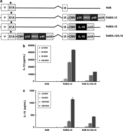
Three oncolytic Ads expressing IL-12 alone, IL-18 alone, or IL-12 and IL-18 were generated in the E1B-deleted and E1A-mutated oncolytic Ad, oncolytic Ad vector, Ad-ΔE1Bmt7 (RdB) (Figure 1a). To confirm IL-12 and IL-18 secretion from these Ads, B16-F10 melanoma cells were infected with RdB, RdB/IL-12, RdB/IL-18, or RdB/IL-12/IL-18 at various multiplicity of infections (MOIs), and culture supernatants were obtained 96 h after infection to examine IL-12 and IL-18 production. A dose (MOI)-dependent increase in IL-12 and IL-18 expression was observed in the cells infected with RdB/IL-12, RdB/IL-18 or RdB/IL-12/IL-18 at different MOIs (Figures 1b and c). Interestingly, the addition of IL-12 or IL-18 seemed to adversely affect the expression of either cytokine induced by oncolytic Ad. Both RdB/IL-12 and RdB/IL-18 induced higher IL-12 and IL-18 expression compared with RdB/IL-12/IL-18. Specifically, the expression of IL-12 and IL-18 from RdB/IL-12 (100 MOI) and RdB/IL-18 (200 MOI) were 43 428±797 and 1150±68, respectively, whereas those from RdB/IL-12/IL-18 were 13 468±48 and 445±8, showing about a threefold decrease in cytokine expression.
Figure 1.
Characterization of adenovirus (Ad) vectors used in this work. (a) Schematic representations of the IL-12 or IL-18 gene inserted into the E3 region of RdB, respectively. RdB/IL-12/IL-18 is comprised of the IL-12 and IL-18 genes in the E1 and E3 region of RdB, respectively (asterisk indicates mutation at Rb binding site of E1A). (b, c) Expression of IL-12 (b) and IL-18 (c). The concentration of cytokines was measured in the culture supernatants 96 h after infection by ELISA. Mean values and s.e.m. of three or more experiments, each performed in triplicate, are shown.
To further investigate whether IL-12, IL-18, or IL-12 plus IL-18 expression would alter viral replication, RdB/IL-12, RdB/IL-18 or RdB/IL-12/IL-18 were evaluated for their capability to induce viral cytopathic effects in a variety of cell lines. Because murine cells tend to be less sensitive or even resistant to Ad infection compared with human cells, we employed three different types of human cancer cell lines (U343, C33A and A549) from varying histological types. Cells were infected with RdB (cognate oncolytic Ad), RdB/IL-12, RdB/IL-18 or RdB/IL-12/IL-18, along with Ad-ΔE1 as a negative control. The cells were then treated with crystal violet to show the relative extent of cell lysis. RdB/IL-12, RdB/IL-18 and RdB/IL-12/IL-18 generated cytopathic effects equivalent to RdB alone in all cell lines tested, showing that IL-12, IL-18, or IL-12 plus IL-18 expression does not decrease the viral replication ability of oncolytic Ad (Supplementary Figure 1).
Enhanced antitumor effect of IL-12- and IL-18-expressing oncolytic Ad
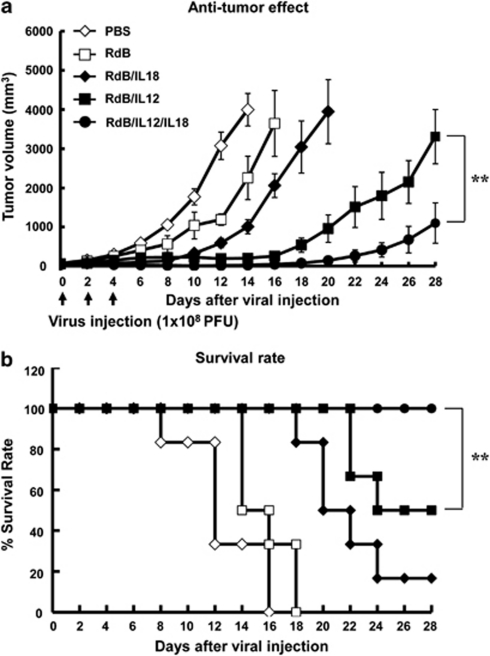
To ascertain whether RdB/IL-12, RdB/IL-18 or RdB/IL-12/IL-18 induced the suppression of tumor growth in syngeneic mice, we injected 5 × 10⁵ cells of B16-F10 melanoma subcutaneously into C57BL/6 mice. When the tumor volume averaged 80 mm3, mice were injected intratumorally with phosphate-buffered saline (PBS), RdB, RdB/IL-12, RdB/IL-18 or RdB/IL-12/IL-18 once every other day for a total of three injections. As presented in Figure 2a, control tumors treated with PBS showed aggressive growth, leading to a tumor volume of 3997.1±412.2 mm3 14 days after viral treatment. In marked comparison, RdB/IL-12-, RdB/IL-18- and RdB/IL-12/IL-18-treated tumors reached an average volume of 202.4±43, 1009±181.7 and 26.5±10.3 mm3, respectively, showing 95% (RdB/IL-12), 75% (RdB/IL-18) and 99% (RdB/IL-12/IL-18) tumor growth inhibition compared with PBS control (P<0.05). By day 20 following treatment, tumor re-growth was observed in RdB/IL-18- and RdB/IL-12-treated tumors, showing 3946.7±817.8 and 960.1±349.3 mm3, respectively, whereas those treated with RdB/IL-12/IL-18 maintained inhibition of tumor growth (145±64.7 mm3) (P<0.01). Moreover, RdB/IL-12/IL-18 elicited more prolonged suppression of tumor growth than RdB/IL-12 (P<0.01) (Supplementary Figure 2). Furthermore, RdB/IL-12/IL-18-treated mice showed higher survival rates compared with either RdB/IL-12 or RdB/IL-18 (P<0.01). All animals treated with RdB/IL-12/IL-18 remained viable 28 days after the beginning of the treatment, whereas only 16.7% of those treated with RdB/IL-18 and 50% of those treated with RdB/IL-12 survived for the same period of time (Figure 2b). Taken together, these results suggest that RdB/IL-12/IL-18 significantly prolonged survival and enhanced antitumor efficacy compared with either RdB/IL-12 or RdB/IL-18 in the B16-F10 murine melanoma model.
Figure 2.
Antitumor effects and survival rate of tumor-bearing mice. Antitumor (a) and survival rate (b) in mice given PBS (⋄), RdB (□), RdB/IL-18 (⧫), RdB/IL-12 (▪) or RdB/IL-12/IL-18 (•). C57BL/6 tumor-bearing mice were treated with intratumoral injections of 1 × 10⁸ plaque-forming unit per 30 μl of Ads on days 0, 2 and 4. Tumor volume was monitored on a 2-day interval until the end of the study. The arrow represents Ad inoculation. Values represent the mean±s.e.m. (6 animals per group). RdB/IL-12/IL-18-treated mice showed significantly enhanced antitumor effects and higher survival rates than did RdB/IL-12-treated mice. **P<0.01 vs RdB/IL-12-treated group.
In vivo oncolytic Ad treatment increased local expression of IL-12, IL-18, IFN-γ and GM-CSF
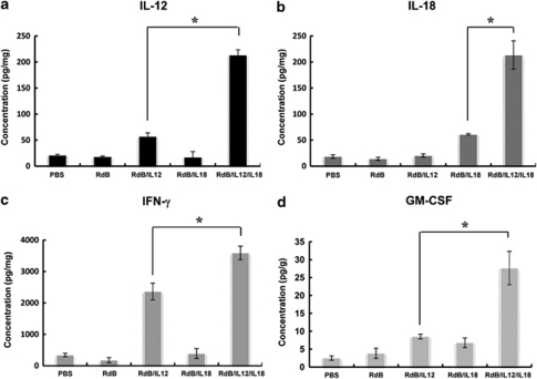
To determine the amount of IL-12 and IL-18 produced in RdB/IL-12-, RdB/IL-18- or RdB/IL-12/IL-18-treated mice, tumor tissues were harvested 3 days following the last viral injection. As seen in Figures 3a and b, a minimal amount of IL-12 was observed in the tumors treated with PBS, RdB or RdB/IL-18. In contrast, tumors treated with RdB/IL-12 or RdB/IL-12/IL-18 showed high concentrations of IL-12 (57.3±6.6 pg mg−1 for RdB/IL-12 and 213.6±9.9 pg mg−1 for RdB/IL-12/IL-18, P<0.05) (Figure 3a). Similarly, RdB/IL-18- or RdB/IL-12/IL-18-treated tumors produced significantly higher levels of IL-18 (61±1.8 pg mg−1 for RdB/IL-18 and 213.4±27.1pg mg−1 for RdB/IL-12/IL-18) than did those treated with PBS, RdB or RdB/IL-12 (18.6±3.3, 14.3±2.9 and 20.4±3.1 pg mg−1, respectively, P<0.05) (Figure 3b). Interestingly, IL-12 and IL-18 levels in tumor tissues treated with RdB/IL-12/IL-18 were synergistically greater than those treated with RdB/IL-12 or RdB/IL-18 in vivo, even though in vitro, IL-12 and IL-18 levels from RdB/IL-12/IL-18-treated cells were much lower than those of either RdB/IL-12- or RdB/IL-18-treated cells.
Figure 3.
RdB/IL-12/IL-18 treatment increased local expression of IL-12, IL-18, IFN-γ and GM-CSF in vivo. Tumor tissue was obtained 3 days after final viral treatment. ELISA was carried out to estimate the level of IL-12 (a), IL-18 (b), IFN-γ (c) and GM-CSF (d) in tumor tissues. Experiments were performed in triplicate and repeated three times. Each data point indicates means±s.e.m. of IL-12, IL-18, IFN-γ and GM-CSF levels for each individual tumor. RdB/IL-12/IL-18 produced a synergistically higher levels of IL-12, IL-18, IFN-γ and GM-CSF than RdB/IL-12 or RdB/IL-18 (Wilcoxon's signed-rank *P<0.05 compared with the sum of RdB/IL-12 or RdB/IL-18 alone).
Given that IL-12 and IL-18 can act synergistically to enhance the production of IFN-γ and GM-CSF from T and NK cells,11, 13, 14 we examined IFN-γ and GM-CSF production in the RdB/IL-12/IL-18-treated tumor tissue. As shown in Figure 3c and d, PBS-, RdB- and RdB/IL-18-injected mice showed relatively low levels of IFN-γ (342.6±54.6, 179.2±80.7 and 391.2±159.7 pg mg−1, respectively). In comparison, RdB/IL-12- or RdB/IL-12/IL-18-injected tissue showed high levels of IFN-γ (2364.4±265.4 pg mg−1 for RdB/IL-12 and 3590.6±213.5 pg mg−1 for RdB/IL-12/IL-18, P<0.05) (Figure 3c). In addition, RdB/IL-12/IL-18-treated tissue showed the highest GM-CSF expression (27.7±4.7 pg g−1) compared with PBS- (2.5±4.7 pg g−1), RdB- (3.9±1.4 pg g−1), RdB/IL-12- (8.5±0.6 pg g−1) and RdB/IL-18-treated tumor tissue (6.8±1.4 pg g−1) (Figure 3d). These results show that RdB/IL-12/IL-18-treated tumors produced significantly higher amounts of IL-12, IL-18, IFN-γ and GM-CSF than any of the other groups.
Increase of Th1/Th2 cytokine ratio
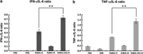
An aberrant Th1/Th2 cytokine balance with elevated Th2 cytokine levels, which favors development and progression of cancers, was found in various tumor-bearing patients, and a correlation between cytokine levels and the efficacy of cancer treatments has also been reported.16, 17 Hence, we further investigated whether RdB/IL-12/IL-18, which produced markedly higher amounts of Th1 cytokines (IFN-γ and GM-CSF) than any of the other groups, could converse from Th2 immunity toward Th1 immunity in tumor tissues. Tumor tissues from mice at 3 days following the final viral injection were harvested and analyzed with Th1/Th2/Th17 cytometric bead assay (CBA) kit (BD Biosciences PharMingen, San Diego, CA, USA). As IL-2, IL-4, IL-10 and IL-17 were not detected in the tumor tissues of all groups (data not shown), the ratio of Th1/Th2 cytokine was estimated from the ratios of IFN-γ/IL-6 and tumor necrosis factor-α (TNF-α)/IL-6. As presented in Figure 4, RdB/IL-12/IL-18-treated tumor tissue exhibited the highest IFN-γ/IL-6 and TNF-α/IL-6 ratio value compared with PBS-, RdB-, RdB/IL-12- or RdB/IL-18-treated tumor tissue (P<0.01), suggesting that RdB/IL-12/IL-18 polarizes T cell responses toward the type 1 pattern by enhancing Th1 cytokine and suppressing Th2 cytokine in the tumor microenvironment.
Figure 4.
Increased Th1/Th2 cytokine ratio in tumor tissue treated with RdB/IL-12/IL-18. Tumor tissue was harvested 3 days after last viral injection. Th1/Th2/Th17 CBA assay was performed to determine the Th1/Th2 cytokine ratio in tumor tissues. (a) IFN-γ/IL-6 ratio. (b) Tumor necrosis factor-α/IL-6 ratio. Each data point indicates mean value±s.e.m. of triplicates of representative of three independent experiments. **P<0.01 vs RdB/IL-12-treated group.
Generation of a tumor-specific immune response
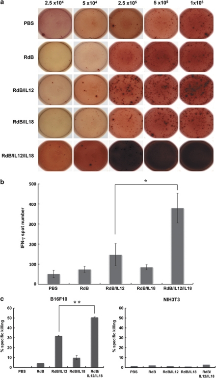
Results from enzyme-linked immunosorbent assay (ELISA) and CBA assay in tumor tissues indicate that RdB/IL-12/IL-18 might generate a tumor microenvironment that is more favorable to activate tumor-specific immune cells than an oncolytic Ad expressing either IL-12 or IL-18 alone. Therefore, we examined whether RdB/IL-12/IL-18 could enhance immune responses in vivo by assessing the number of immune cells expressing IFN-γ, a cytokine secreted by activated T cells. Splenocytes from mice at 3 days following the final viral injection were harvested and co-cultured with irradiated B16-F10 tumor cells for 3 days in the presence of recombinant human IL-2. Co-cultured splenocytes were serially seeded into the IFN-γ ELISPOT plate at concentrations from 2.5 × 10⁴ to 1 × 10⁶ cells per well and the number of IFN-γ-secreting cells was determined. As shown in Figure 5a, the frequency of IFN-γ-secreting immune cells recovered from mice injected with RdB/IL-12/IL-18 was significantly greater than cells from mice given RdB/IL-12 or RdB/IL-18. More specifically, the number of IFN-γ-secreting immune cells isolated from RdB/IL-12/IL-18-treated mice at a concentration of 2.5 × 10⁵ was much higher (380±74) than cells isolated from PBS- (51±18), RdB- (74±15), RdB/IL-12- (147±55) or RdB/IL-18-treated mice (85±13) (Figure 5b).
Figure 5.
Generation of a tumor-specific immune response. Subcutaneous tumors derived from B16-F10 cells were inoculated with RdB, RdB/IL-12, RdB/IL-18 or RdB/IL-12/IL-18 along with PBS as a negative control. Ad-treated mice were killed 3 days following last administration, and IFN-γ ELISPOT and 51Cr-release assays were carried out. (a) Examples of spot-forming cell (SFC) response. (b) ELISPOT assay for IFN-γ. The number of spots counted at a concentration of 2.5 × 10⁵. Each value represents the mean spot number±s.e.m. of triplicates of representative of three independent experiments. *P<0.05 vs RdB/IL-12. (c) 51Cr-release assay. B16-F10 cells were labeled with 51Cr and incubated with activated T cells isolated from mice treated with Ads at 100:1 E:T ratios. Each data point indicates means±s.e.m. of triplicates of representative of three independent experiments. **P<0.01 vs RdB/IL-12.
To further delineate the tumor-specific immune response in vivo, a 51Cr-release assay was carried out. Splenocytes obtained from RdB/IL-12/IL-18-treated mice showed the most potent B16-F10-specific lytic activity on day 3 after exposure. CTL killing of splenocytes from mice treated with RdB/IL-12, RdB/IL-18 or RdB/IL-12/IL-18 was 32.1±0.5, 10±2.1 and 50.8±0.5, respectively, at an effector-to-target (E:T) ratio of 100:1, respectively, with no measurable background (Figure 5c). The CTL activity of splenocytes from mice treated with RdB/IL-12, RdB/IL-18 or RdB/IL-12/IL-18 was specific to a given tumor, as no CTL activity could be detected when NIH3T3 cells were used as target cells. These results show that RdB/IL-12/IL-18-treated mice had a higher tumor-specific adaptive immune response than did RdB/IL-12- or RdB/IL-18-treated mice.
Increased infiltration of CD4+ T, CD8+ T and NK cells in RdB/IL-12/IL-18-treated tumors
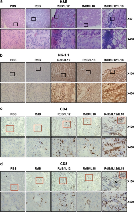
To analyze histological characteristics of tumor tissues following RdB/IL-12/IL-18 treatment, we performed hematoxylin and eosin staining of tumor tissues. Histological evaluation of tumor sections revealed that large areas of tumors treated with RdB/IL-12/IL-18 were necrotic. In particular, tumors treated with RdB/IL-12 or RdB/IL-12/IL-18 were extensively infiltrated with immune cells, whereas tumors treated with RdB or RdB/IL-18 showed sparse infiltration. Moreover, denser immune cell infiltration was observed not only around, but also inside the remaining tumor tissues treated with RdB/IL-12/IL-18.
To obtain more insight into the molecular mechanisms of cytokine-mediated inhibition of tumor growth, we performed immunohistological analysis. Immunohistochemical studies confirmed that the tumor-infiltrating immune cells were CD4+ T cells, CD8+ T cells and NK cells. Higher numbers of all three cell types were detected in wider areas of RdB/IL-12/IL-18-treated tumors than in tumors treated with either RdB/IL-12 or RdB/IL-18 (Figure 6).
Figure 6.
Histological and immunohistochemical analysis of tumor sections from mice treated with Ads. Ads were injected on days 0, 2 and 4, and tumors were collected on day 7 for histological analysis. (a) Frozen sections of tumor tissue were then stained by hematoxylin and eosin. Original magnification: × 40 and × 400. Cryosections of tumor tissue were stained with anti-NK-1.1 (b), anti-CD4 (c) and anti-CD8 (d) monoclonal antibodies. Original magnification: × 100 and × 400.
Increased IL-12Rβ2 or IL-18Rα expression in RdB/IL-12/IL-18-treated tumors
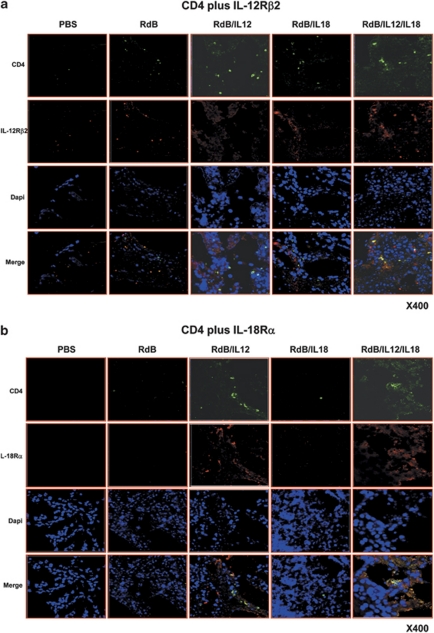
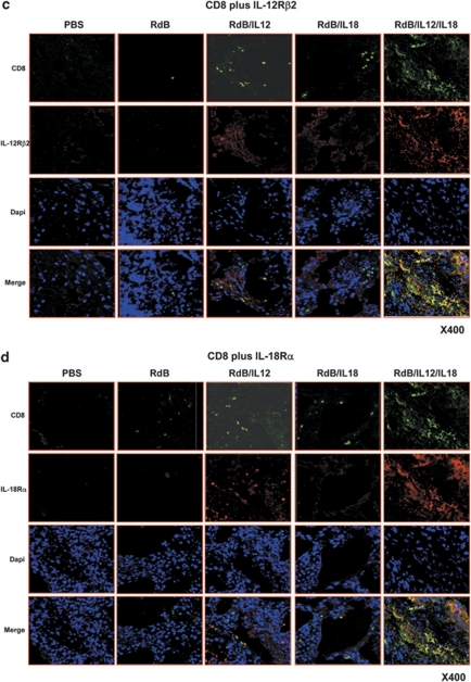
To examine whether expression of IL-12Rβ2 or IL-18Rα of T cells could be enhanced in RdB/IL-12/IL-18-treated tumor tissue, we performed immunofluorescence double staining using anti-CD4, anti-CD8, anti-IL-12Rβ2 and anti-IL-18Rα antibodies. Sections were prepared from C57BL6 mice 3 days after the final virus injection and were analyzed to determine the population of T cells expressing IL-12Rβ2 or IL-18Rα. The analysis revealed an elevation in the number of CD4+ and CD8+ T cells expressing IL-12Rβ2 or IL-18Rα in the tumor tissues of RdB/IL-12/IL-18-treated mice, compared with those of RdB/IL-12- or RdB/IL-18-treated mice (Figure 7). Effector T cells have to maintain both IL-12Rβ2 and IL-18Rα expression for differentiating T cells.18 Therefore, these data indicate that RdB/IL-12/IL-18 treatment resulted in an improved generation of optimally differentiated T cells expressing IL-12Rβ2 or IL-18Rα.
Figure 7.
Enhancement of T cells expressing IL-12Rβ2 or IL-18Rα in tumor tissues. Tumor tissues were harvested from tumor-bearing mice 3 days following final virus treatment. Sections of tumor tissue were incubated with anti-CD4 (Green) plus anti-IL-12Rβ2 (Red) (a), anti-CD4 (Green) plus anti-IL-18Rα (Red) (b), anti-CD8 (Green) plus anti-IL-12Rβ2 (Red) (c), and anti-CD8 (Green) plus anti-IL-18Rα (Red) (d). Original magnification: × 400.
Discussion
Therapeutic manipulation of immune responses by the use of cytokines is potent strategy for cancer immunotherapy and tumor growth suppression.19 However, the systemic administration of cytokines at a therapeutic dose can result in serious toxicity in human cancer-bearing patients as well as in animal cancer models.20, 21 Some patients given IL-12 have even died following treatment.22 Therefore, it is important to understand as to how to induce therapeutic levels of cytokines long enough to generate antitumor immunity effectively within the tumor microenvironment while simultaneously attenuating systemic toxicity. To address this issue, we have used an E1B-deleted and E1A-mutated oncolytic Ad, RdB, which exerts an antitumor effect in a cancer cell-specific manner as an intratumoral delivery system of therapeutic cytokines.23 Moreover, this tumor-selective oncolytic adenoviral vector is competent at killing the tumor cells, as well as amplifying production of transgenes, by continuously replicating in tumor cells until the tumor tissues were eradicated.24
Earlier data from our group showed that locally high expression of IL-12 in the tumor tissues by oncolytic Ad elicited potent antitumor effects without the induction of adverse effects by an increase in IL-12 or IFN-γ in the serum.3 However, combination therapies of IL-12 and IL-18 have a more effective antitumor activity than either cytokine alone.20, 25, 26 Several studies have shown that IL-18 and IL-12 synergistically mediate antitumor immunity in a number of animal tumor models. Oshikawa et al.27 showed that IL-12-, pro-IL-18- and IL-1β-converting enzyme act synergistically to elicit an enhanced antitumor immune response in a murine mammary adenocarcinoma model via induction of cytotoxic CD8+ T cells and IFN-γ. Similarly, recombinant IL-12 and IL-18 proteins were shown to significantly inhibit tumor growth and lymph-node metastasis in hepatoma tumor-bearing guinea-pigs.28 Furthermore, in DC-based tumor vaccine therapy, DCs co-producing IL-12 and IL-18 induced complete tumor rejection through the generation of an effective Th1 immune response in a BALB/c sarcoma model.29
In an effort to augment the efficiency of immune gene therapy in vivo, we explored the potential therapeutic benefit of an oncolytic Ad co-expressing IL-12 and IL-18. We first showed that RdB/IL-12/IL-18 resulted in not only enhanced and prolonged suppression of tumor growth and increased survival compared with either RdB/IL-12 or RdB/IL-18, but also a higher incidence of complete tumor regression (data not shown), suggesting that RdB/IL-12/IL-18 elicited an improved antitumor effect than RdB/IL-12 or RdB/IL-18 alone.
To show the underlying mechanism of this enhanced RdB/IL-12/IL-18-mediated antitumor effect, we next evaluated the characteristics of RdB/IL-12/IL-18-treated tumor tissues. Surprisingly, in contrast to our in vitro results, the expression levels of both IL-12 and IL-18 were markedly elevated in RdB/IL-12/IL-18-treated tumor tissues. More specifically, in vivo, RdB/IL-12/IL-18 elicited a 3.7- and 3.5-fold increase in IL-12 and IL-18 expression, whereas in vitro, RdB/IL-12/IL-18 elicited a 3.2- and 2.6-fold decrease in IL-12 and IL-18 expression.
IL-18, in concert with IL-12, enhances IFN-γ production from activated T and NK cells.11, 14 IFN-γ in turn induces DC maturation and macrophages activation, resulting in the elevation of IL-12 and IL-18 expression in those cells.18, 30 This positive feedback loop could explain the upregulation of IL-12 and IL-18 in RdB/IL-12/IL-18-treated tumor tissues. Furthermore, matured DCs and activated macrophages can themselves produce IFN-γ as well as IL-12 and IL-18.31, 32 In accordance with this positive feedback loop, we found that expression levels of IFN-γ, a Th1 cytokine, were considerably higher in RdB/IL-12/IL-18-treated mice than in RdB/IL-12- or RdB/IL-18-treated mice (Figure 3c). IFN-γ has a significant role in boosting cell-mediated antitumor immune responses by increasing cancer cell immunogenicity through enhancing antigen presentation as well as prompting antigen-presenting cells, NK cells, CTLs and Th1 T cells.18
Yang et al.33 have shown that co-administration of IFN-γ or IL-12 with the recombinant Ad into the airway of C57BL/6 mice prevents the activation of Th2 cells and formation of neutralizing antibody, allowing for efficient re-administration of recombinant Ad. In agreement with such findings, our study shows that RdB/IL-12/IL-18 generates increased Th1/Th2 cytokine ratio with the upregulation of IL-12, IL-18, IFN-γ and GM-CSF in tumor tissues. Taken together, these results suggest that RdB/IL-12/IL-18 administered intratumorally may be competent at killing the tumor cells, as well as amplifying production of cytokines, by continuously replicating in tumor cells regardless of Ad-specific neutralizing antibody levels.
GM-CSF expression was also dramatically increased in tumor tissues treated with RdB/IL-12/IL-18 compared with those treated with RdB/IL-12 or RdB/IL-18, supporting the importance of IFN-γ- and GM-CSF-mediated antitumor immune response. GM-CSF, a Th1 cytokine, stimulates an antitumor cell-mediated immune response by facilitating not only the recruitment, maturation and function of DCs, but also by enhancing the cross-presentation of DCs, which is important for recognizing tumor cells.34, 35, 36 Recently, we showed that intratumoral delivery of GM-CSF can promote antigen presentation and elevate the tumor-specific CTL response.37 Therefore, we suggest that upregulation of GM-CSF in the tumor microenvironment may enhance antigen presentation, leading to the generation of a tumor-specific immune response.
Tumor microenvironment has been shown to establish immunosuppressive cytokine networks that favor the suppression of an antitumor immune response and eventually generate tumor proliferation, angiogenesis and metastasis. Therefore, it is critical to understand the immunosuppressive environment at the tumor site to manipulate and generate antitumor immunity effectively within the tumor microenvironment. To this end, our data suggest that RdB/IL-12/IL-18 generated a tumor microenvironment more favorable to activate tumor-specific immune cells via upregulation of IL-12, IL-18, IFN-γ and GM-CSF in tumor tissues. Moreover, the manipulation of a tumor milieu more favorable to activate tumor-specific immune cells by RdB/IL-12/IL-18 was further supported by the observation that Th1/Th2 cytokine ratio was markedly elevated in RdB/IL-12/IL-18-treated tumor tissues (Figures 4a and b).
To activate tumor-specific immune cells, antitumor adaptive immunity in the tumor milieu should be optimally generated. Here, we showed that mice injected with RdB/IL-12/IL-18 had enhanced CTL activity, as well as an increase in tumor-specific IFN-γ-secreting immune cells, than did those given RdB/IL-12 or RdB/IL-18. In accordance with these results, both histological and immunohistochemical results showed immense necrotic regions as well as infiltration of NK, CD4+ T and CD8+ T cells into the tumor tissues of RdB/IL-12/IL-18-treated mice compared with those of RdB/IL-12- or RdB/IL-18-treated mice. NK cells are known to play an important role in the promotion of Th1 responses by producing IFN-γ and modulating DC activity.38, 39 This observation suggests that IL-12 plus IL-18 activate NK cells that mediate oncolysis and DC maturation that, in turn, enhance antigen presentation to induce tumor-specific T cells to differentiate from naïve T cells, leading to the generation of antitumor immunity.
Biological functionality of RdB/IL-12/IL-18-mediated IL-12 and IL-18 expression in tumor tissues depends on the expression level of IL-12Rβ2 and IL-18Rα. Therefore, upregulation of IL-12Rβ2 and IL-18Rα correlates with the capacity of IL-12 and IL-18 to bind and initiate the development of Th1 immunity. Reports from in vitro studies have shown that IL-18 enhances the production of IL-12Rβ2 on T cells, whereas IL-12 mediates upregulation of IL-18Rα on T cells.40, 41 Here, we showed that the numbers of T cells expressing IL-12Rβ2 or IL-18Rα were increased in tumor tissues of RdB/IL-12/IL-18-treated tumors (Figure 7). Moreover, animals treated with RdB/IL-12/IL-18 showed an elevated number of CD4+ and CD8+ T cells expressing IL-12Rβ2 or IL-18Rα at the tumor tissues. The expression of IL-12Rβ2 or IL-18Rα receptors are necessary for the differentiation of effector T cells.18 Therefore, our finding implies that intratumoral delivery of IL-12 and IL-18 by RdB/IL-12/IL-18 resulted in improved generation of optimally differentiated CD4+ and CD8+ T cells expressing IL-12Rβ2 or IL-18Rα. Our results further support the view put forth by Li et al.13 that IL-12, in combination with IL-18, induces differentiation of the antitumor effector T cells CD4+ and CD8+.
In conclusion, the finding in our report provides a novel underlying mechanism of combination therapies via IL-12 and IL-18 that promotes differentiation of T cells expressing IL-12Rβ2 or IL-18Rα. We also provide the first study where intratumoral expression of both IL-12 and IL-18 via oncolytic Ad vector results in the creation of more potent tumor-specific immunity. These results provide a new insight into therapeutic mechanisms through IL-12 plus IL-18, as well as a potential clinical cancer immunotherapeutic agent for the generation of improved antitumor immunity.
Materials and methods
Cell lines
All cell lines were maintained in Dulbecco's modified Eagle's medium (Gibco BRL, Grand Island, NY, USA) supplemented with 10% fetal bovine serum (Gibco BRL), -glutamine (2 m l−1), penicillin (100 IU ml−1) and streptomycin (50 μg ml−1). All cell lines were cultured at 37 °C in a humidified atmosphere of 5% CO2 and 95% air.
Animal studies
Six- to eight-week-old male C57BL/6 mice were obtained from Charles River Laboratories International Inc. (Wilmington, MA, USA). During the experiments, all mice were kept in a laminar air flow cabinet under specific pathogen-free conditions at Yonsei University College of Medicine, an Association for Assessment and Accreditation of Laboratory Animal Care-accredited animal facility. All animal experiments were conducted under the institutional guidelines established by the Animal Research Committee.
Generation of Ads expressing IL-12 and IL-18
To generate an oncolytic Ad expressing IL-12 at the E3 region, we first constructed a pSP72-E3 Ad shuttle vector42 expressing murine IL-12 comprised of p35 and p40. The IL-12 gene (p35/IRES/p40) was digested from pCA14-IL-12 using BglII and subcloned into pSP72-E3 Ad shuttle vector with BamHI, creating a pSP72-E3-IL-12 E3 shuttle vector. The newly constructed pSP72-E3-IL-12 E3 shuttle vector was then linearized with NdeI digestion, and RdB23 was linearized with SpeI digestion. The linearized pSP72-E3-IL-12 E3 shuttle vector was then co-transformed into Escherichia coli BJ5183 along with the SpeI-digested RdB for homologous recombination, resulting in RdB/IL-12 oncolytic Ad. To generate an oncolytic Ad expressing IL-18 at the E3 region, we first constructed an E3 shuttle vector expressing murine IL-18. BglII fragments containing IL-18 expression cassette were excised from pCA14-IL-18, and subcloned into pSP72-E3 Ad shuttle vector predigested with BamHI, resulting in a pSP72-E3-IL-18. The newly constructed pSP72-E3-IL-18 shuttle vector was then linearized with XmnI digestion, and then co-transformed into E. coli BJ5183 together with the SpeI-digested RdB for homologous recombination, creating an RdB/IL-18 oncolytic Ad. To construct an oncolytic Ad-expressing IL-12 and IL-18 at the E1 and E3 region, respectively, pSP72-E3-IL-18 E3 shuttle vector was then linearized with XmnI digestion, and the Ad vector RdB/IL-12 was linearized with SpeI digestion. The linearized pSP72-E3-IL-18 E3 shuttle vector was then co-transformed into E. coli BJ5183 along with the SpeI-digested RdB/IL-123 for homologous recombination, generating an RdB/IL-12/IL-18 oncolytic Ad. The ratio of plaque-forming unit to viral particle for RdB, RdB/IL-12, RdB/IL-18 and RdB/IL-12/IL-18 used in this study is 1:42, 1:46, 1:39 and 1:47, respectively.
ELISA for IL-12 and IL-18 expression
To assess IL-12 and IL-18 levels, B16-F10 melanoma cells were plated onto six-well plates at 5 × 10⁴ cells per well, and then infected with RdB, RdB/IL-12, RdB/IL-18 or RdB/IL-12/IL-18 Ad at MOIs of 10–200. At 96 h post-infection, the supernatants were harvested. IL-12 and IL-18 expression were determined using an ELISA according to the manufacturer's specifications (IL-12p70 ELISA kit: Endogen, Woburn, MA, USA; IL-18 ELISA kit: MBL, Nagoya, Japan).
Antitumor effects of oncolytic Ad co-expressing IL-12 plus IL-18
Tumors were implanted subcutaneously on the left abdomen of C57BL/6 mice by inoculating viable B16-F10 murine melanoma cells (5 × 10⁵) in 50 μl of Hank's balanced salt solution (Gibco BRL). When the average tumor volume reached 80 mm3, mice were randomly divided into one of five groups (six animals per group) receiving PBS, RdB, RdB/IL-12, RdB/IL-18 or RdB/IL-12/IL-18 mixed with Lipofectamine and plus reagent (Gibco BRL) at a 2:6 ratio. Ads or PBS were injected intratumorally (1 × 10⁸ plaque-forming unit per tumor in 30 μl of PBS) on days 0, 2 and 4. Control mice were injected with PBS only. Tumor volume was evaluated once every 2 days by electronic caliper measure: volume=0.523LW2 (L=the length of tumors; W=the width of tumors). Tumor responses to each treatment were compared using a log-rank test analysis (StatView software; Abacus Concepts Inc., Berkeley, CA, USA). The percentage of surviving mice was determined by monitoring the tumor growth-related events (tumor size >2500 mm3). The survival curve was then plotted against time after treatment. Differences in survival were considered statistically significant when P-values were <0.01.
Quantification of cytokines and Th1/Th2/Th17 profile in tumor tissue
Tumors were harvested 3 days after the last viral inoculation of the B16-F10 tumor-bearing mice and snap-frozen in liquid nitrogen. Tumor tissues were homogenized (ART-MICCRA D-8; ART moderne Labortechnik, Munchen, Germany) in ice-cold radio immunoprecipitation assay buffer (Elipis Biotech, Taejeon, South Korea) with a proteinase inhibitor cocktail (Sigma, St Louis, MO, USA). Homogenates were then centrifuged in a high-speed microcentrifuge for 10 min and determined for total protein content using a BCA protein assay reagent kit (Pierce, Rockford, IL, USA). Levels of IL-12, IL-18, IFN-γ and GM-CSF in the tumor tissue extract were determined by ELISA according to the supplier's recommendations (IL-12 ELISA kit; Endogen, IL-18 ELISA kit; MBL, IFN-γ ELISA kit; Endogen, GM-CSF ELISA kit; Endogen). In addition, Th1/Th2/Th17 type cytokine expression profiles in the tumor tissue extract were estimated using Th1/Th2/Th17 CBA kit (BD Biosciences PharMingen). ELISA and CBA results were normalized relative to the total protein concentration in each tumor and were calculated as picograms per milligram of total protein.
IFN-γ ELISPOT assay in splenocytes
At 3 days following the last viral treatment, spleens were obtained aseptically from tumor-bearing mice, and unicellular splenocytes were prepared as described previously.3 Prepared spleen cells were co-cultured with irradiated B16-F10 (5000 rad) tumor cells for 3 days in the presence of recombinant human IL-2 (100 U ml−1; R&D Systems, Minneapolis, MN, USA). An IFN-γ ELISPOT assay was then carried out as described previously.3 The spots were measured with a computer-based immunospot system (AID Elispot Reader System, version 3.4; Autoimmun Diagnostika GmbH, Strassberg, Germany).
Cytotoxicity assay in splenocytes
The cytolytic activity of tumor-specific CTL was evaluated by 4 h 51Cr-release assay. Co-cultured spleen cells were prepared as described for the IFN-γ ELISPOT assay. After co-culture, splenocytes as effector cells were recovered and incubated with 51Cr-labeled B16-F10 or NIH3T3 cells as target cells at 100:1 E:T ratios for 4 h at 37 °C and 5% CO2. Thereafter, the supernatant was obtained and 51Cr release was assessed. The percentage of specific lysis was calculated by the following formula: ((c.p.m. experimental release−c.p.m. spontaneous release)/(c.p.m. maximum release−c.p.m. spontaneous release)) × 100. Spontaneous release was determined by incubation of the labeled target cells without effector cells. For maximum release, labeled target cells were treated with detergent.
Histology and immunohistochemistry for NK-1.1, CD4 and CD8
For histological study, tumor tissues were snap-frozen in OCT compound (Sakura Finetec, Torrance, CA, USA), and 10-μm sections were cut on a cryostat. Representative sections were stained with hematoxylin and eosin, and then investigated using light microscopy. To detect lymphocytes, tumor tissues were snap-frozen in OCT compound (Sakura Finetec), and 10-μm sections were cut, subjected to immunohistochemistry, and then counterstained with Meyer's hematoxylin. The sections were incubated with purified rat anti-mouse CD4 monoclonal antibody (BD Biosciences PharMingen), purified rat anti-mouse CD8 monoclonal antibody (BD Biosciences PharMingen) or purified mouse anti-mouse NK-1.1 monoclonal antibody (Biolegend, San Diego, CA, USA) as a primary antibodies, and then with goat anti-rat IgG (BD Biosciences PharMingen) or goat-anti mouse IgG (Southern Biotech, Birmingham, AL, USA) as a secondary antibody. Diaminobenzidine/hydrogen peroxidase (DAKO, Carpinteria, CA, USA) was used as the chromogen substrate.
Immunofluorescence double staining for T cells expressing IL-12Rβ2 or IL-18Rα
For immunofluorescence double staining for CD4+ and CD8+ T cells expressing IL-12Rβ2 or IL-18Rα, cryosections were treated with rat anti-mouse CD4 monoclonal antibody (BD Biosciences PharMingen) or rat anti-mouse CD8 monoclonal antibody (BD Biosciences PharMingen) and incubated overnight at 4 °C, followed by Alexa flour 488 (Green)-labeled goat anti-rat IgG (Invitrogen, Carlsbad, CA, USA) at room temperature for 1 h. The cryosections were then incubated with goat anti-IL-12Rβ2 polyclonal antibody (Santa Cruz Biotechnology, Santa Cruz, CA, USA) or rabbit anti-mouse IL-18Rα polyclonal antibody (Santa Cruz Biotechnology) at 4 °C overnight and at room temperature for 1 h with Alexa flour 568 (Red)-labeled rabbit anti-goat IgG (Invitrogen) or Alexa flour 568 (Red)-labeled goat anti-rabbit IgG (Invitrogen), respectively. For counterstaining, the samples were incubated with 4,6-diamidino-2-phenylindole (Sigma), and then observed by a laser scanning confocal imaging system (LSM 510-META; Carl Zeiss, Jena, Germany).
Statistical analysis
The data are expressed as mean±s.e. of the mean (s.e.m.). Statistical comparison was made using Stat View software (Abacus Concepts Inc., Berkeley, CA, USA) and the Mann–Whitney test (non-parametric method). The criterion for statistical significance was P<0.05.
Acknowledgments
This work was supported by grants from the Ministry of Knowledge Economy (10030051, Dr C-O Yun), the Korea Science and Engineering Foundation (R15-2004-024-02001-0, 2009K001644, 2010-0029220, Dr C-O Yun), the Korea Food and Drug Administration (KFDA-10172-332 to Dr C-O Yun), and Yonsei University College of Medicine faculty research grant (6-2010-0052, Dr C-O Yun). I-K Choi is a graduate student sponsored by KOSEF through National Core Research Center for Nanomedical Technology. J-S Lee and S-N Zhang are graduate students sponsored by the Brain Korea 21 Project for Medical Science, Yonsei University College of Medicine, Seoul, South Korea.
The authors declare no conflict of interest.
Footnotes
Supplementary Information accompanies the paper on Gene Therapy website (http://www.nature.com/gt)
Supplementary Material
References
- Colombo MP, Trinchieri G. Interleukin-12 in anti-tumor immunity and immunotherapy. Cytokine Growth Factor Rev. 2002;13:155–168. doi: 10.1016/s1359-6101(01)00032-6. [DOI] [PubMed] [Google Scholar]
- Wigginton JM, Wiltrout RH. IL-12/IL-2 combination cytokine therapy for solid tumours: translation from bench to bedside. Expert Opin Biol Ther. 2002;2:513–524. doi: 10.1517/14712598.2.5.513. [DOI] [PubMed] [Google Scholar]
- Lee YS, Kim JH, Choi KJ, Choi IK, Kim H, Cho S, et al. Enhanced antitumor effect of oncolytic adenovirus expressing interleukin-12 and B7-1 in an immunocompetent murine model. Clin Cancer Res. 2006;12:5859–5868. doi: 10.1158/1078-0432.CCR-06-0935. [DOI] [PubMed] [Google Scholar]
- Dao T, Ohashi K, Kayano T, Kurimoto M, Okamura H. Interferon-gamma-inducing factor, a novel cytokine, enhances Fas ligand-mediated cytotoxicity of murine T helper 1 cells. Cell Immunol. 1996;173:230–235. doi: 10.1006/cimm.1996.0272. [DOI] [PubMed] [Google Scholar]
- Kohno K, Kataoka J, Ohtsuki T, Suemoto Y, Okamoto I, Usui M, et al. IFN-gamma-inducing factor (IGIF) is a costimulatory factor on the activation of Th1 but not Th2 cells and exerts its effect independently of IL-12. J Immunol. 1997;158:1541–1550. [PubMed] [Google Scholar]
- Tomura M, Zhou XY, Maruo S, Ahn HJ, Hamaoka T, Okamura H, et al. A critical role for IL-18 in the proliferation and activation of NK1.1+ CD3− cells. J Immunol. 1998;160:4738–4746. [PubMed] [Google Scholar]
- Tsutsui H, Nakanishi K, Matsui K, Higashino K, Okamura H, Miyazawa Y, et al. IFN-gamma-inducing factor up-regulates Fas ligand-mediated cytotoxic activity of murine natural killer cell clones. J Immunol. 1996;157:3967–3973. [PubMed] [Google Scholar]
- Okamura H, Nagata K, Komatsu T, Tanimoto T, Nukata Y, Tanabe F, et al. A novel costimulatory factor for gamma interferon induction found in the livers of mice causes endotoxic shock. Infect Immun. 1995;63:3966–3972. doi: 10.1128/iai.63.10.3966-3972.1995. [DOI] [PMC free article] [PubMed] [Google Scholar]
- Micallef MJ, Yoshida K, Kawai S, Hanaya T, Kohno K, Arai S, et al. In vivo antitumor effects of murine interferon-gamma-inducing factor/interleukin-18 in mice bearing syngeneic Meth A sarcoma malignant ascites. Cancer Immunol Immunother. 1997;43:361–367. doi: 10.1007/s002620050345. [DOI] [PMC free article] [PubMed] [Google Scholar]
- Marshall DJ, Rudnick KA, McCarthy SG, Mateo LR, Harris MC, McCauley C, et al. Interleukin-18 enhances Th1 immunity and tumor protection of a DNA vaccine. Vaccine. 2006;24:244–253. doi: 10.1016/j.vaccine.2005.07.087. [DOI] [PubMed] [Google Scholar]
- Tominaga K, Yoshimoto T, Torigoe K, Kurimoto M, Matsui K, Hada T, et al. IL-12 synergizes with IL-18 or IL-1beta for IFN-gamma production from human T cells. Int Immunol. 2000;12:151–160. doi: 10.1093/intimm/12.2.151. [DOI] [PubMed] [Google Scholar]
- Nakanishi K, Yoshimoto T, Tsutsui H, Okamura H. Interleukin-18 is a unique cytokine that stimulates both Th1 and Th2 responses depending on its cytokine milieu. Cytokine Growth Factor Rev. 2001;12:53–72. doi: 10.1016/s1359-6101(00)00015-0. [DOI] [PubMed] [Google Scholar]
- Li Q, Carr AL, Donald EJ, Skitzki JJ, Okuyama R, Stoolman LM, et al. Synergistic effects of IL-12 and IL-18 in skewing tumor-reactive T-cell responses towards a type 1 pattern. Cancer Res. 2005;65:1063–1070. [PubMed] [Google Scholar]
- Ahn HJ, Maruo S, Tomura M, Mu J, Hamaoka T, Nakanishi K, et al. A mechanism underlying synergy between IL-12 and IFN-gamma-inducing factor in enhanced production of IFN-gamma. J Immunol. 1997;159:2125–2131. [PubMed] [Google Scholar]
- Coughlin CM, Salhany KE, Wysocka M, Aruga E, Kurzawa H, Chang AE, et al. Interleukin-12 and interleukin-18 synergistically induce murine tumor regression which involves inhibition of angiogenesis. J Clin Invest. 1998;101:1441–1452. doi: 10.1172/JCI1555. [DOI] [PMC free article] [PubMed] [Google Scholar]
- Gadducci A, Cosio S, Carpi A, Nicolini A, Genazzani AR. Serum tumor markers in the management of ovarian, endometrial and cervical cancer. Biomed Pharmacother. 2004;58:24–38. doi: 10.1016/j.biopha.2003.11.003. [DOI] [PubMed] [Google Scholar]
- Smyth MJ, Cretney E, Kershaw MH, Hayakawa Y. Cytokines in cancer immunity and immunotherapy. Immunol Rev. 2004;202:275–293. doi: 10.1111/j.0105-2896.2004.00199.x. [DOI] [PubMed] [Google Scholar]
- Szabo SJ, Sullivan BM, Peng SL, Glimcher LH. Molecular mechanisms regulating Th1 immune responses. Annu Rev Immunol. 2003;21:713–758. doi: 10.1146/annurev.immunol.21.120601.140942. [DOI] [PubMed] [Google Scholar]
- Dranoff G. Cytokines in cancer pathogenesis and cancer therapy. Nat Rev Cancer. 2004;4:11–22. doi: 10.1038/nrc1252. [DOI] [PubMed] [Google Scholar]
- Osaki T, Peron JM, Cai Q, Okamura H, Robbins PD, Kurimoto M, et al. IFN-gamma-inducing factor/IL-18 administration mediates IFN-gamma- and IL-12-independent antitumor effects. J Immunol. 1998;160:1742–1749. [PubMed] [Google Scholar]
- Marshall E. Sciencescope. Science. 1995;268:1555. doi: 10.1126/science.268.5217.1555. [DOI] [PubMed] [Google Scholar]
- Leonard JP, Sherman ML, Fisher GL, Buchanan LJ, Larsen G, Atkins MB, et al. Effects of single-dose interleukin-12 exposure on interleukin-12-associated toxicity and interferon-gamma production. Blood. 1997;90:2541–2548. [PubMed] [Google Scholar]
- Kim J, Kim JH, Choi KJ, Kim PH, Yun CO. E1A- and E1B-double mutant replicating adenovirus elicits enhanced oncolytic and antitumor effects. Hum Gene Ther. 2007;18:773–786. doi: 10.1089/hum.2006.167. [DOI] [PubMed] [Google Scholar]
- Ganly I, Kirn D, Eckhardt G, Rodriguez GI, Soutar DS, Otto R, et al. A phase I study of Onyx-015, an E1B attenuated adenovirus, administered intratumorally to patients with recurrent head and neck cancer. Clin Cancer Res. 2000;6:798–806. [PubMed] [Google Scholar]
- Yamanaka K, Hara I, Nagai H, Miyake H, Gohji K, Micallef MJ, et al. Synergistic antitumor effects of interleukin-12 gene transfer and systemic administration of interleukin-18 in a mouse bladder cancer model. Cancer Immunol Immunother. 1999;48:297–302. doi: 10.1007/s002620050578. [DOI] [PMC free article] [PubMed] [Google Scholar]
- Hara I, Nagai H, Miyake H, Yamanaka K, Hara S, Micallef MJ, et al. Effectiveness of cancer vaccine therapy using cells transduced with the interleukin-12 gene combined with systemic interleukin-18 administration. Cancer Gene Ther. 2000;7:83–90. doi: 10.1038/sj.cgt.7700083. [DOI] [PubMed] [Google Scholar]
- Oshikawa K, Shi F, Rakhmilevich AL, Sondel PM, Mahvi DM, Yang NS. Synergistic inhibition of tumor growth in a murine mammary adenocarcinoma model by combinational gene therapy using IL-12, pro-IL-18, and IL-1beta converting enzyme cDNA. Proc Natl Acad Sci USA. 1999;96:13351–13356. doi: 10.1073/pnas.96.23.13351. [DOI] [PMC free article] [PubMed] [Google Scholar]
- Shiratori I, Suzuki Y, Oshiumi H, Begum NA, Ebihara T, Matsumoto M, et al. Recombinant interleukin-12 and interleukin-18 antitumor therapy in a guinea-pig hepatoma cell implant model. Cancer Sci. 2007;98:1936–1942. doi: 10.1111/j.1349-7006.2007.00614.x. [DOI] [PubMed] [Google Scholar]
- Tatsumi T, Huang J, Gooding WE, Gambotto A, Robbins PD, Vujanovic NL, et al. Intratumoral delivery of dendritic cells engineered to secrete both interleukin (IL)-12 and IL-18 effectively treats local and distant disease in association with broadly reactive Tc1-type immunity. Cancer Res. 2003;63:6378–6386. [PubMed] [Google Scholar]
- Boehm U, Klamp T, Groot M, Howard JC. Cellular responses to interferon-gamma. Annu Rev Immunol. 1997;15:749–795. doi: 10.1146/annurev.immunol.15.1.749. [DOI] [PubMed] [Google Scholar]
- Darwich L, Coma G, Pena R, Bellido R, Blanco EJ, Este JA, et al. Secretion of interferon-gamma by human macrophages demonstrated at the single-cell level after costimulation with interleukin (IL)-12 plus IL-18. Immunology. 2009;126:386–393. doi: 10.1111/j.1365-2567.2008.02905.x. [DOI] [PMC free article] [PubMed] [Google Scholar]
- Fricke I, Mitchell D, Mittelstadt J, Lehan N, Heine H, Goldmann T, et al. Mycobacteria induce IFN-gamma production in human dendritic cells via triggering of TLR2. J Immunol. 2006;176:5173–5182. doi: 10.4049/jimmunol.176.9.5173. [DOI] [PubMed] [Google Scholar]
- Yang Y, Trinchieri G, Wilson JM. Recombinant IL-12 prevents formation of blocking IgA antibodies to recombinant adenovirus and allows repeated gene therapy to mouse lung. Nat Med. 1995;1:890–893. doi: 10.1038/nm0995-890. [DOI] [PubMed] [Google Scholar]
- Hung K, Hayashi R, Lafond-Walker A, Lowenstein C, Pardoll D, Levitsky H. The central role of CD4(+) T cells in the antitumor immune response. J Exp Med. 1998;188:2357–2368. doi: 10.1084/jem.188.12.2357. [DOI] [PMC free article] [PubMed] [Google Scholar]
- Mach N, Gillessen S, Wilson SB, Sheehan C, Mihm M, Dranoff G. Differences in dendritic cells stimulated in vivo by tumors engineered to secrete granulocyte–macrophage colony-stimulating factor or Flt3-ligand. Cancer Res. 2000;60:3239–3246. [PubMed] [Google Scholar]
- Jinushi M, Tahara H. Cytokine gene-mediated immunotherapy: current status and future perspectives. Cancer Sci. 2009;100:1389–1396. doi: 10.1111/j.1349-7006.2009.01202.x. [DOI] [PMC free article] [PubMed] [Google Scholar]
- Huang JH, Zhang SN, Choi KJ, Choi IK, Kim JH, Lee M, et al. Therapeutic and tumor-specific immunity induced by combination of dendritic cells and oncolytic adenovirus expressing IL-12 and 4-1BBL. Mol Ther. 2010;18:264–274. doi: 10.1038/mt.2009.205. [DOI] [PMC free article] [PubMed] [Google Scholar]
- Scharton TM, Scott P. Natural killer cells are a source of interferon gamma that drives differentiation of CD4+ T cell subsets and induces early resistance to Leishmania major in mice. J Exp Med. 1993;178:567–577. doi: 10.1084/jem.178.2.567. [DOI] [PMC free article] [PubMed] [Google Scholar]
- Moretta A. Natural killer cells and dendritic cells: rendezvous in abused tissues. Nat Rev Immunol. 2002;2:957–964. doi: 10.1038/nri956. [DOI] [PubMed] [Google Scholar]
- Yoshimoto T, Takeda K, Tanaka T, Ohkusu K, Kashiwamura S, Okamura H, et al. IL-12 up-regulates IL-18 receptor expression on T cells, Th1 cells, and B cells: synergism with IL-18 for IFN-gamma production. J Immunol. 1998;161:3400–3407. [PubMed] [Google Scholar]
- Chang JT, Segal BM, Nakanishi K, Okamura H, Shevach EM. The costimulatory effect of IL-18 on the induction of antigen-specific IFN-gamma production by resting T cells is IL-12 dependent and is mediated by up-regulation of the IL-12 receptor beta2 subunit. Eur J Immunol. 2000;30:1113–1119. doi: 10.1002/(SICI)1521-4141(200004)30:4<1113::AID-IMMU1113>3.0.CO;2-P. [DOI] [PubMed] [Google Scholar]
- Yun CO, Kim E, Koo T, Kim H, Lee YS, Kim JH. ADP-overexpressing adenovirus elicits enhanced cytopathic effect by induction of apoptosis. Cancer Gene Ther. 2005;12:61–71. doi: 10.1038/sj.cgt.7700769. [DOI] [PubMed] [Google Scholar]
Associated Data
This section collects any data citations, data availability statements, or supplementary materials included in this article.


