



MYCN oncogene overview
The MYCN oncogene plays a major role in human oncogenesis. Previous data indicate that the primary function of MYCN is as a transcription factor, known to bind to the specific DNA E-box sequence, CACGTG (1, 2). Recent evidence supports a dual role for MYCN. Murphy et al recently showed that MYCN more commonly binds to the CATGTG E-box sequence, and MYCN binding correlates with DNA hypermethylation, thereby also functioning as a mediator of chromatin structure (3). Normal expression of MYCN is limited to embryonic brain and kidney tissues, and lymphocytes early in their differentiation (4–6). The importance of MYCN in embryogenesis is demonstrated by the fact that in mice, homozygous MYCN null is an embryonic lethal mutation, with these embryos showing a significant decrease in mature neurons of neural crest origin (7). Abnormal expression of MYCN is associated with neuroblastoma, the most common extracranial solid tumor of childhood. The exact function and gene targets of MYCN have not been completely defined (8), and a thorough investigation of which target genes are required for the proliferation and tumorigenesis of neuroblastoma have not been published (9). However, there is significant interest in this arena and some progress is being made with recent studies showing in vitro (10–12), and in vivo (9, 13) binding of MYCN to the genes that it regulates. For example, MYCN has been shown to bind to the gene promoters for various oncogenes involved in cellular proliferation, differentiation, and survival and to increase the expression of these targets. These genes include the proto-oncogenes high mobility group A1 (HMGA1) (14) and Pax-3 (11), the multidrug resistance-associated protein gene (MRP1) (12), ATP-binding cassette (ABC) transporters that mediate efflux of chemotherapeutic agents from cancer cells (15), and the proteins nestin (10) and livin (16). There are also a number of negative transcriptional targets for N-Myc in neuroblastoma including the growth-inhibitory gene Ndrg1 (17) and leukemia inhibitory factor (LIF) (18). The MYCN protein has a number of functional regions including a basic helix-loop-helix leucine zipper motif and two highly conserved areas, Myc box I and II. These regions are important for the function of the MYCN protein however, the specific regions required for transcriptional regulation are segregated and appear to vary with different target genes (19).
Neuroblastoma and MYCN oncogene
The strongest adverse prognostic indicator in human neuroblastoma is gene amplification of greater than ten copies of the MYCN oncogene (20). Amplification of this gene occurs in about 20% of neuroblastomas (8, 20) and is associated with both increased recurrence of disease and decreased survival. Numerous studies have demonstrated the importance of MYCN in neuroblastoma tumorigenicity. The level of MYCN expression has been shown to correlate with the growth and proliferation of neuroblastoma cells in vitro (4, 21–23). Interruption of MYCN induces cellular differentiation in neuroblastoma cells. Nara and colleagues showed that after blocking MYCN, neuroblastoma cells developed multidirectional neurite extension and increased cellular and nuclear size, findings consistent with differentiation (24). Downregulation of MYCN with antisense oligonucleotides resulted in a decrease in both cellular proliferation and anchorage independent growth in the cells (4, 23). Other authors have utilized small interfering RNAs (siRNAs) to silence MYCN. Woo et al demonstrated that MYCN interference resulted in a decrease in the number of neuroblastoma cells in the S-phase of the cell cycle (25). In addition, other investigators have utilized this method to silence MYCN and have shown decreased neuroblastoma cell growth and an increase in neuroblastoma cell apoptosis (26). Finally, transgenic mice with MYCN over-expression develop spontaneous neuroblastomas (27) that have the same histological features seen in human tumors (28).
MYCN, neuroblastoma, and cell adhesion
An association between MYCN amplification and tumorigenicity through cell adhesion, motility, and invasiveness has been implied in some studies. MYCN amplified human neuroblastoma cells have both increased cellular motility and invasiveness (29) and decreased attachment (30) compared to MYCN non-amplified cell lines, but no mechanistic explanation for these observations has been elucidated. To this end, Ma and others recently demonstrated that MYCN amplification correlates with levels of microRNA-9 (miR-9) which is associated with increased cell motility and invasiveness by regulating E-cadherin in breast cancer cells (31). More specifically to neuroblastoma, Akeson and Bernards showed that rat neuroblastoma cells transfected with a MYCN expression vector have significant reductions in mRNA and protein expression of neural cell adhesion molecule, a specific cell-cell adhesion molecule (32). In addition, other researchers have shown that the expression of integrin subunits α3 and β1 are inversely related to overexpression of the N-Myc protein in neuroblastoma (33, 34). MYCN overexpression in neuroblastoma cells in vitro also results in decreased expression of α1 integrin, leading to decreased attachment and increased migratory activity (35). In addition, Wu and others have demonstrated that FAK activity is required to promote integrin stimulated neuroblastoma motility through α5β1 but not for α4β1 integrin (36). These data provide evidence that MYCN is involved in neuroblastoma cell adhesion.
MYCN, neuroblastoma, and focal adhesion kinase (FAK)
The association between MYCN amplification and cellular motility and invasiveness in neuroblastoma implies a potential relationship between focal adhesion kinase (FAK) and MYCN, since FAK is a key protein involved in cellular motility. Early studies relating neuroblastoma and FAK primarily involved the use of SH-SY5Y neuroblastoma cells (non-amplified MYCN) to investigate the relationship between insulin-like growth factor -1 (IGF-1) and FAK activation in the role of neuronal morphology (37, 38). These investigators showed that IGF-1 stimulated cell motility was mediated through the phosphorylation of FAK. In these studies, the researchers utilized SH-SY5Y cells not because of their cancer properties, but because these cells can be induced to differentiate, and are thereby a good experimental model for neuronal differentiation studies. In more recent studies, Kim and Feldman began investigating the effects of FAK dephosphorylation in neuroblastoma cell survival (39, 40). In the first study they showed that treatment of SH-EP neuroblastoma cells with mannitol resulted in the loss of FAK phosphorylation which was reversible by IGF-1 (39). In the subsequent study, they reported that okadaic acid treatment of SH-EP cells resulted in the loss of FAK phosphorylation and apoptosis that was not reversible with the treatment of IGF-1 (40). Until recently, however, there were no data demonstrating the expression of FAK in human neuroblastoma specimens, and none to clearly show that FAK and MYCN were related. An examination of 70 human neuroblastoma specimens with various INSS stage and MYCN amplification status revealed an increase in the expression of FAK protein, as detected by immunohistochemistry, in MYCN amplified human neuroblastomas compared to those tumors that were not MYCN amplified (Fig. 1, panels A, B) (41), although FAK expression was not correlated with overall patient survival.
Figure 1. FAK protein detection in formalin-fixed, paraffin embedded human neuroblastoma specimens.
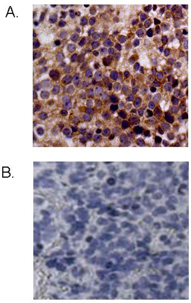
A. Antibody to p125FAK was utilized to detect FAK staining in 70 human neuroblastoma specimens. This photomicrograph (40×) shows a MYCN amplified INSS stage 4 neuroblastoma specimen with significant staining for p125FAK. B. This photomicrograph (40×) shows a MYCN non-amplified INSS stage 4 neuroblastoma specimen with no significant p125FAK staining.
Investigations into the mechanisms of FAK regulation in neuroblastoma have been ongoing. Recently, the FAK promoter has been cloned (42) and evaluations have demonstrated a number of binding sites on this promoter for various oncogenes, such as p53. In addition, studies have revealed E-box sequences on the FAK promoter that are potential binding sites for MYCN (Fig. 2, panel A). Further studies including electrophoretic mobility shift, chromatin immunoprecipitation (ChIP), and dual luciferase assays have shown that MYCN does bind to the FAK promoter in vitro and in vivo, resulting in an upregulation of FAK expression (43) (Fig. 2, panel B). Understanding of the effects of MYCN upon the FAK promoter in neuroblastoma impacts our understanding of the role of FAK in tumorigenesis in other tumor types. MYCN is reported to be amplified in human melanoma and sarcomas (44) and is associated with poor outcomes in these tumor types (45). FAK has also been shown to be overexpressed in human sarcoma and melanoma tumors (46, 47), and that downregulation of FAK results in decreased survival in human melanoma cells (48). These data provide evidence that MYCN is potentially a transcriptional regulator of FAK in these tumor types as well, but as of yet the role of FAK here has not been fully investigated.
Figure 2. MYCN regulates the FAK promoter.
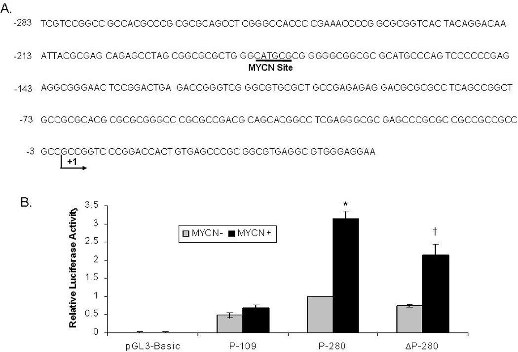
A. Nucleotide sequence (GenBank Accession Number AY323812) of the 5’-flanking region of the FAK gene. The major transcription initiation site is marked as +1. The putative E-box binding site for MYCN is underlined. B. Dual luciferase assays are utilized to detect FAK promoter activity. Graph showing fold change in FAK promoter activity in MYCN− and MYCN+ isogenic neuroblastoma cell lines. There is a significant increase in FAK promoter activity with the P-280 construct in the MYCN+ cell line compared to the MYCN− cells (*P<0.01). When the E-box binding site is mutated (ΔP-280), the FAK promoter activity in the MYCN+ neuroblastoma cell lines significantly decreases compared to the wild type (P-280) promoter construct (†P<0.01).
Studies to determine whether the increased expression of FAK by MYCN amplified neuroblastoma cell lines is biologically relevant have been performed. To more fully determine the biological significance of the effects of MYCN upon FAK, an isogenic MYCN neuroblastoma cell line (SHEP Tet-21/N) (49) was utilized. Treatment of these cell lines with siRNA to FAK resulted in a significant increase in cellular apoptosis in the MYCN overexpressing cells compared to their non-expressing MYCN counterparts (43). Additionally, FAK inhibition using AdFAK-CD, an adenoviral construct that functions as a dominant-negative of FAK, in the same isogenic MYCN neuroblastoma cell lines resulted in decreased cellular attachment and proliferation and increased cellular apoptosis in the neuroblastoma cells that had MYCN overexpression compared to the non-overexpressing cell line (50).
Recent advances have been made in the development of small molecule inhibitors of FAK. For instance, TAE226, a small molecule FAK inhibitor, has been reported to decrease cell survival in human glioma cells (51) and to decrease metastasis and enhance survival in xenograft models of pancreatic and breast cancer (52, 53). FAK inhibition with TAE226 treatment of MYCN amplified neuroblastoma cells [SK-N-BE(2)] results in significantly decreased cell viability at low concentrations with minimal effects upon MYCN non-amplified cells (SK-N-AS) (Fig. 3). Golubovskaya et al have also published their results with another small molecule FAK inhibitor, 1,2,4,5-benzenetetraamine tetrahydrochloride (54). These authors reported that FAK inhibition using this small molecule resulted in decreased breast cancer cell survival both in vitro and in vivo. Other investigators have shown the efficacy of this small molecule in treating pancreatic cancer cell xenografts (55). Use of this small molecule inhibitor in human neuroblastoma has also been reported (56). Neuroblastoma cell lines with MYCN amplification were more sensitive to this small molecule treatment than those that were not MYCN amplified. Treatment with 1,2,4,5-benzenetetraamine tetrahydrochloride resulted in decreased cellular attachment, viability and increased apoptosis in vitro, and decreased neuroblastoma tumor growth in vivo, and these effects were more pronounced in the MYCN amplified neuroblastoma cell lines (56). Of note, 1,2,4,5-benzenetetraamine tetrahydrochloride treatment of normal ganglion cells had no effect upon the viability of these non-cancer cells (Fig. 4).
Figure 3. FAK inhibition leads to decreased cell viability in MYCN amplified neuroblastoma cell lines.
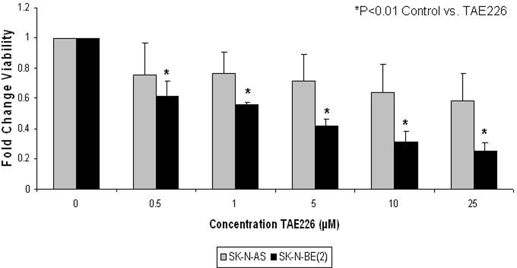
FAK inhibition is achieved with TAE226, a specific FAK kinase inhibitor. Neuroblastoma cells, SK-N-BE(2) (amplified MYCN) and SK-N-AS (non-amplified MYCN) are treated with TAE226 for 24 hours and viability is measured using Alamar Blue Assay. After 24 hours, cellular viability is significantly decreased in the MYCN amplified neuroblastoma cell line, SK-N-BE(2), compared to that in the non-amplified, SK-N-AS, cell line, demonstrating the biologic importance of FAK in MYCN amplified neuroblastoma cells.
Fig. 4. FAK inhibition has minimal effects upon normal ganglion cells.
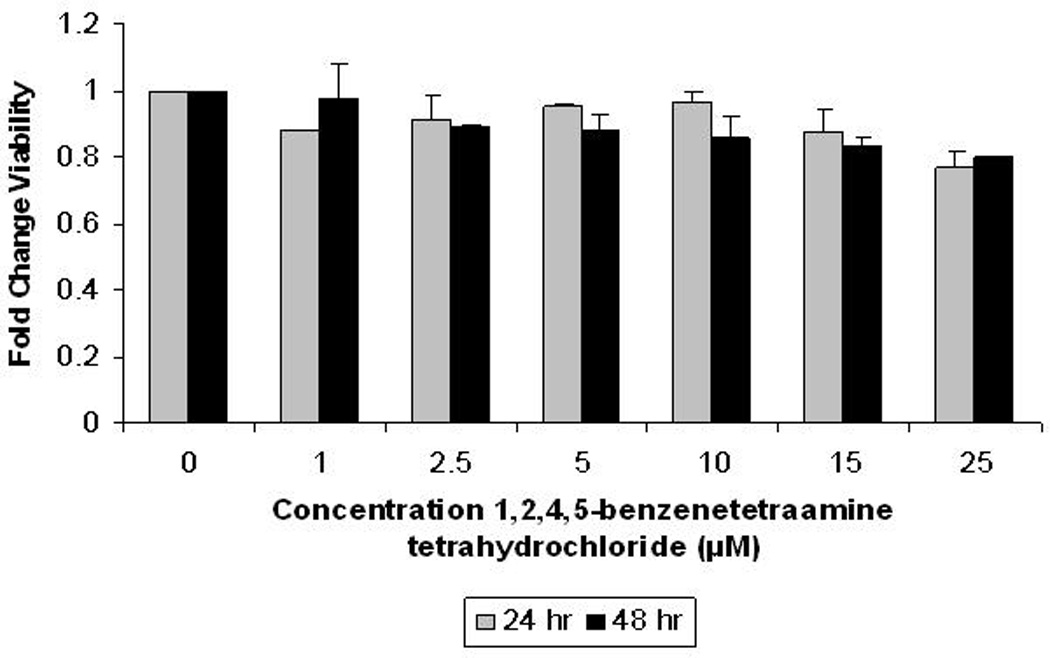
FAK is inhibited with a small molecule inhibitor, 1,2,4,5-benzenetetraamine tetrahydrochloride, for 24 and 48 hours in normal ganglion cells. Viability is measured using Alamar Blue Assay. The cellular viability in these normal cells is essentially unaffected, even after 48 hours of treatment, by FAK inhibition with 1,2,4,5-benzenetetraamine tetrahydrochloride.
Footnotes
This is an, un-copyedited, author manuscript that has been accepted for publication in the Frontiers in Bioscience". Cite this article as appearing in the Journal of Frontiers in Bioscience. Full citation can be found by searching the Frontiers in Bioscience (http://bioscience.org/search/authors/htm/search.htm) following publication and at PubMed (http://www.ncbi.nlm.nih.gov/entrez/query.fcgi?CMD=search&DB=pubmed) following indexing. This article may not be duplicated or reproduced, other than for personal use or within the rule of "Fair Use of Copyrighted Materials" (section 107, Title 17, U.S. Code) without permission of the copyright holder, the Frontiers in Bioscience. From the time of acceptance following peer review, the full final copy edited article of this manuscript will be made available at http://www.bioscience.org/. The Frontiers in Bioscience disclaims any responsibility or liability for errors or omissions in this version of the un-copyedited manuscript or in any version derived from it by the National Institutes of Health or other parties.
References Cited
- 1.Alex R, Sozeri O, Meyer S, Dildrop R. Determination of the DNA sequence recognized by the bHLH-zip domain of the N-myc protein. Nucleic Acids Res. 1992;20:2257–2263. doi: 10.1093/nar/20.9.2257. [DOI] [PMC free article] [PubMed] [Google Scholar]
- 2.Packham G, Cleveland JL. C-myc and apoptosis. Biochim Biophys Acta. 1995;1242:11–28. doi: 10.1016/0304-419x(94)00015-t. [DOI] [PubMed] [Google Scholar]
- 3.Murphy DM, Buckley PG, Bryan K, Das S, Alcock L, Foley NH, Prenter S, Bray I, Watters KM, Higgins D, Stallings RL. Global MYCN transcription factor binding analysis in neuroblastoma reveals association with distinct E-box motifs and regions of DNA hypermethylation. PLoS One. 2009 Dec 4;:e8154. doi: 10.1371/journal.pone.0008154. [DOI] [PMC free article] [PubMed] [Google Scholar]
- 4.Negroni A, Scarpa S, Romeo A, Ferrari S, Modesti A, Raschella G. Decrease of proliferation rate and induction of differentiation by MYCN antisense DNA oligomer in a human neuroblastoma cell line. Cell Growth Diff. 1991;2 511518. [PubMed] [Google Scholar]
- 5.Whitesell L, Rosolen A, Neckers LM. Episome-generated N-myc antisense RNA restricts the differentiation potential of primitive neuroectodermal cell lines. Mol Cell Biol. 1991;11:1360–1371. doi: 10.1128/mcb.11.3.1360-1371.1991. [DOI] [PMC free article] [PubMed] [Google Scholar]
- 6.Hurlin PJ. N-myc functions in transcription and development. Birth Defects Res. 2005;75:340–352. doi: 10.1002/bdrc.20059. [DOI] [PubMed] [Google Scholar]
- 7.Sawai S, Shimono A, Wakamatsu Y, Palmes C, Hanaoka K, Kondoh H. Development. 1993;117:1445–1455. doi: 10.1242/dev.117.4.1445. [DOI] [PubMed] [Google Scholar]
- 8.Stanton LW, Schwab M, Bishop JM. Nucleotide sequence of the human N-myc gene. Proc Natl Acad Sci USA. 1986;83:1772–1776. doi: 10.1073/pnas.83.6.1772. [DOI] [PMC free article] [PubMed] [Google Scholar]
- 9.Adhikary S, Eilers M. Transcriptional regulation and transformation by MYC proteins. Nature. 2005;6:635–645. doi: 10.1038/nrm1703. [DOI] [PubMed] [Google Scholar]
- 10.Thomas DK, Messam CA, Spengler BA, Biedler JL, Ross RA. Nestin is a potential mediator of malignancy in human neuroblastoma cells. J Biol Chem. 2004;279:27994–27999. doi: 10.1074/jbc.M312663200. [DOI] [PubMed] [Google Scholar]
- 11.Harris RG, White E, Phillips ES, Lillycrop KA. The expression of developmentally regulated proto-oncogene Pax-3 is modulated by N-myc. J Biol Chem. 2002;277:34815–34825. doi: 10.1074/jbc.M109609200. [DOI] [PubMed] [Google Scholar]
- 12.Manohar CF, Bray JA, Salwen HR, Madafiglio J, Cheng A, Flemming C, Marshall GM, Norris MD, Haber M, Cohn SL. MYCN-mediated regulation of the MRP1 promoter in human neuroblastoma. Oncogene. 2004;23:753–762. doi: 10.1038/sj.onc.1207151. [DOI] [PubMed] [Google Scholar]
- 13.West AB, Kapatos G, O’Farrell C, Gonzalez-de-Chavez F, Chiu K, Farrer MJ, Maidment NT. N-myc regulates parkin expression. J Biol Chem. 2004;279:28896–28902. doi: 10.1074/jbc.M400126200. [DOI] [PubMed] [Google Scholar]
- 14.Giannini G, Cerignoli F, Mellone M, Massimi I, Ambrosi C, Rinaldi C, Dominici C, Frati L, Screpanti I, Gulino A. High mobility group A1 is a molecular target for MYCN in human neuroblastoma. Cancer Res. 2005;65:8308–8316. doi: 10.1158/0008-5472.CAN-05-0607. [DOI] [PubMed] [Google Scholar]
- 15.Porro A, Haber M, Diolaiti D, Iraci N, Henderson M, Gherardi S, Valli E, Munoz MA, Xue C, Flemming C, Schwab M, Wong JH, Marshall GM, Della Valle G, Norris MD, Perini G. Direct and coordinate regulation of ATP-binding cassette transporter genes by Myc factors generates specific transcription signatures that significantly affect the chemoresistance phenotype of cancer cells. J Biol Chem. 2010;285:19532–19543. doi: 10.1074/jbc.M109.078584. [DOI] [PMC free article] [PubMed] [Google Scholar]
- 16.Dasgupta A, Peirce SK, Findley HW. MycN is a transcriptional regulator of livin in neuroblastoma. Oncol Rep. 2009;22:831–835. doi: 10.3892/or_00000506. [DOI] [PubMed] [Google Scholar]
- 17.Li J, Kretzner L. The growth-inhibitory Ndrg1 gene is a Myc negative target in human neuroblastomas and other cell types with overexpressed N- or c-myc. Molec Cell Biochem. 2003;250:91–105. doi: 10.1023/a:1024918328162. [DOI] [PubMed] [Google Scholar]
- 18.Hatzi E, Murphy C, Zoephel A, Ahorn H, Tontsch U, Bamberger AM, Yamauchi-Takihara K, Schweigerer L, Fotsis T. N-myc oncogene overexpression down-regulates leukemia inhibitory factor in neuroblastoma. Oncogene. 2002;13:803–812. doi: 10.1046/j.1432-1033.2002.03066.x. [DOI] [PubMed] [Google Scholar]
- 19.Oster SK, Mao DYL, Kennedy J, Penn LZ. Functional analysis of the N-terminal domain of the Myc protein. Oncogene. 2003;22:1998–2010. doi: 10.1038/sj.onc.1206228. [DOI] [PubMed] [Google Scholar]
- 20.Brodeur GM, Seeger RC, Schwab M, Varmus HE, Bishop JM. Amplification of N-myc in untreated human neuroblastomas correlates with advanced stage disease. Science. 1984;224:1121–1124. doi: 10.1126/science.6719137. [DOI] [PubMed] [Google Scholar]
- 21.Schweigerer L, Breit S, Wenzel A, Tsunamoto K, Ludwig R, Schwab M. Augmented MYCN expression advances the malignant phenotype of human neuroblastoma cells: evidence for induction of autocrine growth factor activity. Cancer Res. 1990;15:4411–4416. [PubMed] [Google Scholar]
- 22.Gross N, Miescher G, Beck D, Favre S, Beretta C. Altered growth and phenotype in clonal mycN transfectants of the SK-N-SH neuroblastoma cell line. Int J Cancer. 1994;59:141–148. doi: 10.1002/ijc.2910590124. [DOI] [PubMed] [Google Scholar]
- 23.Schmidt ML, Salwen HR, Manohar CF, Ikegaki N, Cohn SL. The biological effects of antisense N-myc expression in human neuroblastoma. Cell Growth Differ. 1994;5:171–178. [PubMed] [Google Scholar]
- 24.Nara K, Kusafuka T, Yoneda A, Oue T, Sangkhathat S, Fukuzawa M. Silencing of MYCN by RNA interference induces growth inhibition, apoptotic activity, and cell differentiation in a neuroblastoma cell line with MYCN amplification. Int J Oncol. 2007;30:1189–1196. [PubMed] [Google Scholar]
- 25.Woo CW, Tan F, Cassano H, Lee JH, Lee KC, Thiele CJ. Use of RNA interference to elucidate the effect of MYCN on cell cycle in neuroblastoma. Pediatr Blood Cancer. 2008;50:208–212. doi: 10.1002/pbc.21195. [DOI] [PubMed] [Google Scholar]
- 26.Kang JH, Rychahou PG, Ishola TA, Qiao J, Evers BM, Chung DH. MYCN silencing induces differentiation and apoptosis in human neuroblastoma cells. Biochem Biophys Res Commun. 2006;351:192–197. doi: 10.1016/j.bbrc.2006.10.020. [DOI] [PMC free article] [PubMed] [Google Scholar]
- 27.Weiss WA, Aldape K, Mohapatra G, Feuerstein BG, Bishop JM. Targeted expression of MYCN causes neuroblastoma in transgenic mice. EMBO J. 1997;16:2985–2995. doi: 10.1093/emboj/16.11.2985. [DOI] [PMC free article] [PubMed] [Google Scholar]
- 28.Moore HC, Wood KM, Jackson MS, Lastowska MA, Hall D, Imrie H, Redfern CP, Lovat PE, Ponthan F, O'Toole K, Lunec J, Tweddle DA. Histological profile of tumours from MYCN transgenic mice. J Clin Pathol. 2008;61:1098–1103. doi: 10.1136/jcp.2007.054627. [DOI] [PubMed] [Google Scholar]
- 29.Zaizen Y, Taniguchi S, Suita S. The role of cellular motility in the invasion of human neuroblastoma cells with or without N-myc amplification and expression. J Pediatr Surg. 1998;33:1765–1770. doi: 10.1016/s0022-3468(98)90281-0. [DOI] [PubMed] [Google Scholar]
- 30.Goodman LA, Liu BC, Thiele CJ, Schmidt ML, Cohn SL, Yamashiro JM, Pai DS, Ikegaki N, Wada RK. Modulation of N-myc expression alters the invasiveness of neuroblastoma. Clin Exp Metastasis. 1997;15:130–139. doi: 10.1023/a:1018448710006. [DOI] [PubMed] [Google Scholar]
- 31.Ma L, Young J, Prabhala H, Pan E, Mestdagh P, Muth D, Teruya-Feldstein J, Reinhardt F, Onder TT, Valastyan S, Westermann F, Speleman F, Vandesompele J, Weinberg RA. miR-9, a MYC/MYCN-activated microRNA, regulates E-cadherin and cancer metastasis. Nat Cell Biol. 2010;12:247–256. doi: 10.1038/ncb2024. [DOI] [PMC free article] [PubMed] [Google Scholar]
- 32.Akeson R, Bernards R. N-myc down regulates neural cell adhesion molecule expression in rat neuroblastoma. Mol Cell Biol. 1990;10:2012–2016. doi: 10.1128/mcb.10.5.2012-2016.1990. [DOI] [PMC free article] [PubMed] [Google Scholar]
- 33.Judware R, Culp LA. Over-expression of transfected N-myc oncogene in human SKNSH neuroblastoma cells down-regulates expression of beta 1 integrin subunit. Oncogene. 1995;11:2599–2607. [PubMed] [Google Scholar]
- 34.Judware R, Culp LA. N-myc over-expression downregulates alpha3beta1 integrin expression in human Saos-2 osteosarcoma cells. Clin Exp Metastasis. 1997;15:228–238. doi: 10.1023/a:1018417330479. [DOI] [PubMed] [Google Scholar]
- 35.Tanaka N, Fukuzawa M. MYCN downregulates integrin alpha1 to promote invasion of human neuroblastoma cells. Int J Oncol. 2008;33:815–821. [PubMed] [Google Scholar]
- 36.Wu L, Bernard-Trifilo JA, Lim Y, Lim ST, Mitra SK, Uryu S, Chen M, Pallen CJ, Cheung NKV, Mikolon D, Mielgo A, Stupack DG, Schlaepfer DD. Distinct FAK-Crc activation events promote α5β1 and α4β1 integrin-stimulated neuroblastoma cell motility. Oncogene. 2008;27:1439–1448. doi: 10.1038/sj.onc.1210770. [DOI] [PMC free article] [PubMed] [Google Scholar]
- 37.Leventhal PS, Shelden EA, Kim B, Feldman EL. Tyrosine phosphorylation of paxillin and focal adhesion kinase during insulin-like growth factor-I-stimulated lamellipodial advance. J Biol Chem. 1997;272:5214–5218. doi: 10.1074/jbc.272.8.5214. [DOI] [PubMed] [Google Scholar]
- 38.Kim B, Feldman EL. Differential regulation of focal adhesion kinase and mitogen-activated protein kinase tyrosine phosphorylation during insulin-like growth factor-I-mediated cytoskeletal reorganization. J Neurochem. 1998;71:1333–1336. doi: 10.1046/j.1471-4159.1998.71031333.x. [DOI] [PubMed] [Google Scholar]
- 39.Kim B, Feldman EL. Insulin-like growth factor I prevents mannitol-induced degradation of focal adhesion kinase and Akt. J Biol Chem. 2002;277:27393–27400. doi: 10.1074/jbc.M201963200. [DOI] [PubMed] [Google Scholar]
- 40.Kim B, van Golen CM, Feldman EL. Degradation and dephosphorylation of focal adhesion kinase during okadaic acid-induced apoptosis in human neuroblastoma cells. Neoplasia. 2003;5:405–416. doi: 10.1016/s1476-5586(03)80043-x. [DOI] [PMC free article] [PubMed] [Google Scholar]
- 41.Beierle EA, Massoll NA, Hartwich J, Kurenova EV, Golubovskaya VM, Cance WG, McGrady P, London WB. Focal adhesion kinase expression in human neuroblastoma: immunohistochemical and real-time PCR analyses. Clin Cancer Res. 2008 doi: 10.1158/1078-0432.CCR-07-1511. in press. [DOI] [PubMed] [Google Scholar]
- 42.Golubovskaya VM, Kaur A, Cance WG. Cloning and characterization of the promoter region of human focal adhesion kinase gene: nuclear factor kappa B and p53 binding sites. Biochim Biophys Acta. 1678;2004:111–125. doi: 10.1016/j.bbaexp.2004.03.002. [DOI] [PubMed] [Google Scholar]
- 33.Beierle EA, Trujillo A, Nagaram A, Kurenova EV, Finch R, Ma X, Vella J, Cance WG. N-myc regulates focal adhesion kinase expression in human neuroblastoma. J Biol Chem. 2007;282:12503–12516. doi: 10.1074/jbc.M701450200. [DOI] [PubMed] [Google Scholar]
- 44.Calalb MB, Polte TR, Hanks SK. Tyrosine phosphorylation of focal adhesion kinase at site in the catalytic domain regulates kinase activity: a role for Src family kinases. Mol Cell Biol. 1995;15:954–963. doi: 10.1128/mcb.15.2.954. [DOI] [PMC free article] [PubMed] [Google Scholar]
- 45.Sonoda Y, Matsumoto Y, Funakoshi M, Yamamoto D, Hanks SK, Kasahara T. Anti-apoptotic role of focal adhesion kinase (FAK). Induction of inhibitor of apoptosis proteins and apoptosis suppression by the overexpression of Fak in a human leukemic cell line, HL60. J Biol Chem. 2000;275:16309–16315. doi: 10.1074/jbc.275.21.16309. [DOI] [PubMed] [Google Scholar]
- 46.Chen HC, Guan JL. Association of focal adhesion kinase with its potential substrate phosphatidylinositol 3-kinase. Proc Natl Acad Sci USA. 1994;91:10148–10152. doi: 10.1073/pnas.91.21.10148. [DOI] [PMC free article] [PubMed] [Google Scholar]
- 47.Hanks SK, Ryzhova L, Shin NY, Brabek J. Focal adhesion kinase signaling activities and their implications in the control of cell survival and motility. Front Biosci. 2003;8:d982–d996. doi: 10.2741/1114. [DOI] [PubMed] [Google Scholar]
- 48.Schlaepfer DD, Mitra SK. Multiple connections link FAK to cell motility and invasion. Curr Opin Genet Dev. 2004;14:92–101. doi: 10.1016/j.gde.2003.12.002. [DOI] [PubMed] [Google Scholar]
- 49.Fulda S, Lutz W, Schwab M, Debatin KM. MycN sensitizes neuroblastoma cells for drug-induced apoptosis. Oncogene. 1999;18:1479–1486. doi: 10.1038/sj.onc.1202435. [DOI] [PubMed] [Google Scholar]
- 50.Beierle EA, Ma X, Trujillo A, Kurenova EV, Cance WG, Golubovskaya VM. Inhibition of focal adhesion kinase and src increases detachment and apoptosis in human neuroblastoma cell lines. Mol Carcinog. 2010;49:224–234. doi: 10.1002/mc.20592. [DOI] [PMC free article] [PubMed] [Google Scholar]
- 51.Shi Q, Hjelmeland AB, Keir ST, Song L, Wickman S, Jackson D, Ohmori O, Bigner DD, Friedman HS, Rich JN. A novel low-molecular weight inhibitor of focal adhesion kinase, TAE226, inhibits glioma growth. Mol Carcinog. 2007 doi: 10.1002/mc.20297. Epub, PMID: 17219439. [DOI] [PubMed] [Google Scholar]
- 52.Hatakeyama S, Tomioka D, Kawahara E, Matsuura N, Masuya K, Miyake T, Umemura I, Kanazawa T, Honda T, Ohmori O. Anti-cancer activity of NVP-TAE226, a potent dual FAK/IGF-IR kinase inhibitor, against pancreatic cancer. J Clin Oncol. 2006;18S:13162. [Google Scholar]
- 53.Kawahara E, Ohmori O, Nonomura K, Murakami Y, Tomioka D, Niwa S, Meyer T, Mestan J, Honda T, Hatakeyama S. NVPTAE226, a potent dual FAK/IGF-IR kinase inhibitor, prevents breast cancer metastasis in vivo. J Clin Oncol. 2006;18S:13163. [Google Scholar]
- 54.Golubovskaya VM, Nyberg C, Zheng M, Kweh F, Magis A, Ostrov D, Cance WG. A small molecule inhibitor, 1,2,4,5-benzenetetraamine tetrahydrochloride, targeting the y397 site of focal adhesion kinase decreases tumor growth. J Med Chem. 2008;51:7405–7416. doi: 10.1021/jm800483v. [DOI] [PMC free article] [PubMed] [Google Scholar]
- 55.Zheng D, Golubovskaya V, Kurenova E, Wood C, Massoll NA, Ostrov D, Cance WG, Hochwald SN. A novel strategy to inhibit FAK and IGF-1R decreases growth of pancreatic cancer xenografts. Mol Carcinog. 2010;49:200–209. doi: 10.1002/mc.20590. [DOI] [PMC free article] [PubMed] [Google Scholar]
- 56.Beierle EA, Ma X, Stewart J, Nyberg C, Trujillo A, Cance WG, Golubovskaya VM. Inhibition of focal adhesion kinase decreases tumor growth in human neuroblastoma. Cell Cycle. 2010;9:1005–1015. doi: 10.4161/cc.9.5.10936. [DOI] [PMC free article] [PubMed] [Google Scholar]