Sir,
Full-thickness macular holes (FTMHs) have been rarely documented in type 2 idiopathic macular telangiectasia (IMT2); and are generally considered poor candidates for surgery because of the underlying neurosensory degeneration.1, 2, 3 I present the evolution and successful management of an FTMH secondary to IMT2.
Case report
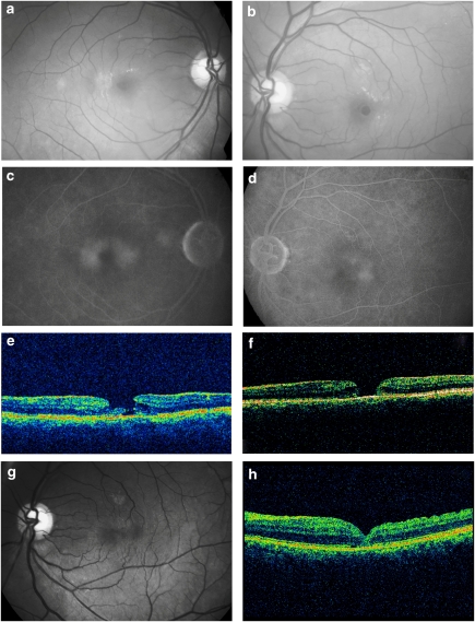
A 60-year-old diabetic and hypertensive man presented for a routine eye check-up. His best-corrected visual acuity (BCVA) was 6/9 OU. On ocular examination, anterior segment was unremarkable OU. Fundus examination revealed a loss of foveal transparency and intra-retinal crystalline deposits typical of IMT2 OU; no diabetic/hypertensive changes were seen (Figure 1a). Fluorescein angiography showed late leakage from the telangiectasia OU (Figures 1c and d). Optical coherence tomography (OCT) revealed foveal atrophy OD (not shown) and a lamellar macular hole OS (Figure 1e). The patient next followed up after 14 months with a drop in vision OS. His BCVA was 6/24 OS; fundus examination revealed an FTMH (448 μ Figure 1a and f). With informed consent of the patient and under guarded visual prognosis, the patient underwent vitrectomy, internal limiting membrane peeling with Brilliant Blue G 0.05% (Ocublue Plus, Aurolab, Madurai, India), and sulphur hexafluoride (20%) tamponade. At 2 months postoperatively, the macular hole had closed; BCVA had improved to 6/18. By the last follow-up at 11 months, BCVA was 6/9 OS (Figures 1g and h).
Figure 1.
(a and b) Fundus examination of both the eyes reveals a loss of foveal transparency and intra-retinal crystals suggestive of idiopathic macular telangiectasia type 2. The left eye also shows a FTMH, which was preceded by an inner lamellar defect. (c and d) Late-phase fluorescein angiogram shows typical parafoveal hyperfluorescence from the telangiectasia in both eyes. (e and f) Horizontal OCT scan of the left eye initially showed an lamellar macular hole, which dehisced into a FTMH after 14 months. (g and h) Fundus view and OCT scan at 11 months after surgery show complete closure of the macular hole without any residual neurosensory degeneration.
Comment
Inner-retinal cystic degeneration and vitreoretinal traction act in tandem to cause a macular hole.4 Olson and Mandava1 reported probably the first FTMH in IMT2, and hypothesised the pathogenesis to be similar to more frequently described lamellar macular hole. The pre-operative course of our case endorses this view as the lamellar hole dehisced into a full-thickness hole. Koizumi et al3 proposed Muller cell loss and dysfunction as the cause for FTMH in IMT2. These holes were not operated. More recently, Charbel Issa et al2 described four eyes with FTMH in three patients with IMT2. They proposed vitreomacular traction as an additional aetiology, and attempted vitrectomy in two cases without anatomical success, though vision improved marginally in one case. To the best of my knowledge, this is the first report to document anatomical closure and functional improvement in a macular hole in IMT2. The pre-operative OCT configuration of the hole which predicted success in this case could be the oedematous edges typical of an idiopathic macular hole, unlike the irregular, moth-eaten edges suggestive of tissue loss in the previous reports. I propose that vitreomacular traction may contribute to macular hole formation in some cases of IMT2; and such macular holes, with configuration similar to idiopathic macular hole, may be good surgical candidates.
The author declares no conflict of interest.
References
- Olson JL, Mandava N. Macular hole formation associated with idiopathic parafoveal telangiectasia. Graefes Arch Clin Exp Ophthalmol. 2006;244:411–412. doi: 10.1007/s00417-005-0057-9. [DOI] [PubMed] [Google Scholar]
- Charbel Issa P, Scholl HP, Gaudric A, Massin P, Kreiger AE, Schwartz S, et al. Macular full-thickness and lamellar holes in association with type 2 idiopathic macular telangiectasia. Eye. 2009;23:435–441. doi: 10.1038/sj.eye.6703003. [DOI] [PubMed] [Google Scholar]
- Koizumi H, Slakter JS, Spaide RF. Full-thickness macular hole formation in idiopathic parafoveal telangiectasis. Retina. 2007;2:473–476. doi: 10.1097/01.iae.0000246678.93495.2f. [DOI] [PubMed] [Google Scholar]
- Smiddy WE, Flynn HW. Pathogenesis of macular holes and therapeutic implications. Am J Ophthalmol. 2004;137:525–537. doi: 10.1016/j.ajo.2003.12.011. [DOI] [PubMed] [Google Scholar]


