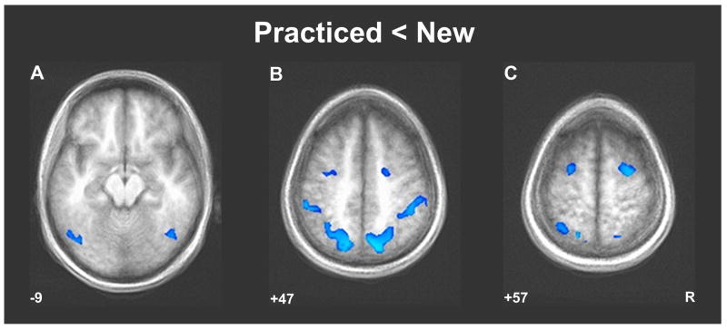
Figure 2.
Decreased activity across a distributed bilateral cortical network during performance of the practiced sequence relative to the transfer conditions. The Practiced-New contrast shows that there was a decrease in neural activity in the extrastriate occipital (A), parietal (B), and premotor (C) cortices while participants were performing the practiced sequence relative to the SO and ST conditions (“New”). Significantly deactivated clusters were those consisting of deactivated voxels (t > 4.5) that passed a minimum cluster volume threshold of V > 327 mm3.

