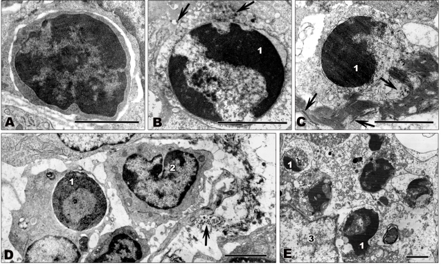
Figure 4.
Transmission electron microscopy of ultrathin sections of lymph node tissue from filovirus-infected African green monkeys infected with ZEBOV (A,C–E) or MARV (B). Tissues were fixed at 4 days p.i. (A) Small lymphocyte showing normal nuclear morphology with large areas of heterochromatin. (B,C) Apoptotic lymphocytes. Typical signs of apoptosis such as chromatin condensation and marginal location of chromatin are visible. (D,E) Apoptotic lymphocytes being engulfed by macrophages. (D) shows initial stages of phagocytosis. The lymphocyte is engulfed by a macrophage. The macrophage shown in (E) contains several destroyed apoptotic lymphocytes. Arrows show filoviral particles. 1—highly condensed heterochromatin; 2—monocyte showing normal nuclear morphology; 3—nucleus of macrophage. Bars correspond to 2 μm.

