Abstract
There is still considerable controversy as to whether or not the inflamed margins of a cuff tear should be excised during surgical suture. We have tried to discover whether anti-inflammatory drugs used before surgical treatment could resolve this issue. Thirty-eight patients were randomly either treated with an anti-inflammatory drug for 2 weeks or not. During the subsequent arthroscopic repair, a few fragments of supraspinatus edge were excised and examined microscopically. No significant differences emerged among samples belonging to the two groups. In all cases, we observed inflammatory infiltrate-lined tear edges. Fibrocytes and newly formed vessels were detected near the margin. Dystrophic calcifications were observed in both groups. Away from the edge, the tendon appeared hypocellular; containing areas with myxoid or fatty degeneration. Our study demonstrates that an anti-inflammatory drug is unable to resolve the inflammatory infiltrate. This failure is probably related to the poor blood supply to the cuff, which, in cases of rupture, is deprived of vessels coming from the humeral periosteum. Further studies are needed to understand how to eliminate the inflammatory process and clarify whether it might inhibit cuff healing and give rise to re-tearing of the sutured cuff.
Résumé
Quelle est la réponse à la controverse: faut-il exciser les tissus inflammatoires dans les lésions de la coiffe des rotateurs? Nous avons vérifié ce problème en invitant les patients à prendre un traitement inflammatoire avant le traitement chirurgical. Trente-huit patients ont été randomisés avec ou sans traitement anti-inflammatoire dans les quinze jours précédant l’intervention. Au cours de la réparation arthroscopique, de petits fragments sous épineux ont été excisés et étudiés microscopiquement sans différence significative entre les deux groupes. Dans tous les cas nous observons une infiltration inflammatoire des berges des lésions. Des fibrocytes et des néo-vaisseaux ont été détectés au niveau des marges, des calcifications ont été également observées dans les deux groupes. A distance des bords le tendon apparaît avec des lésions hypo-cellulaires, une dégénérescence mixoide ou graisseuse. Notre étude démontre que le traitement anti-inflammatoire n’est pas capable de résoudre le problème de l’infiltration inflammatoire, ceci est dû au fait que les lésions cicatricielles sont privées de vaisseaux venant du périoste huméral. Des études ultérieures seront nécessaires pour comprendre comment nous pouvons éliminer ce processus inflammatoire et permettre une meilleure cicatrisation de la coiffe après suture.
Introduction
Many problems relating to the surgical treatment of rotator cuff tears remain unsolved. Although many papers have been published on histological studies of tear edges, it remains unclear whether they should be excised during surgical treatment. Sonnabend et al. [10] observed that the margins of the tears are represented by an inflammatory and, in cases of large tears, by a synovial-like cell edge. They also observed a delamination of the margins that might compromise tendon healing. Therefore, they suggested completely resecting the tear margins. Conversely, Humme and Nelimarkka [7] studied rotator cuff tear edges histologically and did not observe any severe changes capable of hindering the healing process. In fact, in their patients with large or massive tears, a mild inflammatory reaction was observed and delamination was not detected. On the basis of this outcome, excision of the margins was not recommended. Finally, Goodmurphy et al. [4] suggested that only minimal debridement of the tendon edges is required to maximise the healing of the rotator cuff tendon at the time of repair; while Craig [3] and Hawkins [5] reported that the edges of the torn cuff should be trimmed or freshened.
Since the real controversy is whether or not the inflammatory edge should be removed during surgical cuff repair, we invited patients to take an anti-inflammatory drug in 2 weeks before the operation to see if we could resolve the issue.
Materials and methods
From a group of patients with rotator cuff tears treated during 2004, we selected a number of patients presenting suitable characteristics for our research. This group was formed by selecting patients with similar history, clinical features and background. Patients with angina pectoris, peptic ulcers, and cervical nerve root syndrome were excluded from the study.
We considered right-handed patients suffering for no more than 1 year, with an average age of 67 years (ranging from 62 to 70 years), who were not engaged in any manual or heavy work. In all cases, the right shoulder was involved. Thirty-eight patients (22 men and 16 women) represent the study cohort. At the clinical examination, forward flexion, abduction, external and internal rotation were: 131° (range 100–170°); 122° (range 90–160°); 32° (range 20–55° with the arm in the side position), and T9 (range T12–T4) respectively. The mean Constant score was 64.
Magnetic resonance imaging revealed large supero-posterior rotator cuff tears (3–5 cm) and a decrease in the subacromial space.
Each patient in the study group was randomly treated or not with oxaprozin (4,5-diphenyl-2-oxazole propionic acid; 600 mg ;once daily) in the 2 weeks before operation). Those treated with oxaprozin (19 patients: 13 men and 6 women) represent Group I, and the others (19 patients: 9 men and 10 women) Group II. Informed consent was obtained from each patient in Group I. Other analgesic and anti-inflammatory, sedative, hypnotic, tranquilising, and muscle-relaxing agents were prohibited to both groups in the 2 weeks before surgery.
Oxaprozin was chosen because the literature indicates that it is efficient in treating painful shoulders [1, 6] thanks to its elective concentration in the synovial fluid and membrane. Furthermore, the risk of side effects at the gastrointestinal level is considered low [6].
During the arthroscopic surgical treatment, a fragment of the posterior edge of the supraspinatus tendon was excised. Biopsies were fixed in 10% buffered formalin and embedded in paraffin for histological (H&E) and histochemical (colloidal iron, periodic acid Schiff [PAS], Von Kossa) examination. Histological examination was performed by two different observers for all the specimens and the inflammatory infiltrate was evaluated by a semiquantitative score as mild, moderate, and high. We have considered as mild an inflammatory infiltrate with less than 25 lymphocytes for 10 HPF (high power fields); moderate when lymphocytes were 25–50 and high when they were more than 50. Classifications that include other inflammatory cells are used only for chronic intestinal diseases. Data were subjected to statistical analysis using the Chi-squared test. The level of significance was set at p<0.001.
Results
The results are summarized in Table 1.
Table 1.
Microscopic observations of the edges of torn rotator cuffs belonging to the study cohort
| Number | Sex | Age | Drug | Inflammatory infiltrate | Fatty degeneration | Myxoid degeneration | Dystrophic calcification |
|---|---|---|---|---|---|---|---|
| 1 | Man | 62 | Yes | ++ | No | No | No |
| 2 | Woman | 65 | Yes | ++ | No | No | No |
| 3 | Man | 70 | No | + | No | No | Yes |
| 4 | Woman | 66 | No | ++ | Yes | No | No |
| 5 | Man | 66 | Yes | + | No | Yes | No |
| 6 | Woman | 63 | Yes | ++ | No | No | No |
| 7 | Woman | 68 | Yes | ++ | No | No | Yes |
| 8 | Woman | 67 | Yes | ++ | No | Yes | No |
| 9 | Woman | 69 | No | ++ | Yes | No | No |
| 10 | Man | 67 | Yes | ++ | No | No | No |
| 11 | Man | 63 | No | ++ | No | Yes | No |
| 12 | Man | 64 | No | ++ | No | Yes | No |
| 13 | Man | 66 | Yes | ++ | No | No | Yes |
| 14 | Man | 69 | Yes | ++ | No | No | No |
| 15 | Man | 65 | No | ++ | No | No | Yes |
| 16 | Woman | 70 | No | ++ | Yes | Yes | No |
| 17 | Woman | 62 | Yes | ++ | No | No | No |
| 18 | Man | 70 | Yes | ++ | No | No | No |
| 19 | Woman | 66 | No | ++ | Yes | No | No |
| 20 | Woman | 68 | Yes | ++ | No | Yes | No |
| 21 | Man | 67 | Yes | ++ | No | No | Yes |
| 22 | Woman | 70 | No | ++ | Yes | No | No |
| 23 | Man | 70 | Yes | ++ | No | Yes | No |
| 24 | Woman | 62 | No | ++ | Yes | No | No |
| 25 | Woman | 65 | No | ++ | No | No | Yes |
| 26 | Man | 67 | Yes | ++ | No | No | No |
| 27 | Man | 68 | No | ++ | No | No | No |
| 28 | Woman | 69 | No | ++ | No | No | No |
| 29 | Woman | 70 | No | ++ | No | No | No |
| 30 | Man | 62 | No | ++ | No | No | No |
| 31 | Man | 64 | No | ++ | Yes | Yes | No |
| 32 | Man | 65 | No | ++ | No | No | No |
| 33 | Man | 64 | No | ++ | No | Yes | No |
| 34 | Woman | 69 | No | ++ | No | No | Yes |
| 35 | Man | 62 | Yes | ++ | Yes | No | No |
| 36 | Man | 70 | Yes | ++ | No | No | No |
| 37 | Man | 70 | Yes | ++ | No | No | No |
| 38 | Man | 68 | Yes | ++ | Yes | Yes | No |
Inflammatory infiltrate: mild (+) and moderate (++)
The samples belonging to patients treated with oxaprozin did not present histological and histochemical differences compared with the patients who were not pharmacologically treated (p>0.001). Paradoxically, the only two samples with a relatively mild inflammatory infiltrate belonged to patients to whom the drug was not administered.
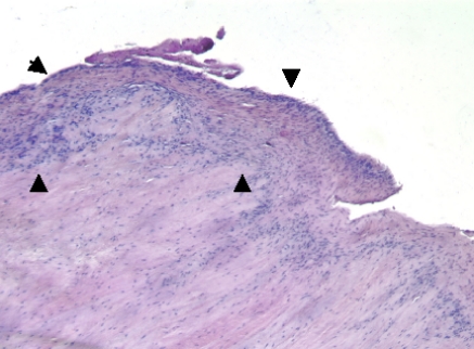
A chronic inflammatory infiltrate, consisting of synovial-like cells, lymphocytes, macrophages, plasmacytes, and young fibrocytes formed a lip bordering the tear margin in all samples (Fig. 1). Fibrocytes and newly formed vessels, bordered by intumescent endothelial cells, were detected not far from the margin tear. Dystrophic calcifications have occasionally been observed in both groups distant from the tear margin, often not far from the fibrocartilaginous areas with regenerating features. Adjacent to the inflammatory infiltrate, the tendon appears hypocellular and disorganized with microfragmentation of the normal collagenous architecture. Areas with myxoid or fatty degeneration were often observed.
Fig. 1.
Hematoxylin and eosin staining of the edges of the torn rotator cuff (×10). Chronic inflammatory infiltrate (arrowheads)
The tear margin and the remaining tissue were found to be inconsistently positive for the colloidal iron and for PAS. In contrast, a more intense staining for colloidal iron was detected in the surrounding regenerating areas.
Discussion
Our study showed no histological or histochemical differences among samples belonging to patients who were or were not pharmacologically treated. In fact, the tear edge excised in both groups was represented by a mild or moderate chronic inflammatory infiltrate. Again, independent of drug intake, we constantly observed newly formed vessels and collagen fibres, dystrophic calcification, fibrocartilaginous areas, and different types of tissue degeneration. The reason for the lack of observable effect from the drug is debatable. It is plausible that the concentration of the drug in the tendon tissue was low because blood only reaches the torn cuff from the subacromial bursa and the musculotendinous junction [2, 9]; therefore, the cuff tear margin, deprived of the vascular supply from the greater tuberosity, would be hypoxic. This would explain the presence of fibrocartilaginous areas observed not far from the inflammatory infiltrate. Although the molecule involved was found in the synovial fluid of patients with rheumatoid arthritis a few hours after the last drug intake [8], there is no experimental evidence that the molecule may penetrate into the cuff tissue through a diffusion mechanism. Furthermore, the supposed low pharmacological concentration cannot be ascribed to a short period (2 weeks) of administration since previous studies have demonstrated a high drug presence in synovial membrane, synovial fluid, and plasma only 5 days after the beginning of oral treatment [8].
Our findings suggest that preoperative oral administration of an anti-inflammatory drug is not able to remove the inflammatory infiltrate along the cuff tear margin; therefore, controversy regarding whether or not to excise the tear edge remains. However, we believe that if there is no delaminating tear, a wide excision of the margin might remove the newly formed vessels too; this might hinder granulation, prevent fibrous scar tissue from forming and consequently tissue healing. Therefore, extensive excision should be avoided. On the other hand, when margins are delaminated and therefore lined with a cellular layer that has an appearance suggestive of synovium, excision is recommended because, as previously reported [10], synovium does not usually adhere to synovium.
In conclusion, further studies are needed to understand how to suppress the inflammatory process and clarify whether it hinders cuff healing and gives rise to re-tearing of the sutured cuff.
References
- 1.Bono RF, Finkel S, Goodman HF, Hanna CB, Rabinowitz SR, Sharon E, Hubsher JA. A multicenter, double blind comparison of oxaprozin, phenylbutazone, and placebo therapy in patients with tendonitis and bursitis. Clin Ther. 1983;6:79–85. [PubMed] [Google Scholar]
- 2.Chansky HA, Iannotti JP. The vascularity of the rotator cuff. Clin Sports Med. 1991;2:204–211. [PubMed] [Google Scholar]
- 3.Craig E. Open anterior acromioplasty for full thickness rotator cuff tears. In: Craig E, editor. The shoulder. New York: Rowan; 1995. pp. 21–27. [Google Scholar]
- 4.Goodmurphy CW, Osborn J, Akesson EJ, Johnson S, Stanescu V, Regan WD. An immunocytochemical analysis of torn rotator cuff tendon taken at the time of repair. J Shoulder Elbow Surg. 2003;12:368–374. doi: 10.1016/S1058-2746(03)00034-X. [DOI] [PubMed] [Google Scholar]
- 5.Hawkins RH. The rotator cuff and biceps tendon. In: Evarts M, editor. Surgery of the musculoskeletal system. New York: Churchill Livingstone; 1983. pp. 128–132. [Google Scholar]
- 6.Heller B, Tarricone R. Oxaprozin versus diclofenac in NSAID-refractory periarthritis pain of the shoulder. Curr Med Res Opin. 2004;20:1279–1290. doi: 10.1185/030079904125004411. [DOI] [PubMed] [Google Scholar]
- 7.Humme, Nelimarkka Histologic changes in a ruptured rotator cuff. Finn J Orthop Trauma. 1993;16:258–259. [Google Scholar]
- 8.Kurowski M, Thabe H. The transsynovial distribution of oxaprozin. Agents Actions. 1989;27:458–460. doi: 10.1007/BF01972852. [DOI] [PubMed] [Google Scholar]
- 9.Lohr JF, Uhthoff HK. The microvascular pattern of the supraspinatus tendon. Clin Orthop. 1990;254:35–38. [PubMed] [Google Scholar]
- 10.Sonnabend DH, Yu Y, Howlett CR, Harper GD, Walsh WR. Laminated tears of the human rotator cuff: a histologic and immunochemical study. Shoulder Elbow Surg. 2001;10:109–115. doi: 10.1067/mse.2001.112882. [DOI] [PubMed] [Google Scholar]


