Abstract
Purpose
The purpose of this study was to assess accurately the three-dimensional movements of the scapula and humerus relative to the thorax during internal/external rotation motion with abduction of the shoulder joint.
Methods
Ten right shoulders of ten healthy volunteers were examined using a wide-gantry open magnetic resonance imaging (MRI) system. MRI was performed every 30° from 90° external rotation to 90° internal rotation of the shoulder joint.
Results
The contribution ratio of the scapulothoracic joint was 12.5% about the long axis of the humerus during internal/external rotation motion. With arm position changes from 90° external rotation to 60° internal rotation, most movement was performed by the glenohumeral joint. Conversely, at internal rotation of ≥60°, the scapula began to markedly tilt in the anterior direction. At 90° internal rotation, the scapula was significantly tilted anteriorly (p < 0.05) when compared with the other positions.
Conclusions
We clarified the existence of a specific scapulohumeral motion pattern, whereby the glenohumeral joint moves with internal rotation and the scapulothoracic joint moves with anterior tilt together with internal rotation motion of the shoulder joint.
Electronic supplementary material
The online version of this article (doi:10.1007/s00264-011-1219-5) contains supplementary material, which is available to authorised users.
Introduction
The shoulder joint has the largest range of motion of any of the major diarthrodial joints in the human body. Motion in the shoulder involves the glenohumeral joint and the scapulothoracic joint. Simultaneous analysis of these movements is important for the identification of variable clinical conditions and the planning of treatment methods. Analysis of shoulder joint motion during internal and external rotation of the shoulder joint, with the humerus in 90° of abduction, has already been performed by McClure et al. [1]. In that study, using bone pins in the scapula, and using skin markers on the humerus and thorax, three-dimensional (3-D) motion of the shoulder joint was analysed. However, large discrepancies are said to exist between skin markers and bones, and a lack of accuracy has been noted in studies using skin markers [2–6]. The insertion of bone pins directly into bones is clearly invasive [1, 7, 8], and this added invasiveness may result in movements that are not necessarily physiological in nature. Furthermore, in studies that utilise both skin markers and bone pins, obtaining accurate information on where the bony landmark being used has been placed on the humerus or the glenoid fossa is impossible. Although the 3-D positions and kinematics of bony landmarks can be analysed relative to each other, the positions and kinematics of the component bones overall cannot be analysed. The purpose of this study was to assess accurately the 3-D movements of the scapula and humerus relative to the thorax during internal/external rotation motion with abduction of the shoulder joint. We performed magnetic resonance imaging (MRI) in multiple limb positions during internal and external rotation of the shoulder joint with the humerus in 90° of abduction. After extracting the 3-D position data, by calculating the rotation angle and movement distance between each position for each bone, we analysed 3-D motion of each joint.
Materials and methods
Subjects
The right shoulders of ten healthy adults (two men and eight women; mean age 27.8 ± 3.5 years; range 22–32 years) were examined. None of the subjects had shoulder pain or a medical history of shoulder joint disorders. All study protocols were approved by the Ethics Committee of our institution, and informed consent was obtained from all subjects prior to enrolment.
Methods
Image acquisition of 3-D MRI
Three-dimensional MRI images were acquired using a wide-gantry open MRI scanner (1.5 T MAGNETOM Espree, Siemens, Erlangen, Germany) with an open-bore system (internal bore 70 cm, length 125 cm). MRI scanning was performed using the 3-D FLASH method [repetition time (TR) 12 ms, echo time (TE) 5.8 ms, flip angle 20°, thickness 0.8 mm, field of view (FOV) 240 × 240 mm2, 452 × 512 matrix]. Each subject was examined in a supine position, wearing a soft coil around the shoulder. Fixation of the arm and body without any restriction of scapular motion was required to minimise motion artefacts when acquiring images. The arm was fixed with Velcro tape using urethane wedges at 30° intervals. A wooden board was made with the area in the region of the scapula removed, to avoid obstructing physiological movements of the scapula. The design allowed free movement of the scapula while preserving a comfortable position for the volunteer. MRI was performed every 30° from 90° external rotation to 90° internal rotation of the shoulder joint at 90° abduction of the shoulder. Scan time was five minutes and 25 seconds for each arm position (Fig. 1).
Fig. 1.
Image acquisition for 3-D MRI. a We created a special device designed to allow free movement of the scapula while maintaining a comfortable posture for the volunteer. b Scanning images at the neutral position
Creation of a surface model by segmentation
The images were transferred to a computer running Virtual Place M® software (AZE, Tokyo, Japan), which was developed at our institution. Segmentation was defined as extracting bone contours and associating each contour with the individual bone. Contours of the humerus, scapula and thoracic cage including the lungs were automatically segmented from MR volume images in the neutral position, and contours were then manually modified including the cortical bones. For each subject, 3-D surface bone models were reconstructed from the segmented area using the marching cubes algorithm [9]. The bone model was visualised using original software based on the Visualization Toolkit (Kitware, Clifton Park, NY, USA).
Image matching (voxel-based registration)
The voxel-based registration technique is a method for superimposing images by minimising the sum of squared intensity differences for segmented voxels [10]. Using this technique, a segmented bone in the neutral position was superimposed on an image of the same bone in another position. Transformation matrices (registration matrices) from the neutral position to other positions were calculated for each bone in the MR scanner coordinate system. Using this method, we can analyse six degrees of freedom in vivo kinematics of the bones respectively at the shoulder joint. The original validation study for this voxel-based registration technique using phantoms revealed that rotational error was 0.43° and translational error was 0.52 mm [11].
Definition of the local coordinate systems (LCSs)
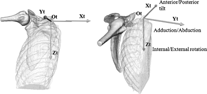
To evaluate the scapula motion about the long axis of the humerus, the LCSs of the thorax at the neutral position were defined according to the definitions in Fig. 2. The LCS matrices were defined in the global coordinate system (GCS), which was the MR scanner coordinate system at the neutral position. The Xg axis was the lateral-medial direction, the Yg axis was the posterior-anterior direction and the Zg axis was the cranial-caudal direction. The LCS of the thorax was defined as having its origin at the apical portion of the lung (Ot). The direction of the axis was the same with the GCS.
Fig. 2.
Definition of the local coordinate system (LCS) of the thorax and rotational description of the scapula. The LCS of the thorax was defined as having its origin at the apical portion of the lung (Ot). To express movement of the scapula around each axis, the Xt axis was used for anterior/posterior tilt, the Yt axis for adduction/abduction and the Zt axis for internal/external rotation
Motion analysis
Using our system, any bone can be selected as a standard and the relative motions of its surrounding bones can be analysed. The relative motions of the scapula and humerus relative to the thorax from the neutral position to other positions were calculated from the registration matrices of each bone, which were obtained by superimposition of MR images from the neutral position to other positions using the voxel-based registration technique. For instance, when analysing the transformation of motion of the scapula relative to the thorax from the neutral position to 30° external rotation, the relative matrix can be calculated as follows:
 |
In this equation Rs is the registration matrix of the scapula created by superimposition of MR images from the neutral position to 30° external rotation, Rt is the registration matrix of the thorax created by superimposition of MR images from the neutral position to 30° external rotation and tRs is the matrix of the scapula relative to the thorax from the neutral position to 30° external rotation. We analysed the movements of the scapula and humerus relative to the thorax using the LCS of the thorax. To express movement of the scapula around each axis, the X t axis was used for anterior/posterior tilt, the Yt axis for adduction/abduction and the Zt axis for internal/external rotation. The decomposition order of the scapula relative to the thorax was Z-Y-X in Euler angles [12]. To express movement of the humerus, the Xt axis was used for rotation. The decomposition order of the humerus relative to the thorax was Z-Y-X in Euler angles [12]. We investigated the following points, which were evaluated during internal and external rotation motion:
The contribution ratio of the scapulothoracic joint in internal/external rotation motion, which was calculated from the mean rotation angles of the scapula and the humerus relative to the thorax respectively around the Xt axis.
The incremental contribution ratio of the scapulothoracic rotation to humerothoracic rotation between each position. This quantity, known as the scapulohumeral rhythm [15], was defined as the ratio of the mean rotation angles of the scapulothoracic joint to the mean humerothoracic rotation angles. In this section, we calculated the amount of change of the scapulothracic rotation around the Xt axis (Fig. 2). Then, for each amount of change between positions, we expressed the incremental contribution ratio of the scapulothoracic rotation to humerothoracic rotation as a percentage. Thereby we measured the scapulohumeral rhythm between the scapula and humerus during internal/external rotation motion.
Movements of the scapula relative to the thorax for (a) anterior/posterior tilt, (b) adduction/abduction and (c) internal/external rotation.
Statistical analysis
For each rotation of the scapula and humerus, one-way repeated measures analysis of variance (ANOVA) and post hoc tests (Bonferroni-Dunn test) were performed to compare mean rotation angles among the arm positions. A difference of p < 0.05 was considered statistically significant. Statistical analysis was conducted using the Statcel2® software package for personal computers (OMS Publishing Inc., Saitama, Japan). Using data from a pilot study, sample size requirements were determined by a power calculation. The sample size for this study was selected to provide adequate power to detect a statistically significant difference.
Results
Contribution ratio of the scapulothoracic joint during internal/external rotation
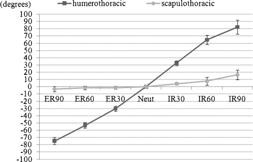
During internal/external rotation at 90° abduction of the humerus, the mean total range of internal/external rotation of the shoulder joint was 157.0°, which included a mean 19.7° rotation of the scapulothoracic joint. The total contribution ratio of the scapulothoracic joint was 12.5% during total rotation motion from 90° internal rotation to 90° external rotation (Fig. 3).
Fig. 3.
The mean rotation angles of the scapula and humerus relative to the thorax around the Xt axis during internal/external rotation. The horizontal axis indicates rotational arm positions and the vertical axis indicates mean rotation angles
Incremental contribution ratio of the scapulothoracic rotation to humerothoracic rotation between each position (scapulohumeral rhythm)
Except for the incremental contribution ratio from 90° internal rotation to 60° internal rotation, the contribution ratios of the scapulothoracic rotations were very low, ranging between 0.8 and 12.8%. The incremental contribution ratio from 90° internal rotation to 60° internal rotation was 50.1% and the highest for the others. Overall, the change in the contribution ratio was not constant and did not increase in a uniform pattern (Fig. 4).
Fig. 4.
The ratio of the mean rotation angles of the scapulothoracic joint relative to the mean rotation angles of the humerothoracic around the Xt axis for each position. There was no constant ratio across the range of positions
Relative movement of the scapula over the thorax
Anterior/posterior tilt
The scapula was slightly tilted anteriorly from 90° external rotation to the neutral position. It increased from −3.1 ± 3.8° at 90° external rotation to 0° at neutral rotation; between these positions the scapula tilted 3.1° anteriorly (Fig. 5a). From the neutral position to 60° internal rotation, the scapula was gradually tilted anteriorly and then rapidly tilted anteriorly from 60° internal rotation to 90° internal rotation. It increased from 0° at the neutral position to 16.5 ± 6.7° at 90° internal rotation; between these positions the scapula tilted 16.5° anteriorly.
Fig. 5.
a The mean rotation angles of anterior/posterior tilt of the scapula. Asterisks indicate a significant difference. There was a significant effect of the rotation position on the anterior-posterior tilt angle of the scapula. At 90° internal rotation, the scapula was significantly tilted anteriorly when compared with the other positions (p < 0.05). At 60° internal rotation, the scapula was significantly tilted anteriorly when compared with 90° external rotation, 60° external rotation, 30° external rotation and the neutral position (p < 0.05). At 30° internal rotation, the scapula was significantly tilted anteriorly when compared with 90° external rotation, 60° external rotation and 30° external rotation (p < 0.05). b The mean rotation angles of adduction/abduction of the scapula. Asterisks indicate a significant difference. At 90° internal rotation, the scapula was significantly adducted when compared with 60° external rotation, 30° external rotation, the neutral position and 60° internal rotation (p < 0.05). c The mean rotation angles of internal/external rotation of the scapula. No significant differences were observed
At 30° internal rotation, the scapula was significantly tilted anteriorly when compared with 90° external rotation, 60° external rotation and 30° external rotation (p < 0.05).
At 60° internal rotation, the scapula was significantly tilted anteriorly when compared with 90° external rotation, 60° external rotation, 30° external rotation and the neutral position (p < 0.05).
At 90° internal rotation the scapula was significantly tilted anteriorly when compared with the other positions (p < 0.05).
Adduction/abduction
At 90° internal rotation, the scapula was significantly adducted compared with 60° external rotation, 30° external rotation, neutral position and 60° internal rotation (p < 0.05). Adduction increased from 0° at the neutral position to 7.2 ± 7.4° at 90° internal rotation; the scapula rotated 7.2° between these positions (Fig. 5b).
Internal/external rotation
For scapular movement relative to the thorax, no consistent trend was seen, and no significant differences were observed. External rotation increased from −1.3 ± 3.9° at 90° external rotation to 1.2 ± 2.1° at 60° internal rotation; the scapula rotated 2.5° between these positions (Fig. 5c).
Discussion
Various methods have previously been used for kinematic analysis of the shoulder joint. However, each of these methods has limitations and disadvantages that have not yet been resolved. Cadaveric experiments possess several limitations in terms of methodology [13, 14]. In addition to skin and soft tissues, outer muscles are excised, as are the clavicle and ribs, but these muscles may exert major influences on the kinematics of the shoulder joint. Little evidence has suggested that physiological arm positions and movements can be analysed in cadaveric studies. Movements of the humerus and scapula are not in the 2-D plane but are instead 3-D; hence, X-ray studies are not feasible [15–18]. Meanwhile, studies using biplane fluoroscopy combined with computed tomography (CT) [19, 20] or MRI [21] images provide extremely accurate analysis, but in the shoulder joint it is impossible to depict the glenohumeral and scapulothoracic joints simultaneously because of limits on the region of interest. Such procedures would not have enabled the simultaneous depiction of the 3-D kinematics of the scapula and humerus relative to the thorax. Fluoroscopy also raises issues regarding radiation exposure. Studies using skin markers are highly susceptible to skin movement artefacts [2–6]. In particular, the scapula moves substantially beneath the skin during internal/external rotation of the arm; thus, calculating the movement of the scapula by means of skin markers placed on the skin is regarded as problematic. Studies using bone pins are clearly invasive and pin insertion may restrict physiological joint motion [1, 7, 8].
To overcome these problems, we have developed our own accurate, noninvasive in vivo joint 3-D kinematic analysis system and have used this to perform various joint analyses [11, 22–25]. This method enables the computation of parameters such as angles of rotation of each bone and transformation distances, by performing MRI in different arm positions and using the voxel-based registration method on the resulting data to superimpose images of the same bone in different arm positions. Use of our method enables detailed, highly accurate 3-D kinematic analysis of the shoulder joint in vivo in a noninvasive manner. In order to carry out kinematic analysis of the scapula and humerus with respect to internal/external rotation motion during arm abduction, it is important to elucidate the kinematics of both the scapula and humerus relative to the thorax, but this requires a wide-gantry MRI to scan the part of the thorax alongside the scapula and humerus. Only with this method does it become possible to accurately evaluate the movements of the scapula and humerus relative to the thorax in different arm positions. Conversely, a disadvantage of our study is that these measurements were taken under sequential static conditions, which may not accurately represent what actually occurs during dynamic activities. We also used a custom-made device to remove as much restraint on the scapula as possible, but the study was performed with the subject in a supine position, and the results of kinematic analysis may therefore differ from those involving load-bearing in the upright position. The results of this study showed that in a position of 90° abduction of the shoulder joint, from 90° external rotation to 60° internal rotation, the scapula changed position slightly to an anterior tilt relative to the thorax. Meanwhile, from 60° internal rotation to 90° internal rotation, however, the position changed drastically to an anterior tilt. At 90° internal rotation, the scapula showed significant adduction to the position at 60° internal rotation, whereas other arm positions showed no significant changes in position observed, toward either adduction or abduction. No significant changes in position in terms of internal/external rotation were observed in any arm position. McClure et al. [1] used bone pins and skin markers in a report on shoulder joint internal/external rotation with the humerus in 90° of abduction. In that study, the scapula moved with anterior tilt, adduction and internal rotation during the movement from 80° external rotation to 50° internal rotation. Our analysis revealed similar results despite the fact that different axes were used, with changes in position in the anterior/posterior tilt and adduction/abduction directions. However, with arm position changes from 50 to 90° of internal rotation, the scapula clearly began to markedly tilt in the anterior direction. In addition, we found that in this position, movement of the glenohumeral joint was minor and barely perceptible. From 90° external rotation to 60° internal rotation of the shoulder joint, laxity of the articular capsule and the glenohumeral ligaments meant that most movement was performed by the glenohumeral joint. Meanwhile, at internal rotation of ≥60°, tension on the articular capsule and the glenohumeral ligaments increased, decreasing the movement of the glenohumeral joint and resulting in increased movement of the scapulothoracic joint. We clarified the existence of a specific scapulohumeral motion pattern, whereby the glenohumeral joint moves with internal rotation and the scapulothoracic joint moves with anterior tilt together with internal rotation motion of the shoulder joint. We believe that an analysis of normal scapular and humeral kinematics in healthy people could provide extremely important information for future research on various shoulder disorders.
Electronic supplementary materials
Below is the link to the electronic supplementary material.
(MPEG 2373 kb)
Acknowledgments
We wish to thank Mr. Y. Sakaguchi, a radiological technologist, for performing MRI at Matsumoto medical clinic and Mr. R. Nakao, a programmer, for developing the software.
References
- 1.McClure PW, Michener LA, Sennett BJ, et al. Direct 3-dimensional measurement of scapular kinematics during dynamic movements in vivo. J Shoulder Elbow Surg. 2001;10(3):269–277. doi: 10.1067/mse.2001.112954. [DOI] [PubMed] [Google Scholar]
- 2.Ludewig PM, Cook TM, Nawoczenski DA. Three-dimensional scapular orientation and muscle activity at selected positions of humeral elevation. J Orthop Sports Phys Ther. 1996;24(2):57–65. doi: 10.2519/jospt.1996.24.2.57. [DOI] [PubMed] [Google Scholar]
- 3.Helm FC, Pronk GM. Three-dimensional recording and description of motions of the shoulder mechanism. J Biomech Eng. 1995;117(1):27–40. doi: 10.1115/1.2792267. [DOI] [PubMed] [Google Scholar]
- 4.McQuade KJ, Hwa Wei S, Smidt GL. Effects of local muscle fatigue on three-dimensional scapulohumeral rhythm. Clin Biomech (Bristol, Avon) 1995;10(3):144–148. doi: 10.1016/0268-0033(95)93704-W. [DOI] [PubMed] [Google Scholar]
- 5.Rettig O, Fradet L, Kasten P, et al. A new kinematic model of the upper extremity based on functional joint parameter determination for shoulder and elbow. Gait Posture. 2009;30(4):469–476. doi: 10.1016/j.gaitpost.2009.07.111. [DOI] [PubMed] [Google Scholar]
- 6.Lovern B, Stroud LA, Evans RO, et al. Dynamic tracking of the scapula using skin-mounted markers. Proc Inst Mech Eng H. 2009;223(7):823–831. doi: 10.1243/09544119JEIM554. [DOI] [PubMed] [Google Scholar]
- 7.Bourne DA, Choo AM, Regan WD, et al. Three-dimensional rotation of the scapula during functional movements: an in vivo study in healthy volunteers. J Shoulder Elbow Surg. 2007;16(2):150–162. doi: 10.1016/j.jse.2006.06.011. [DOI] [PubMed] [Google Scholar]
- 8.Ludewig PM, Phadke V, Braman JP, et al. Motion of the shoulder complex during multiplanar humeral elevation. J Bone Joint Surg Am. 2009;91(2):378–389. doi: 10.2106/JBJS.G.01483. [DOI] [PMC free article] [PubMed] [Google Scholar]
- 9.Lorensen WE, Cline HE. Marching cubes: a high resolution 3D surface construction algorithm. Comput Graph. 1987;21(4):163–170. doi: 10.1145/37402.37422. [DOI] [Google Scholar]
- 10.Hill DL, Batchelor PG, Holden M, et al. Medical image registration. Phys Med Biol. 2001;46:R1–R45. doi: 10.1088/0031-9155/46/3/201. [DOI] [PubMed] [Google Scholar]
- 11.Ishii T, Mukai Y, Hosono N, et al. Kinematics of the upper cervical spine in rotation: in vivo three-dimensional analysis. Spine. 2004;29(7):E139–E144. doi: 10.1097/01.BRS.0000116998.55056.3C. [DOI] [PubMed] [Google Scholar]
- 12.Wu G, Helm FC, Veeger HE, et al. ISB recommendation on definitions of joint coordinate systems of various joints for the reporting of human joint motion—Part II: shoulder, elbow, wrist and hand. J Biomech. 2005;38(5):981–992. doi: 10.1016/j.jbiomech.2004.05.042. [DOI] [PubMed] [Google Scholar]
- 13.Karduna AR, Williams GR, Williams JL, et al. Kinematics of the glenohumeral joint: influences of muscle forces, ligamentous constraints, and articular geometry. J Orthop Res. 1996;14(6):986–993. doi: 10.1002/jor.1100140620. [DOI] [PubMed] [Google Scholar]
- 14.Huffman GR, Tibone JE, McGarry MH, et al. Path of glenohumeral articulation throughout the rotational range of motion in a thrower’s shoulder model. Am J Sports Med. 2006;34(10):1662–1669. doi: 10.1177/0363546506287740. [DOI] [PubMed] [Google Scholar]
- 15.Inman VT, Saunders JB, Abbott LC. Observations of the function of the shoulder joint. J Bone Joint Surg Am. 1944;26(1):1–30. [Google Scholar]
- 16.Freedman L, Munro RR. Abduction of the arm in the scapular plane: scapular and glenohumeral movements. A roentgenographic study. J Bone Joint Surg Am. 1966;48(8):1503–1510. [PubMed] [Google Scholar]
- 17.Poppen NK, Walker PS. Normal and abnormal motion of the shoulder. J Bone Joint Surg Am. 1976;58(2):195–201. [PubMed] [Google Scholar]
- 18.Paletta GA, Jr, Warner JJ, Warren RF, et al. Shoulder kinematics with two-plane X-ray evaluation in patients with anterior instability or rotator cuff tearing. J Shoulder Elbow Surg. 1997;6(6):516–527. doi: 10.1016/S1058-2746(97)90084-7. [DOI] [PubMed] [Google Scholar]
- 19.Bey MJ, Kline SK, Zauel R, et al. Measuring dynamic in-vivo glenohumeral joint kinematics: technique and preliminary results. J Biomech. 2008;41(3):711–714. doi: 10.1016/j.jbiomech.2007.09.029. [DOI] [PMC free article] [PubMed] [Google Scholar]
- 20.Nishinaka N, Tsutsui H, Mihara K, et al. Determination of in vivo glenohumeral translation using fluoroscopy and shape-matching techniques. J Shoulder Elbow Surg. 2008;17(2):319–322. doi: 10.1016/j.jse.2007.05.018. [DOI] [PubMed] [Google Scholar]
- 21.Boyer PJ, Massimini DF, Gill TJ, et al. In vivo articular cartilage contact at the glenohumeral joint: preliminary report. J Orthop Sci. 2008;13(4):359–365. doi: 10.1007/s00776-008-1237-3. [DOI] [PubMed] [Google Scholar]
- 22.Goto A, Moritomo H, Murase T, et al. In vivo elbow biomechanical analysis during flexion: three-dimensional motion analysis using magnetic resonance imaging. J Shoulder Elbow Surg. 2004;13(4):441–447. doi: 10.1016/j.jse.2004.01.022. [DOI] [PubMed] [Google Scholar]
- 23.Moritomo H, Murase T, Goto A, et al. In vivo three-dimensional kinematics of the midcarpal joint of the wrist. J Bone Joint Surg Am. 2006;88(3):611–621. doi: 10.2106/JBJS.D.02885. [DOI] [PubMed] [Google Scholar]
- 24.Sahara W, Sugamoto K, Murai M, et al. 3D kinematic analysis of the acromioclavicular joint during arm abduction using vertically open MRI. J Orthop Res. 2006;24(9):1823–1831. doi: 10.1002/jor.20208. [DOI] [PubMed] [Google Scholar]
- 25.Sahara W, Sugamoto K, Murai M, et al. Three-dimensional clavicular and acromioclavicular rotations during arm abduction using vertically open MRI. J Orthop Res. 2007;25(9):1243–1249. doi: 10.1002/jor.20407. [DOI] [PubMed] [Google Scholar]
Associated Data
This section collects any data citations, data availability statements, or supplementary materials included in this article.
Supplementary Materials
Below is the link to the electronic supplementary material.
(MPEG 2373 kb)


