Abstract
The osteonecrosis of the femoral head implies significant disability partly due to pain. After conventional core decompression using a 10-mm drill, patients normally are requested to be non-weight bearing for several weeks due to the risk of fracture. After core decompression using multiple small drillings, patients were allowed 50% weight bearing. The alternative of simultaneous implantation of a tantalum implant has the supposed advantage of unrestricted load bearing postoperatively. However, these recommendations are mainly based on clinical experience. The aim of this study was to perform a finite element analysis and confirm the results by clinical data after core decompression and after treatment using a tantalum implant. Postoperatively, the risk of fracture is lower after core decompression using multiple small drillings and after the implantation of a tantalum rod according to finite element analysis compared to core decompression of one 10-mm drill hole. According to the results of this study, a risk of fracture exists only during extreme loading. The long-term results reveal a superior performance for core decompression presumably due to the lack of complete bone ingrowth of the tantalum implant. In conclusion, core decompression using small drill holes seems to be superior compared to the tantalum implant and to conventional core decompression.
Introduction
Core decompression (CD) represents an established technique for treatment of early stages of osteonecrosis of the femoral head (ONFH). This therapeutic principle was developed by Ficat and Arlet in 1962 during diagnostic “functional exploration of bone” [6]. CD is supposed to reduce the intraosseous pressure and accomplish reperfusion [21]. However, other studies have not confirmed the initially promising results. Thus, alternative methods have been developed to prevent destruction of the joint. Modifications such as additional implantation of bone-marrow cells, growth factors, fibular grafting or implantation of a tantalum implant are described [4, 8, 10, 13–15, 17, 19, 21, 23, 24]. However, these additional procedures are thought to be associated with an increased donor-side morbidity [21]. Isolated CD on the other hand is associated with a lack of structural support. This lack after CD using a 10-mm drill leads to the recommendation of non-weight bearing postoperatively for several weeks, restricting patients’ daily activities for several weeks. The treatment of CD combined with the implantation of a tantalum osteonecrosis intervention implant (TI) in addition to CD by multiple 3.2-mm drill holes has been recommended to reduce the risk of fracture after surgery. For both techniques the advantages of conventional CD were ensured. However, the postoperative recommendations are mainly based on clinical experience.
Thus, this study performed a finite element analysis (FEM) of CD using a 10-mm drill and CD via multiple small 3.2-mm drill holes as well as CD combined with the insertion of a TI. Thereby, the postoperative risk of fracture and the long-term behaviour of bone ingrowth after the different techniques was simulated by FEM. Clinical experience and MRI were used to confirm the FEM data.
Methods
Finite element analysis
Geometric modelling
A computer aided design (CAD) model of a left human femur was generated from computed tomography data. Segmentation and shape reconstruction techniques were used. A necrotic area of 90° in antero-posterior and lateral views within the stress-bearing area of the femoral head were added according to Kerboul et al. [11]. For the simulation of the TI, necessary dimensions were gained from the manufacturer (Zimmer Germany GmbH, Freiburg, Germany). A length of 95 mm with a 10 mm diameter and 14 mm threads was chosen. The entrance point was located in the subtrochanteric lateral cortex of the femur. The apex of the implant was placed 5 mm within the necrotic area. This situation without the implant represents the situation for CD using a 10-mm drill. For CD via multiple drill holes a model was created according to Mont et al. [14]. This technique was simulated with five drill holes of 3.2 mm entering the lateral femur at the same point as the tantalum implant.
Finite element model
Based on the CAD models, FEM models were constructed using linear tetrahedrons. The models consist of 43,500 elements with 8,637 nodes (TI model) and 82,666 elements with 14,418 nodes (CD model). All models were meshed with linear tetrahedrons.
Loads and boundary conditions
This model was fixed at its distal end, which represents equivalent physiological conditions. A so-called static-equivalent load case was chosen. This long-term load collective was obtained by an inverse simulation approach, proposed and executed previously [7, 12]. This case comprised nine major muscle forces and the joint force of the femur.
A macroscopic continuum approach with a smeared bone mass density was applied for describing the bone material. The strain energy density for the linear elastic case represented the stimulus for bone remodelling.
Computational study
Several situations were simulated. The preoperative state was defined by physiological bone mass density. For simulation of the postoperative state after insertion of the TI, the region of the final implant was clarified by the characteristics of tantalum. For the CD models the bone mass for these drill regions was set close to zero. An initial stage of ONFH was assumed so that for simulation no changes of bone characteristics in the femoral head were made. These conditions were presumed in order to evaluate the risk of subtrochanteric fractures.
An analysis of postoperative stage, extreme loads during stumbling and peak loads during walking down stairs were simulated. Von Mises stresses were determined in order to compare multidirectional stresses. The von Mises stress is a scalar equivalent stress, which is used to compare the results to the tensile strength of bone [22]. It can be used to predict yielding of materials under any loading condition from results of simple uniaxial tensile tests. Furthermore, the simulation of remodelling and ingrowth behaviour for the long-term state after the different treatments was performed.
Clinical analysis
Tantalum osteonecrosis intervention implant
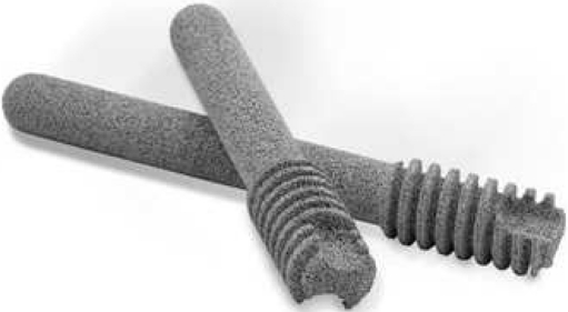
In the Department of Orthopaedic Surgery of the Hanover Medical School between 2003 and 2007, 23 ONFH in 19 patients were treated with CD combined with the implantation of a TI (Zimmer Trabecular Metal Technology, Warsaw, IN, USA) (Fig. 1). This TI has a porous structure with stiffness similar to physiological bone. The implant shows a good biocompatibility. Details were described in previous studies [2]. The diagnosis of ONFH was established by clinical and MRI diagnosis. A follow-up was performed by MRI and clinical assessment including the Harris hip score [8]. Furthermore, the MRIs were analysed to gain information about possible bone ingrowth in the implant and such radiological phenomena as a liquid seam around the implant were determined. The advantage of the TI was supposed to be a structural support allowing full postoperative weight bearing as tolerated. High-impact loading such as jumping was not permitted for 12 months. During clinical follow-up the rate of complications including fractures was assessed.
Fig. 1.
A tantalum osteonecrosis intervention implant (Zimmer Germany GmbH, Freiburg, Germany)
Core decompression via multiple drill holes
In addition, in the same department CD by multiple drill holes using a K-wire (3.2 mm, Aesculap AG, Tuttlingen, Germany) was performed over several years. This type of treatment was replaced temporarily because of the initially encouraging idea of the TI. From 2007 the postoperative procedure included partial load bearing at approximately 50% weight for five to six weeks using crutches according to the recommendation of Mont et al. [14]. In case of bilateral CD, two crutches were used for a four-point gait. After five to six weeks, the patients were advanced to full weight bearing as tolerated. High-impact loading such as jumping was not permitted for 12 months. Since then, 25 patients with 29 ONFH were treated by CD via multiple drill holes followed by the Tantalum Implant procedure.
A clinical follow-up including a questionnaire was performed, which included information about complications such as fractures. MRI follow-ups were conducted in 13 patients in order to determine possible effects of the CD.
Results
FEM analysis
Preoperative simulation
The preoperative simulation determined a von Mises stress of approximately 10 MPa.
Postoperative simulation
After simulation of CD and insertion of a TI, all models revealed an increase of stress at the regions of drill holes and the implant. While the von Mises stress for CD using a 10-mm drill was 22.2 MPa and via multiple small drilling 21.5 MPa, the stress for the insertion of the TI was slightly higher (22.6 MPa).
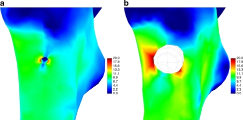
Furthermore, the peak loads while walking down stairs were analysed. The maximum von Mises stresses were 73.5 MPa for the CD via single 10-mm drill hole, 59.7 MPa for the CD model via multiple small drilling and 74.6 MPa for the TI model (Fig. 2a, b).
Fig. 2.
Lateral view on the proximal femur illustrating the von Mises stresses (MPa) after core decompression via multiple drills holes (a) and after insertion of an osteonecrosis intervention implant (b) for postoperative simulation
In case of stumbling, von Mises stresses increased to 88.8 MPa for CD using a single 10-mm drill and to 86.0 MPa using multiple small drillings. The stresses for the TI were slightly greater at 90.4 MPa.
Long-term simulation
The long-term remodelling revealed a completely recovered subtrochanteric region after CD of a single 10-mm drill hole and after multiple small drillings of 3.2 mm to an almost preoperative status. Thus, the von Mises stresses were similar to the preoperative stresses (∼10 MPa). This differed from the results of the TI simulation showing only partial bone ingrowth. The long-term simulation revealed stresses of about 25.1 MPa at the former entrance point of the drill hole. The simulation of walking downstairs in the long-term state investigated for both CD models gave von Mises stresses of about 28 MPa at the drilling entrance rising to about 40 MPa in the ventral direction. The von Mises stresses for the TI were 72.9 and 62.6 MPa in the ventral and the dorsal part of the drilling entrance, respectively.
Clinical findings
Tantalum osteonecrosis intervention implant
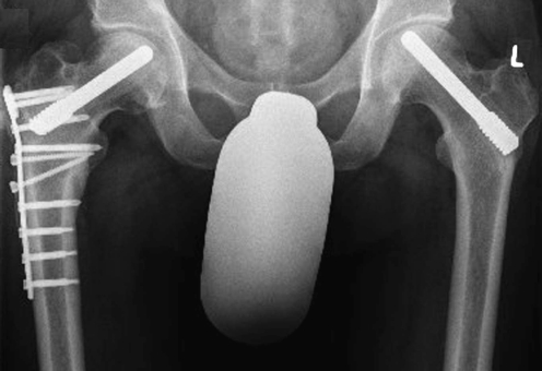
The postoperative treatment of the TI patients allowed full load bearing as tolerated. No fractures occurred apart from one patient who suffered a subtrochanteric fracture due to an adequate trauma (Fig. 3).
Fig. 3.
Postoperative X-ray after osteosynthesis and increasing consolidation of a subtrochanteric fracture after insertion of the tantalum implant
Analysing the MRIs with regard to the ingrowth behaviour, in some cases a slight seam of liquid was visible surrounding the implant (Fig. 4).
Fig. 4.
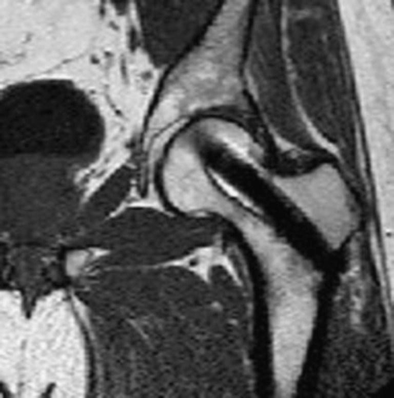
MRI of a patient 25 months after insertion of the tantalum implant revealing a slight liquid seam around the implant
Core decompression via multiple drill holes
After CD via multiple 3.2-mm drill holes patients were only restricted to 50% load bearing for five to six weeks. Afterwards they were allowed unrestricted load bearing. In no case did a subtrochanteric fracture occurred.
In MRI, former drill holes were hardly detectable because of the bony ingrowth (Fig. 5).
Fig. 5.
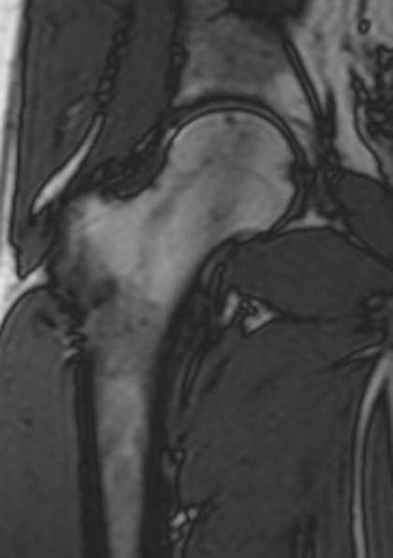
MRI of a patient two years after core decompression using multiple drill holes without residues
Discussion
CD represents an established technique for ONFH in its early stages. Variations of these methods exist. All of them necessitate at least partial weight bearing to allow regeneration of the necrotic area. Nevertheless, depending on the risk of postoperative fracture, differences in the postoperative recommendations exist. The aim of this study was to compare FEM data with clinical experiences and MRI of these different techniques: CD combined with TI and CD using a single 10-mm drill and via multiple small 3.2-mm drill holes. The FEM determined a lower risk of fracture for CD via multiple small drill holes, which may allow earlier partial or full load bearing. Furthermore, the analysis of the assumed long-term bone ingrowth behaviour showed differences between CD and CD combined with the TI. The long-term remodelling revealed a completely recovered subtrochanteric region after CD. The simulation of the implant showed only partial bony ingrowth. Due to difficulties in transfering information from FEM analysis directly to clinical application, the theoretical investigations from the FEM were compared to clinical experience and MRI. This study confirmed the FEM data regarding load bearing and long-term bone ingrowth.
Load bearing
The FEM revealed that CD using multiple 3.2-mm drill holes allowed postoperative unrestricted load bearing. For activities such as walking and climbing stairs, neither for the solitary CD via multiple drill holes nor for the TI do the determined stresses imply a risk of fracture. Only in cases of stumbling, did the stress peaks result in values of the lower range of tensile strength so that a fracture could occur. In the literature, variation within the ultimate stress exists between 78 MPa and 150 MPa [3, 5, 16, 25]. Furthermore, for extreme loads during stumbling, the loads were about four times greater than during normal walking. The FEM simulated stumbling with a corresponding increase to 800% body weight according to Bergmann et al. [1]. This would lead to maximum von Mises stresses of 86.0 MPa for CD via multiple small drill holes, 88.8 MPa for CD using a 10-mm drill and 90.4 MPa for the TI model. Thus, these stresses were on the lower range of what in the literature may be considered as ultimate stress.
The postoperative recommendation after insertion of TI included unrestricted weight bearing as tolerated. Furthermore, the weakening of the femoral neck by CD via multiple drill holes was estimated to be small enough to allow full load bearing. In clinical routine patients were maintained at approximately 50% weight bearing for five to six weeks postoperatively using crutches according to the recommendation of Mont et al. [14]. In case of bilateral CD, two crutches were used for a four-point gait. After five to six weeks, the patients were advanced to full weight bearing as tolerated. High-impact loading such as jogging or jumping was not allowed for 12 months.
In clinical observation of 23 ONFH treated with a TI, one subtrochanteric fracture occurred after an adequate trauma, which required treatment with an osteosynthesis plate. In all other cases after insertion of the TI no fracture was visible although the patients were allowed full-weight bearing after two weeks. Similar experiences exist for CD via multiple drill holes. No fracture in the subtrochanteric region or the femoral neck occurred.
Thus, the FEM can be confirmed by clinical results. It can be concluded that full-load bearing postoperatively is possible for both methods of treatment without relevant increase of the risk of fracture. Nevertheless, to encourage regeneration of the necrotic area partial-weight bearing postoperatively should be recommended for the initial weeks.
Comparing the complications after insertion of the TI with previous literature, Nadeau et al. determined one subtrochanteric fracture four weeks postoperatively within a group of 18 ONFH [15]. A similar case of a subtrochanteric fracture after insertion of the TI was described by Fung et al. [9]. Two other studies analysing the treatment of TI revealed no fracture [17, 21].
The risk of fracture after CD using a 10-mm drill ranges between 0 and 10%. In most studies only rarely inter- or subtrochanteric fracture were described (less than 2.5%) [18–20]. Camp and Cowell [4] mentioned an incidence of intra- and postoperative fracture (4 of 40 ONFH) exceeding previously published reports. However, a study analysing CD using multiple small drill holes revealed no fracture [14], which described the supposed lower comorbidity of this modified procedure.
Thus, summarising the results of this study, the TI and CD via multiple small drill holes is associated with a lower risk of fracture, so that earlier load-bearing is possible compared to CD using a 10-mm drill. Thus, especially in patients with bilateral ONFH, CD via multiple small drills holes should be preferred to CD using a 10-mm drill.
Long-term evaluation
For long-term evaluation the CD, especially via multiple small drill holes, seems to be superior compared to the TI regarding the ultimate stress determined by FEM. The reason for this is the complete replenishment of the drill holes with new bone (Fig. 5). This is based on the defect of the subtrochanteric corticalis which does not regenerate completely in contrast to CD. Furthermore, the ingrowth behaviour of TI is questionable. The FEM does not support the assumed complete bony ingrowth. Thus, increased stresses emerge throughout the treatment. This can be supported by clinical data. In a few cases in the MRI after implantation of the implant a slight seam of fluid could be detected surrounding the implant (Fig. 5). This gives a hint of absence of complete bone ingrowth. Previously, FEM was used to evaluate treatments of ONFH (e.g. intertrochanteric osteotomy, rotational osteotomy or bone grafting). However, up to now no simulation was performed to determine the stresses after CD or after insertion of a TI. Up to now it was uncertain whether FEM analysis can be directly transferred to clinical practice. This study contributes insights because it compares the simulation by FEM with clinical experience and MRI. However, it must be noted that for the FEM analysis only mechanical and no biological considerations were considered. The number of cases treated with CD using multiple small drill holes and the implant procedure is small, because the recommendation of earlier load bearing was indicated by the results of the FEM data.
Conclusion
In conclusion, the FEM can be confirmed by clinical experience and MRI. CD using multiple small drill holes is preferable compared to CD using a 10-mm drill and compared to the insertion of the TI for to two reasons:
For CD via multiple drills holes as well as for the insertion of a TI postoperative load bearing is possible. Thus, this technique seems to have a benefit especially in patients with bilateral ONFH. (Nevertheless, a restriction should be considered to allow regeneration of the ONFH.)
Regarding long-term results CD seems to be superior regarding the risk of fracture due to the probable lack of complete bone ingrowth of the TI.
References
- 1.Bergmann G, Graichen F, Rohlmann A. Hip joint contact forces during stumbling. Langenbecks Arch Surg. 2004;389:53–59. doi: 10.1007/s00423-003-0434-y. [DOI] [PubMed] [Google Scholar]
- 2.Bobyn JD, Poggie RA, Krygier JJ, Lewallen DG, Hanssen AD, Lewis RJ, Unger AS, O’Keefe TJ, Christie MJ, Nasser S, Wood JE, Stulberg SD, Tanzer M. Clinical validation of a structural porous tantalum biomaterial for adult reconstruction. J Bone Joint Surg Am. 2004;86-A(Suppl 2):123–129. doi: 10.2106/00004623-200412002-00017. [DOI] [PubMed] [Google Scholar]
- 3.Burstein AH, Reilly DT, Frankel VH (1973) Failure characteristics of bone and bone tissue. Perspect Biomed Eng 131–134
- 4.Camp JF, Colwell CW., Jr Core decompression of the femoral head for osteonecrosis. J Bone Joint Surg Am. 1986;68:1313–1319. [PubMed] [Google Scholar]
- 5.Dempster WT, Liddicoat RT. Compact bone as a non-isotropic material. Am J Anat. 1952;91:331–362. doi: 10.1002/aja.1000910302. [DOI] [PubMed] [Google Scholar]
- 6.Ficat RP. Idiopathic bone necrosis of the femoral head. Early diagnosis and treatment. J Bone Joint Surg Br. 1985;67:3–9. doi: 10.1302/0301-620X.67B1.3155745. [DOI] [PubMed] [Google Scholar]
- 7.Fischer KJ, Jacobs CR, Carter DR. Computational method for determination of bone and joint loads using bone density distributions. J Biomech. 1995;28:1127–1135. doi: 10.1016/0021-9290(94)00182-4. [DOI] [PubMed] [Google Scholar]
- 8.Floerkemeier T, Thorey F, Daentzer D, Lerch M, Klages P, Windhagen H, Lewinski G. Clinical and radiological outcome of the treatment of osteonecrosis of the femoral head using the osteonecrosis intervention implant. Int Orthop. 2010 doi: 10.1007/s00264-009-0940-9. [DOI] [PMC free article] [PubMed] [Google Scholar]
- 9.Fung DA, Frey S, Menkowitz M, Mark A. Subtrochanteric fracture in a patient with trabecular metal osteonecrosis intervention implant. Orthopedics. 2008;31:614. [PubMed] [Google Scholar]
- 10.Gao YS, Zhang CQ. Cytotherapy of osteonecrosis of the femoral head: a mini review. Int Orthop. 2010;34:779–782. doi: 10.1007/s00264-010-1009-5. [DOI] [PMC free article] [PubMed] [Google Scholar]
- 11.Kerboul M, Thomine J, Postel M, Merle dR. The conservative surgical treatment of idiopathic aseptic necrosis of the femoral head. J Bone Joint Surg Br. 1974;56:291–296. [PubMed] [Google Scholar]
- 12.Lutz A, Nackenhorst U (2008) Computation of static-equivalent load sets for bone remodeling simulation. (GENERIC) Conference Proceeding, Sixth International Congress on Industrial Applied Mathematics (ICIAM07) and GAMM Annual Meeting. ETH Zürich, Switzerland
- 13.Mont MA, Jones LC, Einhorn TA, Hungerford DS, Reddi AH (1998) Osteonecrosis of the femoral head. Potential treatment with growth and differentiation factors. Clin Orthop Relat Res 355S:314–335 [PubMed]
- 14.Mont MA, Ragland PS, Etienne G (2004) Core decompression of the femoral head for osteonecrosis using percutaneous multiple small-diameter drilling. Clin Orthop Relat Res 429:131–138 [DOI] [PubMed]
- 15.Nadeau M, Seguin C, Theodoropoulos JS, Harvey EJ. Short term clinical outcome of a porous tantalum implant for the treatment of advanced osteonecrosis of the femoral head. Mcgill J Med. 2007;10:4–10. [PMC free article] [PubMed] [Google Scholar]
- 16.Sedlin ED, Hirsch C. Factors affecting the determination of the physical properties of femoral cortical bone. Acta Orthop Scand. 1966;37:29–48. doi: 10.3109/17453676608989401. [DOI] [PubMed] [Google Scholar]
- 17.Shuler MS, Rooks MD, Roberson JR. Porous tantalum implant in early osteonecrosis of the hip: preliminary report on operative, survival, and outcomes results. J Arthroplasty. 2007;22:26–31. doi: 10.1016/j.arth.2006.03.007. [DOI] [PubMed] [Google Scholar]
- 18.Smith SW, Fehring TK, Griffin WL, Beaver WB. Core decompression of the osteonecrotic femoral head. J Bone Joint Surg Am. 1995;77:674–680. doi: 10.2106/00004623-199505000-00003. [DOI] [PubMed] [Google Scholar]
- 19.Steinberg ME, Larcom PG, Strafford B, Hosick WB, Corces A, Bands RE, Hartman KE (2001) Core decompression with bone grafting for osteonecrosis of the femoral head. Clin Orthop Relat Res 386:71–78 [DOI] [PubMed]
- 20.Tooke SM, Nugent PJ, Bassett LW, Nottingham P, Mirra J, Jinnah R (1988) Results of core decompression for femoral head osteonecrosis. Clin Orthop Relat Res 228:99–104 [PubMed]
- 21.Veillette CJ, Mehdian H, Schemitsch EH, McKee MD. Survivorship analysis and radiographic outcome following tantalum rod insertion for osteonecrosis of the femoral head. J Bone Joint Surg Am. 2006;88(Suppl 3):48–55. doi: 10.2106/JBJS.F.00538. [DOI] [PubMed] [Google Scholar]
- 22.von Mises R (1913) Mechanik der Festen Korper im plastisch deformablen Zustand. Göttin Nachr Math Phys 1:582–592
- 23.Wang BL, Sun W, Shi ZC, Zhang NF, Yue DB, Guo WS, Shi SH, Li ZR. Treatment of nontraumatic osteonecrosis of the femoral head using bone impaction grafting through a femoral neck window. Int Orthop. 2009;34(5):635–639. doi: 10.1007/s00264-009-0822-1. [DOI] [PMC free article] [PubMed] [Google Scholar]
- 24.Wang CJ, Wang FS, Huang CC, Yang KD, Weng LH, Huang HY. Treatment for osteonecrosis of the femoral head: comparison of extracorporeal shock waves with core decompression and bone-grafting. J Bone Joint Surg Am. 2005;87:2380–2387. doi: 10.2106/JBJS.E.00174. [DOI] [PubMed] [Google Scholar]
- 25.Wirtz DC, Schiffers N, Pandorf T, Radermacher K, Weichert D, Forst R. Critical evaluation of known bone material properties to realize anisotropic FE-simulation of the proximal femur. J Biomech. 2000;33:1325–1330. doi: 10.1016/S0021-9290(00)00069-5. [DOI] [PubMed] [Google Scholar]


