Abstract
A diagnosis of an aberrant right subclavian artery was made in a 3-month-old Boston terrier. Surgical correction was performed after confirming adequate collateral circulation. Reports of surgical correction and evaluation of the perioperative thoracic limb blood pressure are rare in dogs.
Résumé
Correction chirurgicale d’une artère sous-clavière aberrante droite chez un chien. Un diagnostic d’une artère sous-clavière droite aberrante a été posé chez un terrier de Boston âgé de 3 mois. La correction chirurgicale a été réalisée après confirmation d’une circulation collatérale adéquate. Les rapports de correction chirurgicale et d’évaluation périopératoire de la pression artérielle des membres thoraciques sont rares chez les chiens.
(Traduit par Isabelle Vallières)
Vascular ring anomalies are congenital malformations of the great vessels and associated structures that cause constriction of the thoracic esophagus and clinical signs of esophageal obstruction (1,2). Types of vascular rings reported in the dog are persistent right aortic arch with a left ligamentum arteriosum, persistent right aortic arch with an aberrant left subclavian artery, persistent right aortic arch with a combination of a left ligamentum arteriosum and an aberrant left subclavian artery, double aortic arch, persistent right ductus arteriosus, and aberrant right subclavian artery (3). An aberrant right subclavian artery arises distal to the left subclavian or from a bisubclavian trunk instead of arising from the brachiocephalic trunk and crossing beneath the esophagus (4). An aberrant right subclavian artery then passes to the right pectoral limb, dorsal to the esophagus that is constricted on its dorsal aspect (5). Symptomatic aberrant right subclavian arteries typically arise from normal left aortic arches rather than from persistent right aortic arches (4). Some dogs with an aberrant right subclavian artery have postprandial regurgitation resulting in episodes of aspiration pneumonia and in death; however, the anomaly may be an incidental finding not associated with clinical problems (2,5–7).
Reports of surgical correction of aberrant right subclavian arteries are rare (8). This report describes the successful surgical correction of esophageal constriction due to an aberrant right subclavian artery, using intraoperative measurement of pectoral limb blood pressure to ensure adequate perfusion after subclavian artery ligation.
Case description
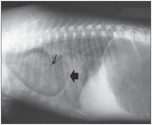
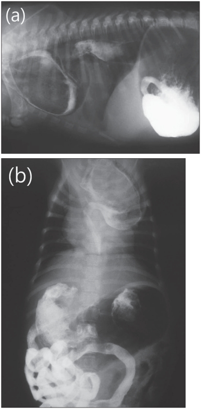
A 3-month-old, sexually intact female Boston terrier weighing 1.2 kg was referred to The University of Missouri-Columbia Veterinary Teaching Hospital for evaluation of regurgitation of undigested food. The owner described onset of regurgitation after eating solid food over the past several weeks. On initial presentation, the dog appeared malnourished. There was no evidence of heart murmurs, coughing, or respiratory distress. Lateral thoracic radiographs revealed soft tissue opacity with mottled air lucency cranial to the heart and the trachea was ventrally displaced within the cranial thorax (Figure 1). Ventrodorsal radiographs revealed widening of the cranial mediastinum. Cardiovascular structures were unremarkable. No definitive radiographic evidence of pneumonia was observed. Positive contrast esophagogram using a barium sulfate liquid (10 mL; E-Z-EM, Lake Success, New York, USA) demonstrated a dilated esophagus within the cranial thorax and esophageal constriction at the base of the heart (Figures 2a, 2b). A tentative diagnosis of vascular ring anomaly was made.
Figure 1.
Radiographic findings in a dog that had been referred because of regurgitation after eating solid food. The wide arrow indicates soft tissue opacity with mottled air lucency cranial to the heart. The narrow arrow indicates the trachea ventrally displaced within the cranial thorax.
Figure 2.
Positive contrast esophagogram. (a), (b) — A dilated esophagus within the cranial thorax is observed cranial to the heart. Esophageal constriction is remarkable at the base of the heart.
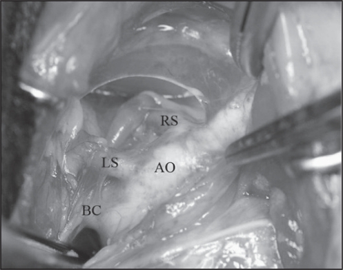
The patient was premedicated for surgery with buprenorphine (Buprenorphine; Bedford Labs, Bedford, Ohio, USA), 0.01 mg/kg body weight (BW), IM, glycopyrrolate (Glycopyrrolate; American Regent, Shirley, New York, USA), 0.01 mg/kg BW, IM, and acepromazine (Promace; Ayerst Laboratories, Rouses Point, New York, USA), 0.05 mg/kg BW, IM, followed by induction of anesthesia with propofol (Diprivan; Astra Zeneca Pharmaceuticals, Wilmington, Delaware, USA), 6 mg/kg BW, IV. The patient was intubated and anesthesia was maintained with isoflurane (Isoflurane; Hospira, Lake Forest, Illinois, USA) and oxygen. Lactated Ringer’s solution was administered intravenously at a rate of 5 mL/kg BW/h until completion of the surgical procedure. The patient received cefazolin (Cefazolin; West Pharmaceutical, Eaton Town, New Jersey, USA), 20 mg/kg BW, IV at the time of induction of anesthesia. Exploratory thoracotomy was performed on the day following admission. The patient was positioned in right lateral recumbency and a left-sided 4th intercostal thoracotomy was performed. Before incising the intercostal muscles, the area around the intercostal nerves was injected with 0.25 mL of 0.25% bupivacaine to provide intra- and postoperative analgesia. The left cranial lung lobe was packed off caudally with a gauze square. Further careful blunt dissection identified 3 vessels coming from the aortic arch (Figure 3). The brachiocephalic artery and left subclavian artery were in normal position with an extraneous artery leaving the aortic arch caudal to the left subclavian artery (Figure 3). The aorta and the esophagus appeared to be in normal relation to each other. The definitive diagnosis of aberrant right subclavian artery was made. The aberrant right subclavian artery was carefully isolated with a right-angle forceps by bluntly dissecting around it. Two circumferential sutures of 2-0 silk (Ethicon, New Jersey, USA) were placed around the aberrant right subclavian artery, first on the aortic side then on the distal side.
Figure 3.
Surgical findings. The brachiocephalic artery (BC) and left subclavian artery (LS) are coming from the aortic arch (AO) with an extraneous artery (RS: right subclavian artery) leaving the aortic arch caudal to the left subclavian artery. Cranial is to the left, and dorsal is to the top of the figure.
To determine if sufficient collateral circulation was provided to the right thoracic limb, the aberrant right subclavian artery was completely occluded using digital pressure with a thumb and index finger. The radial pulse, which had been palpable, could no longer be felt when the right subclavian artery was occluded. To estimate systolic blood pressure, a Doppler flow probe was placed on the palm of each pectoral limb. The occluding cuff was secured proximally to the Doppler flow detector. The measurements of blood pressure were made 3 times and the median values were recorded. The blood pressure before occlusion using digital pressure was 130 mmHg in both thoracic limbs. The blood pressure values after occlusion were 140 mmHg and 120 mmHg in the left and right limbs, respectively, confirming adequate collateral circulation. The 2 circumferential sutures were tightened: the suture closest to the aorta first and then the remaining suture. Two transfixing ligatures were placed between the 2 circumferential sutures with 3-0 poliglecaprone 25 (Monocryl; Ethicon). The aberrant right subclavian artery was transected between the transfixing ligatures. A large esophageal stethoscope was inserted caudally into the esophagus where remaining constricting fibrous bands were located at the caudal extent of the previously constricted site. The fibrous bands were isolated and transected carefully to avoid penetrating the thin-walled esophagus. The large esophageal stethoscope was passed beyond the constricted area and back to ensure adequate dilation.
A thoracostomy tube was placed through the 6th intercostal space and the thorax closed in a routine manner. Air was manually evacuated from the thoracic cavity and the thoracostomy tube was removed. A buprenorphine (0.04 mg/kg BW/day IV; Hospira) constant rate infusion for 18 h was used to control pain. The patient received tramadol (1 mg/kg t.i.d, PO; Pfizer, New York, USA) for 7 d after the surgery.
The dog was fed canned puppy food every 4 h during the day, and was held in an elevated position for 15 min after each feeding. The time between feedings increased over the next 4 wk. Two months after surgery there was no evidence of ventral displacement of the trachea on lateral thoracic radiography. The owner reported that the dog occasionally regurgitated after meals. Patient follow-up by telephone at 6 mo after surgery revealed that the dog was clinically normal, active, and exhibited no evidence of regurgitation.
Discussion
The aberrant right subclavian artery results from abnormal embryological development of the 4th aortic arch developing into the adult aortic arch, descending aorta, brachiocephalic trunk, and right and left subclavian arteries (9,10). Normally, the right subclavian artery arises from the brachiocephalic trunk at the level of the first right intercostal space and is continued by the axillary, internal thoracic, and superficial cervical arteries with blood supply to the right thoracic limb, ventral thorax, and superficial neck, respectively (10). Ligation and transection of the right subclavian artery might result in failure of adequate circulation to these areas (11). The authors recommend measurement of the affected thoracic limb blood pressure before and after ligation of the right subclavian artery using digital pressure to determine if sufficient collateral circulation is provided. In this report, blood pressure measurement using Doppler was performed before and after occluding the aberrant right subclavian artery to confirm sufficient collateral circulation.
Two types of surgical repair for the aberrant right subclavian artery have been described: suture ligature and sectioning of the right subclavian artery in dogs and humans (2,4,11) and surgical revascularization in humans (12,13). In the suture ligature and sectioning of the right subclavian artery, 2 ligatures are used to ligate the vessel, followed by transection between them (2,4,11). The opponents of this method claim that adequacy of collateral vessels around the shoulder in childhood would not be found in adulthood, which would lead to trophic changes in the affected limb. In surgical revascularization, distal anastomosis of the right subclavian artery with the right carotid artery or ascending aorta is performed to provide adequate blood supply to the affected limb (12,13). This technique requires a Dacron graft bypass between the right carotid artery or ascending aorta and the right aberrant subclavian artery, axilloaxillary bypass with a polytetrafluoroethylene graft, or excision of the aberrant artery aneurysm (12,13). This technique is used in humans but has not been used therapeutically in dogs. In this case report, sufficient collateral circulation was confirmed after occlusion of the vessel using digital pressure, and 2 circumferential and 2 transfixing ligatures were used, followed by transection between them. In the veterinary field, the vertebral artery can provide sufficient collateral circulation after ligation of the aberrant right subclavian artery (4). However, surgical revascularization should be considered in cases where adequate collateral circulation is not provided. A study of a large case series is warranted to better determine the adequacy of collateral circulation in the dog. The radial pulse might not be an indicator of sufficient collateral circulation. Herein, the radial pulse was not palpable, but it did not cause ischemic problems. Doppler measurement performed after occlusion of the vessel using digital pressure revealed a sufficient blood flow in the affected limb.
Some surgical techniques may increase the likelihood of a successful surgery. A potential complication of the procedure performed could be slippage of the circumferential sutures placed on an aberrant right subclavian artery resulting in life-threatening hemorrhage. The authors recommend placement of 2 transfixing ligatures between the circumferential sutures to avoid inadvertent slippage of the circumferential sutures.
Radiography is valuable for evaluating obstructive disease and cardiovascular structures as well as indicating the surgical approach. Occasionally on the ventrodorsal view, the normal aortic position might help rule out persistent right aortic arch that is identified on the right side of the esophagus. In this case, it was not possible to identify the aortic position on the ventrodorsal thoracic radiographs due to the dilated esophagus with soft tissue opacity within the mediastinum. Angiography may be valuable in determining the type of vascular ring anomaly for preoperative decision concerning the approach. Anomalies, such as patients with a normal left aortic arch but abnormal right aortic arch, are best approached by using a right thoracotomy rather than the more common left approach. In this case, a left lateral thoracotomy was indicated for the PRAA approach (the most common type of vascular ring anomoly), and was performed as the owner did not consent to angiography.
This report describes the successful surgical correction of an aberrant right subclavian artery and measurement of the thoracic limb blood pressure perioperatively in a dog. Further study is required to explore surgical technique selection, based on age for the treatment of dogs affected by the aberrant right subclavian artery, and measurement of the pectoral limb blood pressure.
Acknowledgments
This work was supported by the National Research Foundation of Korea Grant funded by the Korean Government [NRF-2009-353-E00034]. The authors thank Dr. Dismukes for providing the figures. CVJ
Footnotes
Use of this article is limited to a single copy for personal study. Anyone interested in obtaining reprints should contact the CVMA office (hbroughton@cvma-acmv.org) for additional copies or permission to use this material elsewhere.
References
- 1.Christiansen KJ, Snyder D, Buchanan JW, Holt DE. Multiple vascular anomalies in a regurgitating German shepherd puppy. J Sm Anim Pract. 2007;48:32–35. doi: 10.1111/j.1748-5827.2006.00105.x. [DOI] [PubMed] [Google Scholar]
- 2.Fossum TW. Small Animal Surgery. 3rd ed. St. Louis, Missouri: Mosby; 2007. pp. 405–409. [Google Scholar]
- 3.Ellison GW. Vascular ring anomalies in the dog and cat. Compend Contin Educ Pract Vet. 1980;2:693–706. [Google Scholar]
- 4.Slatter D. Textbook of Small Animal Surgery. 3rd ed. Philadelphia, Pennsylvania: Saunders; 2003. pp. 577–582. [Google Scholar]
- 5.Bezuidenhout AJ. Anomalous origins of the right subclavian and common carotid arteries in the dog. J S Afr Vet Assoc. 1989;60:215–218. [PubMed] [Google Scholar]
- 6.Culau P, de OV, Oliveira JCD, Reckziegel SH, Lindemann T. Ectopic origin of the right subclavian artery and the bicarotid trunk in the dog. Ciencia Rural. 2004;34:1615–1618. [Google Scholar]
- 7.Griffiths D. Three cases of aberrant right subclavian artery in the dog. Acta Vet Scand. 1989;30:355–357. doi: 10.1186/BF03548043. [DOI] [PMC free article] [PubMed] [Google Scholar]
- 8.Park J, Ha D, Lee S, Eom K. A case of aberrant right subclavian artery in a dog. J Vet Clin. 2005;22:281–283. [Google Scholar]
- 9.House AK, Summerfield NJ, German AJ, Noble PJM, Ibarrola P, Brockmann DJ. Unusual vascular ring anomaly associated with a persistent right aortic arch in two dogs. J Sm Anim Pract. 2005;46:585–590. doi: 10.1111/j.1748-5827.2005.tb00291.x. [DOI] [PubMed] [Google Scholar]
- 10.Evans HE. Miller’s Anatomy of the Dog. 3rd ed. Philadelphia, Pennsylvania: WB Saunders; 1993. pp. 602–681. [Google Scholar]
- 11.İğcı A, Kalayci G, Baktiroğlu S, Bozfakıoğlu Y, Ercan E, Műslűmanoğlu M. Dysphagia lusoria. J Thorac Cardiovas Surg. 1993;105:1116–1118. [PubMed] [Google Scholar]
- 12.Gadhinglajkar SV, Sreedhar R, Unnikrishnan M, Varma R. Surgery for dysphagia lusoria caused by right aberrant subclavian artery: Anesthesia perspective. J Cardiothorac Vasc Anesth. 2005;19:86–89. doi: 10.1053/j.jvca.2004.11.017. [DOI] [PubMed] [Google Scholar]
- 13.Kieffer E, Bahnini A, Koskas F. Aberrant subclavian artery: Surgical treatment in thirty-three adult patients. J Vasc Surg. 1994;19:100–109. doi: 10.1016/s0741-5214(94)70125-3. [DOI] [PubMed] [Google Scholar]


