Abstract
The presence of DCs in mycobacterium-containing granulomas, as well as in other granuloma-inducing diseases, is beginning to be appreciated. This review will summarize what is known about DCs with regards to the granuloma and discuss the potential roles DCs may be playing during mycobactieral infection. Potential functions may include mycobacterial dissemination from lesions or sampling of granuloma-containing mycobacterial antigens and migration to the draining lymph nodes to maintain continuous T cell priming. Additionally, the review will discuss the potential outcomes of DC-T cell cross talk within the granuloma and whether it results in boosting the effector functions of newly arrived T cells or anergizing systemic T cells locally. Understanding the DCs complex and changing role during this critical stage may help explain how latency is achieved and maintained. Such knowledge might also lead to improved vaccination strategies.
1. Introduction
The infection of one third of the world’s population with Mycobacterium tuberculosis (Mtb) results in an alarming pandemic. Mutli-drug resistant strains of Mtb and increased susceptibility of HIV infected individuals highlight the evolving threat of the diseases. Importantly, that more than 90% of those infected, amounting to two billion people, are able to adequately control the infection in a latent form [1,2]. The granuloma (also known as a tubercle, which gives rise to the disease name) is the hallmark of mycobacterium infections and is the epicenter for the host immune response and bacterial persistence. Its formation serves the host by containing bacteria, creating a localized immune response, and preventing dissemination; however, the granuloma may also serve the bacteria by ensuring its survival and subsequent disease transmission. As seen in tuberculosis, granulomas are also pathogonomic of several other diseases including sarcoidosis, Chrohn’s disease, Leprosy, syphilis and several autoimmune diseases [1]. Understanding the granuloma microenviornent during latency may not only reveal how Mtb survive for long periods of time, but might also explain why vaccine and therapeutic efforts often fail to result in bacterial clearance.
Initiation of granuloma formation begins with inhalation of a small number of viable bacilli depositing in the alveolar lung space, which are then endocytosed by alveolar macrophage [3]. Infected macrophages begin to aggregate, shown to be partially driven by the RD1-mycobacteria locus in a Mycobacterium marinum zebrafish embryo model, forming early granulomas [4,5]. However, in both murine [6–9] and primate [10] models of Mtb, bacteria are also taken up by dendritic cells (DCs). It is suggested that the mechanism behind DC uptake of Mtb and BCG is mediated by the intercellular adhesion molecule-3 grabbing nonintegrin, DC-SIGN [11]. However, DC cultures without DC-SIGN expression can phagocytose BCG and undergo maturation as well as DCs that express DC-SIGN [12]. This suggests the existence of additional, functionally redundant uptake receptors. In the lung, proinflammatory cytokines, like TNFα, and various chemokines produced by infected macrophage result in the recruitment of additional APCs, monocytes, neutrophils, T cells (NK, CD4+, CD8+, ), B cells and eosinophils [3,13,14]. These cells aggregate around infected macrophage and DCs, ensuring close proximity of their inflammatory cytokines and containment of the pathogen. Despite increasing mycobacterial growth and accumulation of antigen in the lung, protective CD4+ T cells are delayed since they require DC-mediated transport of the mycobacterium to the lung-draining mediastinal lymph nodes [15,16]. In the absence of DCs the CD4+ T cell response is impaired and bacterial load uncontrolled [6,8,17]. The transport of DCs to the draining lymph nodes has been shown to be dependent on IL-12p40 in DCs, which is enhanced by Mtb interaction with Dectin-1 on DCs [18,19]. The production of IFNγ, which coincides with the recruitment of antigen-specific lymphocytes, mediates both mycobacterial killing and down regulation of the inflammatory response within the granuloma.
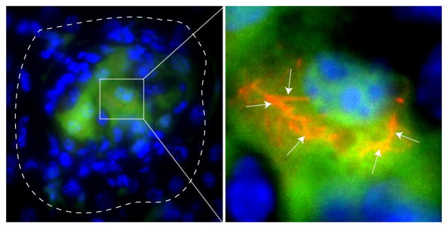
Though recent work has helped uncover DCs role during early mycobacterium infection, little has been done to elucidate their role in chronic granulomas during latent infection. The presence of DCs in both human and murine Mtb and BCG chronic granulomas is somewhat appreciated; however, their exact role during this time is unknown [20–22]. Figure 1 shows CD11c-EYFP cells in chronic granulomatous lesions, with some containing dsRED (Fig. 1). DCs and DC-like cells have also been observed in other granuloma-associated diseases, such as patients or animals with schistosomal egg granulomas, Langerhans cell histiocytosis, Histoplasma capsulatum-induced granulomas, radicular granulomas, intestinal granulomas, those with suppurative granulomas caused by Listeria monocytogenes and Yersinia enterocolitica, and sarcoidosis [23–30]. Ultimately, a better understanding of DC function in chronic lesions may improve vaccine design in a way that would result in sterilizing immunity. DCs are key initiators and manipulators of the immune response and consequently, their role is likely to very throughout infection. This review will summarize and discuss what is known about DCs and multiple functionality during latent mycobacterium infection with a focus on the transition from acute to chronic granuloma.
Figure 1. Dendritic cells in chronic BCG-induced murine granuloma.
Left image, ten-week dsRED BCG-induced liver granuloma in CD11c-EYFP transgenic mouse. CD11c+ cells are green and white-dashed line indicates periphery of granuloma. Image taken at 1000X magnification. Right image, magnified view of white box in left image. White arrows point to dsRED bacilli in CD11c+ YFP cell in the center of the lesion. Image digitally magnified from
2. Dendritic cell migration out of chronic granulomas
Despite progress in our understanding of latent mycobacterial infection, some fundamental questions remain. Does the immune system, both systemically and within the granuloma microenvironment, continuously sample mycobacterial antigen during latency? What is the mechanism of mycobacterial dissemination in latently infected individuals? These questions are likely to be best answered by a thorough investigation of those cells within granulomas that contain either viable mycobacteria and/or bacterial antigen. While macrophages are also professional APCs, DC possess advanced migratory abilities, indispensable for immune surveillance, that uniquely qualifies their candidacy for study [31]. Macrophage, on the other hand, are relatively immobile following mammalian granuloma formation [32], decreasing their chances of facilitating bacterial dissemination or peripheral T cell priming. When considering DC migration either to or from granulomas, it is critical to consider the subset of DCs involved - each subset expresses unique chemokine receptors. The predominating DC subset involved in mycobacterial infections are monocyte-derived DCs, or ‘inflammatory DCs’ [7–9,33]. This lineage differentiates from circulating monocytes (Murine: Ly-6C+, GR-1+, CCR2+, CX3CR1low, L-selectin+ and Human: CD14highCD16−) locally within the inflammatory environment into CD11b+CD11c+ cells [34]. Chemokine receptors in this lineage includes CCR2, CCR5 and CCR6 for recruitment out of the bone marrow into the inflammatory loci [35–37]. CCR2 over-expressing mice have an accelerated adaptive immune response to mycobacteria, highlighting the importance of CD11c+CD11b+ DCs [16]. CCR2 deficient mice are highly susceptible to high doses (2 × 10⁵ CFU i.v.) of Mtb, but resistant to low doses (50–100 CFU aerosol route), which only delayed infection [38,39]. The observation of CCR2−/− mice may be due to compensating effects of CCR5. CCR2 may also compensate for CCR5, as CCR5-deficient mice also showed no significant difference in Mtb control [40]. Furthermore, CCR5−/− mice have more DCs in the lung-draining lymph nodes, suggesting that CCR5 expression helps retain DCs within granulomas [41]. These studies demonstrate that DCs, with the support of multiple chemokine receptors, can migrate to and from granulomas. This movement is well described during acute infection, but poorly understood during chronic infection.
2.1 Dissemination
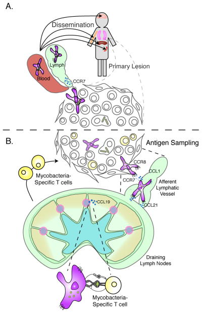
Movement of DCs from granulomas might facilitate dissemination of bacteria, as well as sampling of granuloma-containing mycobacterial antigens and presentation in the draining lymph nodes. Dissemination of Mtb, clinically known as miliary TB, results from lymphatic and then hematogenous dissemination of a few bacilli to the rest of the body [42]. Initially, there is a predilection for dissemination to vascular organs (ie. liver, spleen, bone marrow and brain), and then to additional organs (i.e. skin, aorta, eyes, etc.), resulting in the formation of granulomas within those organs [43–45]. Autopsy reports from individuals with miliary TB have reported dissemination of bacilli into every organ of the body [45]. DCs could act as a vehicle for mycobacterial dissemination during chronic infection since they harbor viable intracellular bacilli and continuously migrate out of the granuloma (Fig. 2A). In comparison to macrophages, DCs are inferior at killing intracellular BCG and Mtb [46–49]. This highlights the possibility that DCs might act as a long-term reservoir for mycobacteria, ensuring their survival in bactericidal environments like the granuloma. However, traffic between mycobacterial phagosomes and the host’s biosynthetic pathway is reduced in DCs, which prohibits mycobacterial access to the necessary nutrients needed for bacterial growth [50,51]. Although infected macrophage were shown to be responsible for dissemination in an early M. marinum infection in zebrafish, in a mammalian model macrophages are likely far less motile [52]. CCR7 and CCR8 expression on monocyte-derived DCs facilitates migration from tissue into the draining lymph nodes [53–55]. In plt mice, which lack CCR7 ligands CCL19 and CCL21ser, 95% less CD11c+CD11b+ DCs are recruited to lung-draining lymph nodes two weeks after Mtb infection in mice [8]. The same lymph nodes contained 95% fewer bacilli, suggesting that CCR7 expression on DCs is responsible for mycobacterial dissemination early in infection. CCR7-deficient mice infected via aerosol with Mtb control infection in the lung, and have decreased bacterial load in the spleen during chronic infection [56]. These studies show the importance of CCR7 and DCs in bacterial dissemination during acute Mtb infection. However, it is still unknown if DCs play a similar role during chronic infection.
Figure 2. Consequences of dendritic cell migration out of chronic granulomas; dissemination and T cell priming.
A, DCs containing viable mycobacteria migrate out of chronic primary lesions. In cases of miliary TB, mycobacterial dissemination first reaches vascular organs liver, spleen bone marrow, and brain. However, dissemination may ultimately reach all organs of the body. B, Dissemination of viable bacilli or mycobacterial antigen from the granuloma to the draining lymph nodes within DCs. During acute infection this process is mediated by chemokine receptor 7, and possible 8 on DCs in response to ligands CCL19 and CCL21, and CCL1, respectively, expressed in the lymphatics and in the lymph nodes. Newly primed mycobacteria-specific T cells would migrate out of the lymph node to chronic lesions to maintain cellular immunity.
2.2 T cell priming
Whether there is continuous antigenic sampling from chronic granulomas and subsequent mycobacterial-specific T cell activation in the draining lymph nodes remains unanswered. Ultimately, answering this question will improve vaccine and therapeutic design (Fig. 2B). To help uncover mechanisms of establishing and maintaining latent infection, Marino and colleagues coupled a nonhuman primate infection model with a mathematical system generated to model human Mtb infection [10]. Theoretically ablating either DC recruitment to the site of infection or DC migration to the draining lymph nodes at 1,000 days post infection eventually resulted in disease reactivation. This suggests that DCs may be necessary in maintaining latent infection by continuous antigenic sampling of the granuloma, followed by migration to the draining lymph nodes. This simulated finding was observed in humans with TB, where mycobacterial antigen was detected in DC-SIGN+ DCs within the lymph nodes [21]. The presence of mycobacterial antigen within DCs in the lymph nodes of humans infected with Mtb is of enormous importance. This suggests that there may be some low-level antigen priming during latency; therefore, the cellular compartment of antigen transport, and the immune response generated demands investigation. Such findings would shed light on both the maintenance of latency and the inability to achieve bacterial clearance.
3. Dendritic cells within chronic granulomas
The granuloma microenvironment has great complexity in architecture and cellular distribution. Different locations within the granuloma represent diverse immunological niches. Theoretically, DCs found in various locations in and around the granuloma may be serving very different purposes. One hypothesis is that chronic granulomas and their local environment can act as a tertiary lymphoid organ. In a murine influenza infection model, T and B cell immune responses were shown to originate from an inducible form of bronchial-associated lymphoid tissue (iBALT) [57]. Mtb induces ectopic lymphoid structures in lung, demonstrated by the formation of B-cell aggregates, homeostatic chemokines CCL19 and CXCL13, follicular DCs, and HEVs [56]. According to these findings, the granuloma and surrounding tissue should be considered an immune-stimulating milieu. This is important because the phenotype and location of DCs in the chronic granuloma could have significant consequences on local T cell immune responses.
3.1 Cross talk with T cells
Egen and colleagues recently demonstrated the motility of T cells in acute granulomas and consequently, their ability to interact with a large number of local cells [32]. This finding warrants a thorough investigation of local APCs, especially DCs during latent infection since they likely shape the response of both resident and newly recruited antigen-specific T cells. These outcomes are hard to predict; however, since the field is dominated by in vitro studies of DC function during early infection, but even these acute studies have conflicting results.
Upon infection with Mtb or BCG in vitro, peripheral monocyte-derived human DCs mature, produce Th1-promoting cytokines, and activate IFNγ-producing T cells [58–61]. Furthermore, Majlessi and colleagues demonstrated that despite DC’s inability to effectively deliver mycobacteria to lysosomes, they could still present antigen by MHC molecules [62]. These findings were recapitulated in vivo by isolating lung DCs shortly after BCG and PPD-Bead challenge in mice and demonstrating their ability to produce IL-12 and stimulate IFNγ Th1 T cells [63,64]. DCs taken from acute granulomatous lungs of rats infected with BCG had increased T cell stimulatory molecules MHCII, CD80, and CD86, as well as the functional ability to stimulate allogeneic T cells [65]. A study by Latchumanan et al. examined the ability of Mtb secretory Ag (MTSA) to differentiate murine bone marrow into DCs. MTSA induced DCs differentiation from bone marrow, high expression of co-stimulatory molecules and nuclear translocation of NF-κB. MTSA also induced secretion of IFNγ over IL-10 in allogeneic T cells [66]. These findings suggest the existence of a possible mechanism of DC generation within the granuloma from recruited precursor cells.
While some studies demonstrate a proinflammatory Th1 IFNγ-producing T cell outcome following DC-mycobacteria interaction, others suggest the opposite. These show that human monocyte-derived DCs infected with Mtb have decreased levels of MHCII and stimulatory molecule CD80− they produced no IL-12, but instead, secrete TNFα and IL-10, and have an impaired ability to activate T cells [67,68]. Similar findings result from infection with BCG [69]. Martino et al. also found that BCG-infected human monocytes produced IL-10 over IL-12, and when cultured with mononuclear cells, they induced IL-4 production, similar to a Th2-like response [70]. Wolf and colleagues showed that myeloid DC population in vivo are responsible for Mtb transport from the lungs to draining lymph nodes in mice, but are poor stimulators of IFNγ from Ag85B-specific CD4+ T cells [8]. It has also been suggested that murine foamy macrophage-like cells in the center of chronic Mtb granulomas have high expression of CD40, MHCII, CD11c, and CD11b− markers typically expressed on DCs [71]. These DC-like cells were characterized as phenotypically mature in early granulomas, and gradually decreased expression of MHCII and CD40 activation markers as disease progressed into chronic phase. These cells also exhibited increased expression of the anti-apoptotic markers TRAF-1, TRAF-2 and TRAF-3. Increased, mycobacterial antigen-induced expression of PD-1 on lymphocytes has been shown to interfere with T cell effector function and shown in patients with Tuberculosis. This highlights the potential inhibitory role of DCs in the granuloma since they express PD-L1 and PD-L2, both ligands for PD-1.
Several mycobacterial products and cytokines in the granuloma have similar effects on DC immunosuppression during Mtb and BCG infection. Unlike MTSA-differentiated DCs, murine bone marrow cultures differentiated into DCs with Mtb 10 kDa Culture Filtrate Protein (CFP-10) resulted in reduced expression of Th1-promoting cytokines when infected with Mtb [72]. A similar effect was observed with Mtb heat-shock protein 70 (TBhsp70) when cultured with murine bone marrow [73]. DCs failed to mature, produced IL-10, and inhibited T cell proliferation. Infection of monocyte-derived human DCs through binding of DC-SIGN by mycobacterial cell wall component ManLAM has been shown to induce IL-10 secretion and interfere with normal TLR-induced activation in DCs [74]. Iyonaga and collegues found that DCs isolated 3 days post infection presented much less antigen than those isolated one day post PPD infection, as determined by T cell proliferation [75]. IL-1β, a proinflammatory cytokine abundant in granulomas, was shown to interfere with human monocyte differentiation into DCs [76]. When infected with M. leprae, human monocyte derived DCs failed to activate antigen-specific T cells in vitro [76–78].
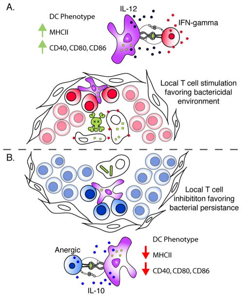
Given the conflicting body of data regarding the immune response generated from DCs in a mycobacterial infection, it is plausible that DCs may both stimulate and suppress the immune response during infection (Fig. 3). This dual function may be the result of a protective mechanism by the host, or an escape mechanism by the bacteria. Regardless, it is certain DCs play an integral part in the overall local immune response within the granuloma during chronic mycobacteria infection.
Figure 3. DC – T cell cross talk within chronic granulomas.
A, DCs with stimulatory phenotype, high expression of MHCII, CD40, CD86, CD80, and secreting IL-12, are likely to support a Th1-IFNγ producing T cell phenotype. This DC phenotype will result in mycobacterial killing within macrophage through the secretion of IFNγ by T cells. B, DCs found within chronic granulomas with low expression of T cell stimulatory molecules and secreting IL-10, will be liable to support an anergic or regulatory T cell response. This would result in less local IFNγ production, and thus, maintain mycobacterial latency inside macrophage.
4. A Complex Response
Considering what is currently known regarding DCs during acute infection and the overall complexity of mycobacterial immunity, it would be logical to hypothesize that DC functionality during the chronic phase might be equally as complex. It is plausible to expect that DC migration in and out of the granuloma can both prime peripheral T cells and help disseminating bacteria, as well as reactivating or down-regulating local T cells within the granuloma. One might expect that the DCs role to very depending on the stage of infection. Recent findings from our group support this complexity. Our data show that DCs within acute granulomas express high costimulatory molecules, and support the reactivation of newly arrived Th1 T cells and priming of naïve T cells to produce IFNγ (Schreiber et al. manuscript in preparation). However, DCs in chronic granulomas have high PD-L expression and do not support the reactivation of newly recruited T cells. We also found PD-L expression on CD11c+ cells in Mtb-induced granulomas in humans, suggesting that the changing DC function observed in mice may also be contributing to latent infection in the human disease. It is especially critical to understand the role DCs play during the chronic phase. Such insight might address key questions about specific immunity against mycobacteria, requirements to maintain latency, and the nature of the immune microenvironment during infection. Additionally, are DCs able to disseminate bacteria or disrupt granulomas integrity in a way that results in active disease? More importantly, an increased knowledge concerning the role DCs play during mycobacterial diseases would ultimately lead to better treatments and vaccines. DC-based vaccines for Mtb have already begun to surface and are proving valuable. The characteristic delay in adaptive immunity observed during Mtb infection is the rationale behind some of the current DC-based vaccines. In theory, observations suggest that a Mtb vaccine should elicit a fast DC turnover in the airway, resulting in an efficient, maximal antigen presentation in the lymph nodes [10]. Vaccine studies have shown that pulsing DCs ex vivo or a bone marrow derived DC culture with either BCG, Mtb, or mycobacterium derivatives, followed by injection back into mice results in potent antigen-specific IFNγ T cell response up to 12 weeks later [79,80]. When challenged with virulent Mtb these DC-based vaccines resulted in greater protection and lower bacterial load compared to control or the current BCG vaccine alone [81,82]. It has also been shown that CD40 stimulation of BCG-infected DCs increased their ability to secrete IL-12 and induce IFNγ-producing CD4+ T cells [83]. A more recent study aimed at generating a DC-based vaccine against Mtb used a GM-CSF secreting strain of BCG [84]. This resulted in an increased number of CD11c+MHCII+ cells in the draining lymph node after vaccination, an increase in anti-mycobacterial IFNγ-secreting T cells and a ~10 fold increase in protection compared to control. A similar study by Triccas et al. engineered a recombinant BCG strain that secrets Flt3L, an Fms-like tyrosine kinase 3 ligand that influences the development of hematopoietic cells, particularly DCs [85]. Vaccination with this strain also resulted in increased stimulation of anti-mycobacterial IFNγ-secreting T cells and was less virulent then conventional BCG in immuno-compromised mice. Collectively, the vaccine efforts mentioned here demonstrate an appreciation of DC function.
The principle behind most tuberculosis vaccine efforts is to generate an increased number of more robust IFNγ-producing CD4+ T cells. Upon recruitment to the granuloma, antigen-specific T cells require a local re-boost to be effective. Based on findings summarized in this review, the functionality of DCs differs depending on the temporal state of infection. In acute lesions, the DC subset, which is highly infected, does not activate T cells well [8,67–69], but other DCs, present in draining lymph nodes and granulomas, have high expression of costimulatory molecules and can present mycobacterial antigen to the recruited T cells, resulting in a high IFNγ antibacterial environment [63–65]. In contrast, chronic granulomas exhibit high inhibitory molecule expression on DCs, which does not support T cell reactivation, likely contributing to latency. The changing phenotype and function of DCs in mycobacterial granulomas is an important feature of the disease course. New approaches that would both induce high systemic Th1 T cell responses, and instruct granuloma DCs to become stimulatory may lead to more efficient control of mycobacteria.
Footnotes
Publisher's Disclaimer: This is a PDF file of an unedited manuscript that has been accepted for publication. As a service to our customers we are providing this early version of the manuscript. The manuscript will undergo copyediting, typesetting, and review of the resulting proof before it is published in its final citable form. Please note that during the production process errors may be discovered which could affect the content, and all legal disclaimers that apply to the journal pertain.
References
- 1.Saunders BM, Britton WJ. Life and death in the granuloma: immunopathology of tuberculosis. Immunol Cell Biol. 2007;85:103–111. doi: 10.1038/sj.icb.7100027. [DOI] [PubMed] [Google Scholar]
- 2.Ulrichs T, Kaufmann SH. New insights into the function of granulomas in human tuberculosis. J Pathol. 2006;208:261–269. doi: 10.1002/path.1906. [DOI] [PubMed] [Google Scholar]
- 3.Russell DG. Who puts the tubercle in tuberculosis? Nat Rev Microbiol. 2007;5:39–47. doi: 10.1038/nrmicro1538. [DOI] [PubMed] [Google Scholar]
- 4.Davis JM, Clay H, Lewis JL, Ghori N, Herbomel P, Ramakrishnan L. Real-time visualization of mycobacterium-macrophage interactions leading to initiation of granuloma formation in zebrafish embryos. Immunity. 2002;17:693–702. doi: 10.1016/s1074-7613(02)00475-2. [DOI] [PubMed] [Google Scholar]
- 5.Volkman HE, Clay H, Beery D, Chang JC, Sherman DR, Ramakrishnan L. Tuberculous granuloma formation is enhanced by a mycobacterium virulence determinant. PLoS Biol. 2004;2:e367. doi: 10.1371/journal.pbio.0020367. [DOI] [PMC free article] [PubMed] [Google Scholar]
- 6.Jiao X, Lo-Man R, Guermonprez P, Fiette L, Deriaud E, Burgaud S, et al. Dendritic cells are host cells for mycobacteria in vivo that trigger innate and acquired immunity. J Immunol. 2002;168:1294–1301. doi: 10.4049/jimmunol.168.3.1294. [DOI] [PubMed] [Google Scholar]
- 7.Humphreys IR, Stewart GR, Turner DJ, Patel J, Karamanou D, Snelgrove RJ, et al. A role for dendritic cells in the dissemination of mycobacterial infection. Microbes Infect. 2006;8:1339–1346. doi: 10.1016/j.micinf.2005.12.023. [DOI] [PubMed] [Google Scholar]
- 8.Wolf AJ, Linas B, Trevejo-Nunez GJ, Kincaid E, Tamura T, Takatsu K, et al. Mycobacterium tuberculosis infects dendritic cells with high frequency and impairs their function in vivo. J Immunol. 2007;179:2509–2519. doi: 10.4049/jimmunol.179.4.2509. [DOI] [PubMed] [Google Scholar]
- 9.Reljic R, Di Sano C, Crawford C, Dieli F, Challacombe S, Ivanyi J. Time course of mycobacterial infection of dendritic cells in the lungs of intranasally infected mice. Tuberculosis (Edinb) 2005;85:81–88. doi: 10.1016/j.tube.2004.09.006. [DOI] [PubMed] [Google Scholar]
- 10.Marino S, Pawar S, Fuller CL, Reinhart TA, Flynn JL, Kirschner DE. Dendritic cell trafficking and antigen presentation in the human immune response to Mycobacterium tuberculosis. J Immunol. 2004;173:494–506. doi: 10.4049/jimmunol.173.1.494. [DOI] [PubMed] [Google Scholar]
- 11.Herrmann JL, Lagrange PH. Dendritic Cells and Mycobacterium tuberculosis: which is the Trojan horse? Pathologie Biologie. 2004;53:35–40. doi: 10.1016/j.patbio.2004.01.004. [DOI] [PubMed] [Google Scholar]
- 12.Gagliardi MC, Teloni R, Giannoni F, Pardini M, Sargentini V, Brunori L, et al. Mycobacterium bovis Bacillus Calmette-Guerin infects DC-SIGN- dendritic cell and causes the inhibition of IL-12 and the enhancement of IL-10 production. J Leukoc Biol. 2005;78:106–113. doi: 10.1189/jlb.0105037. [DOI] [PubMed] [Google Scholar]
- 13.Co DO, Hogan LH, Il-Kim S, Sandor M. T cell contributions to the different phases of granuloma formation. Immunol Lett. 2004;92:135–142. doi: 10.1016/j.imlet.2003.11.023. [DOI] [PubMed] [Google Scholar]
- 14.Algood HM, Lin PL, Flynn JL. Tumor necrosis factor and chemokine interactions in the formation and maintenance of granulomas in tuberculosis. Clin Infect Dis. 2005;41 (Suppl 3):S189–193. doi: 10.1086/429994. [DOI] [PubMed] [Google Scholar]
- 15.Wolf AJ, Desvignes L, Linas B, Banaiee N, Tamura T, Takatsu K, et al. Initiation of the adaptive immune response to Mycobacterium tuberculosis depends on antigen production in the local lymph node, not the lungs. J Exp Med. 2007 doi: 10.1084/jem.20071367. [DOI] [PMC free article] [PubMed] [Google Scholar]
- 16.Schreiber O, Steinwede K, Ding N, Srivastava M, Maus R, Langer F, et al. Mice that overexpress CC chemokine ligand 2 in their lungs show increased protective immunity to infection with Mycobacterium bovis bacille Calmette-Guerin. J Infect Dis. 2008;198:1044–1054. doi: 10.1086/591501. [DOI] [PubMed] [Google Scholar]
- 17.Tian T, Woodworth J, Skold M, Behar SM. In vivo depletion of CD11c+ cells delays the CD4+ T cell response to Mycobacterium tuberculosis and exacerbates the outcome of infection. J Immunol. 2005;175:3268–3272. doi: 10.4049/jimmunol.175.5.3268. [DOI] [PubMed] [Google Scholar]
- 18.Khader SA, Partida-Sanchez S, Bell G, Jelley-Gibbs DM, Swain S, Pearl JE, et al. Interleukin 12p40 is required for dendritic cell migration and T cell priming after Mycobacterium tuberculosis infection. J Exp Med. 2006;203:1805–1815. doi: 10.1084/jem.20052545. [DOI] [PMC free article] [PubMed] [Google Scholar]
- 19.Rothfuchs AG, Bafica A, Feng CG, Egen JG, Williams DL, Brown GD, et al. Dectin-1 interaction with Mycobacterium tuberculosis leads to enhanced IL-12p40 production by splenic dendritic cells. J Immunol. 2007;179:3463–3471. doi: 10.4049/jimmunol.179.6.3463. [DOI] [PubMed] [Google Scholar]
- 20.Uehira K, Amakawa R, Ito T, Tajima K, Naitoh S, Ozaki Y, et al. Dendritic cells are decreased in blood and accumulated in granuloma in tuberculosis. Clin Immunol. 2002;105:296–303. doi: 10.1006/clim.2002.5287. [DOI] [PubMed] [Google Scholar]
- 21.Tailleux L, Schwartz O, Herrmann JL, Pivert E, Jackson M, Amara A, et al. DC-SIGN is the major Mycobacterium tuberculosis receptor on human dendritic cells. J Exp Med. 2003;197:121–127. doi: 10.1084/jem.20021468. [DOI] [PMC free article] [PubMed] [Google Scholar]
- 22.Tsai MC, Chakravarty S, Zhu G, Xu J, Tanaka K, Koch C, et al. Characterization of the tuberculous granuloma in murine and human lungs: cellular composition and relative tissue oxygen tension. Cell Microbiol. 2006;8:218–232. doi: 10.1111/j.1462-5822.2005.00612.x. [DOI] [PubMed] [Google Scholar]
- 23.Rathore A, Sacristan C, Ricklan DE, Flores Villanueva PO, Stadecker MJ. In situ analysis of B7–2 costimulatory, major histocompatibility complex class II, and adhesion molecule expression in schistosomal egg granulomas. Am J Pathol. 1996;149:187–194. [PMC free article] [PubMed] [Google Scholar]
- 24.Hoover KB, Rosenthal DI, Mankin H. Langerhans cell histiocytosis. Skeletal Radiol. 2007;36:95–104. doi: 10.1007/s00256-006-0193-2. [DOI] [PubMed] [Google Scholar]
- 25.Kaneko T, Okiji T, Kaneko R, Nor JE, Suda H. Antigen-presenting cells in human radicular granulomas. J Dent Res. 2008;87:553–557. doi: 10.1177/154405910808700617. [DOI] [PubMed] [Google Scholar]
- 26.Popov A, Abdullah Z, Wickenhauser C, Saric T, Driesen J, Hanisch FG, et al. Indoleamine 2,3-dioxygenase-expressing dendritic cells form suppurative granulomas following Listeria monocytogenes infection. J Clin Invest. 2006;116:3160–3170. doi: 10.1172/JCI28996. [DOI] [PMC free article] [PubMed] [Google Scholar]
- 27.Kojima M, Morita Y, Shimizu K, Yoshida T, Yamada I, Togo T, et al. Immunohistological findings of suppurative granulomas of Yersinia enterocolitica appendicitis: a report of two cases. Pathol Res Pract. 2007;203:115–119. doi: 10.1016/j.prp.2006.10.004. [DOI] [PubMed] [Google Scholar]
- 28.Mizoguchi A, Ogawa A, Takedatsu H, Sugimoto K, Shimomura Y, Shirane K, et al. Dependence of intestinal granuloma formation on unique myeloid DC-like cells. J Clin Invest. 2007;117:605–615. doi: 10.1172/JCI30150. [DOI] [PMC free article] [PubMed] [Google Scholar]
- 29.Heninger E, Hogan LH, Karman J, Macvilay S, Hill B, Woods JP, et al. Characterization of the Histoplasma capsulatum-induced granuloma. J Immunol. 2006;177:3303–3313. doi: 10.4049/jimmunol.177.5.3303. [DOI] [PMC free article] [PubMed] [Google Scholar]
- 30.Ota M, Amakawa R, Uehira K, Ito T, Yagi Y, Oshiro A, et al. Involvement of dendritic cells in sarcoidosis. Thorax. 2004;59:408–413. doi: 10.1136/thx.2003.006049. [DOI] [PMC free article] [PubMed] [Google Scholar]
- 31.Alvarez D, Vollmann EH, von Andrian UH. Mechanisms and consequences of dendritic cell migration. Immunity. 2008;29:325–342. doi: 10.1016/j.immuni.2008.08.006. [DOI] [PMC free article] [PubMed] [Google Scholar]
- 32.Egen JG, Rothfuchs AG, Feng CG, Winter N, Sher A, Germain RN. Macrophage and T cell dynamics during the development and disintegration of mycobacterial granulomas. Immunity. 2008;28:271–284. doi: 10.1016/j.immuni.2007.12.010. [DOI] [PMC free article] [PubMed] [Google Scholar]
- 33.Yoneyama H, Ichida T. Recruitment of dendritic cells to pathological niches in inflamed liver. Med Mol Morphol. 2005;38:136–141. doi: 10.1007/s00795-005-0289-0. [DOI] [PubMed] [Google Scholar]
- 34.Leon B, Lopez-Bravo M, Ardavin C. Monocyte-derived dendritic cells. Semin Immunol. 2005;17:313–318. doi: 10.1016/j.smim.2005.05.013. [DOI] [PubMed] [Google Scholar]
- 35.Serbina NV, Pamer EG. Monocyte emigration from bone marrow during bacterial infection requires signals mediated by chemokine receptor CCR2. Nat Immunol. 2006;7:311–317. doi: 10.1038/ni1309. [DOI] [PubMed] [Google Scholar]
- 36.Tacke F, Randolph GJ. Migratory fate and differentiation of blood monocyte subsets. Immunobiology. 2006;211:609–618. doi: 10.1016/j.imbio.2006.05.025. [DOI] [PubMed] [Google Scholar]
- 37.Leon B, Ardavin C. Monocyte-derived dendritic cells in innate and adaptive immunity. Immunol Cell Biol. 2008;86:320–324. doi: 10.1038/icb.2008.14. [DOI] [PubMed] [Google Scholar]
- 38.Peters W, Scott HM, Chambers HF, Flynn JL, Charo IF, Ernst JD. Chemokine receptor 2 serves an early and essential role in resistance to Mycobacterium tuberculosis. Proc Natl Acad Sci U S A. 2001;98:7958–7963. doi: 10.1073/pnas.131207398. [DOI] [PMC free article] [PubMed] [Google Scholar]
- 39.Scott HM, Flynn JL. Mycobacterium tuberculosis in chemokine receptor 2-deficient mice: influence of dose on disease progression. Infect Immun. 2002;70:5946–5954. doi: 10.1128/IAI.70.11.5946-5954.2002. [DOI] [PMC free article] [PubMed] [Google Scholar]
- 40.Badewa AP, Quinton LJ, Shellito JE, Mason CM. Chemokine receptor 5 and its ligands in the immune response to murine tuberculosis. Tuberculosis (Edinb) 2005;85:185–195. doi: 10.1016/j.tube.2004.10.003. [DOI] [PubMed] [Google Scholar]
- 41.Algood HM, Flynn JL. CCR5-deficient mice control Mycobacterium tuberculosis infection despite increased pulmonary lymphocytic infiltration. J Immunol. 2004;173:3287–3296. doi: 10.4049/jimmunol.173.5.3287. [DOI] [PubMed] [Google Scholar]
- 42.Sharma SK, Mohan A, Sharma A, Mitra DK. Miliary tuberculosis: new insights into an old disease. Lancet Infect Dis. 2005;5:415–430. doi: 10.1016/S1473-3099(05)70163-8. [DOI] [PubMed] [Google Scholar]
- 43.Rietbroek RC, Dahlmans RP, Smedts F, Frantzen PJ, Koopman RJ, van der Meer JW. Tuberculosis cutis miliaris disseminata as a manifestation of miliary tuberculosis: literature review and report of a case of recurrent skin lesions. Rev Infect Dis. 1991;13:265–269. doi: 10.1093/clinids/13.2.265. [DOI] [PubMed] [Google Scholar]
- 44.Felson B, Akers PV, Hall GS, Schreiber JT, Greene RE, Pedrosa CS. Mycotic tuberculous aneurysm of the thoracic aorta. JAMA. 1977;237:1104–1108. [PubMed] [Google Scholar]
- 45.Slavin RE, Walsh TJ, Pollack AD. Late generalized tuberculosis: a clinical pathologic analysis and comparison of 100 cases in the preantibiotic and antibiotic eras. Medicine (Baltimore) 1980;59:352–366. [PubMed] [Google Scholar]
- 46.Buettner M, Meinken C, Bastian M, Bhat R, Stossel E, Faller G, et al. Inverse correlation of maturity and antibacterial activity in human dendritic cells. J Immunol. 2005;174:4203–4209. doi: 10.4049/jimmunol.174.7.4203. [DOI] [PubMed] [Google Scholar]
- 47.Fortsch D, Rollinghoff M, Stenger S. IL-10 converts human dendritic cells into macrophage-like cells with increased antibacterial activity against virulent Mycobacterium tuberculosis. J Immunol. 2000;165:978–987. doi: 10.4049/jimmunol.165.2.978. [DOI] [PubMed] [Google Scholar]
- 48.Bodnar KA, Serbina NV, Flynn JL. Fate of Mycobacterium tuberculosis within murine dendritic cells. Infect Immun. 2001;69:800–809. doi: 10.1128/IAI.69.2.800-809.2001. [DOI] [PMC free article] [PubMed] [Google Scholar]
- 49.Sinha A, Singh A, Satchidanandam V, Natarajan K. Impaired generation of reactive oxygen species during differentiation of dendritic cells (DCs) by Mycobacterium tuberculosis secretory antigen (MTSA) and subsequent activation of MTSA-DCs by mycobacteria results in increased intracellular survival. J Immunol. 2006;177:468–478. doi: 10.4049/jimmunol.177.1.468. [DOI] [PubMed] [Google Scholar]
- 50.Mohagheghpour N, van Vollenhoven A, Goodman J, Bermudez LE. Interaction of Mycobacterium avium with human monocyte-derived dendritic cells. Infect Immun. 2000;68:5824–5829. doi: 10.1128/iai.68.10.5824-5829.2000. [DOI] [PMC free article] [PubMed] [Google Scholar]
- 51.Tailleux L, Neyrolles O, Honore-Bouakline S, Perret E, Sanchez F, Abastado JP, et al. Constrained intracellular survival of Mycobacterium tuberculosis in human dendritic cells. J Immunol. 2003;170:1939–1948. doi: 10.4049/jimmunol.170.4.1939. [DOI] [PubMed] [Google Scholar]
- 52.Davis JM, Ramakrishnan L. The role of the granuloma in expansion and dissemination of early tuberculous infection. Cell. 2009;136:37–49. doi: 10.1016/j.cell.2008.11.014. [DOI] [PMC free article] [PubMed] [Google Scholar]
- 53.Sanchez-Sanchez N, Riol-Blanco L, Rodriguez-Fernandez JL. The multiple personalities of the chemokine receptor CCR7 in dendritic cells. J Immunol. 2006;176:5153–5159. doi: 10.4049/jimmunol.176.9.5153. [DOI] [PubMed] [Google Scholar]
- 54.Geissmann F, Auffray C, Palframan R, Wirrig C, Ciocca A, Campisi L, et al. Blood monocytes: distinct subsets, how they relate to dendritic cells, and their possible roles in the regulation of T-cell responses. Immunol Cell Biol. 2008;86:398–408. doi: 10.1038/icb.2008.19. [DOI] [PubMed] [Google Scholar]
- 55.Qu C, Edwards EW, Tacke F, Angeli V, Llodra J, Sanchez-Schmitz G, et al. Role of CCR8 and other chemokine pathways in the migration of monocyte-derived dendritic cells to lymph nodes. J Exp Med. 2004;200:1231–1241. doi: 10.1084/jem.20032152. [DOI] [PMC free article] [PubMed] [Google Scholar]
- 56.Kahnert A, Hopken UE, Stein M, Bandermann S, Lipp M, Kaufmann SH. Mycobacterium tuberculosis triggers formation of lymphoid structure in murine lungs. J Infect Dis. 2007;195:46–54. doi: 10.1086/508894. [DOI] [PubMed] [Google Scholar]
- 57.Moyron-Quiroz JE, Rangel-Moreno J, Kusser K, Hartson L, Sprague F, Goodrich S, et al. Role of inducible bronchus associated lymphoid tissue (iBALT) in respiratory immunity. Nat Med. 2004;10:927–934. doi: 10.1038/nm1091. [DOI] [PubMed] [Google Scholar]
- 58.Gonzalez-Juarrero M, Orme IM. Characterization of murine lung dendritic cells infected with Mycobacterium tuberculosis. Infect Immun. 2001;69:1127–1133. doi: 10.1128/IAI.69.2.1127-1133.2001. [DOI] [PMC free article] [PubMed] [Google Scholar]
- 59.Giacomini E, Iona E, Ferroni L, Miettinen M, Fattorini L, Orefici G, et al. Infection of human macrophages and dendritic cells with Mycobacterium tuberculosis induces a differential cytokine gene expression that modulates T cell response. J Immunol. 2001;166:7033–7041. doi: 10.4049/jimmunol.166.12.7033. [DOI] [PubMed] [Google Scholar]
- 60.Hickman SP, Chan J, Salgame P. Mycobacterium tuberculosis induces differential cytokine production from dendritic cells and macrophages with divergent effects on naive T cell polarization. J Immunol. 2002;168:4636–4642. doi: 10.4049/jimmunol.168.9.4636. [DOI] [PubMed] [Google Scholar]
- 61.Cheadle EJ, Selby PJ, Jackson AM. Mycobacterium bovis bacillus Calmette-Guerin-infected dendritic cells potently activate autologous T cells via a B7 and interleukin-12-dependent mechanism. Immunology. 2003;108:79–88. doi: 10.1046/j.1365-2567.2003.01543.x. [DOI] [PMC free article] [PubMed] [Google Scholar]
- 62.Majlessi L, Combaluzier B, Albrecht I, Garcia JE, Nouze C, Pieters J, et al. Inhibition of phagosome maturation by mycobacteria does not interfere with presentation of mycobacterial antigens by MHC molecules. J Immunol. 2007;179:1825–1833. doi: 10.4049/jimmunol.179.3.1825. [DOI] [PubMed] [Google Scholar]
- 63.Lagranderie M, Nahori MA, Balazuc AM, Kiefer-Biasizzo H, Lapa e Silva JR, Milon G, et al. Dendritic cells recruited to the lung shortly after intranasal delivery of Mycobacterium bovis BCG drive the primary immune response towards a type 1 cytokine production. Immunology. 2003;108:352–364. doi: 10.1046/j.1365-2567.2003.01609.x. [DOI] [PMC free article] [PubMed] [Google Scholar]
- 64.Chiu BC, Freeman CM, Stolberg VR, Hu JS, Komuniecki E, Chensue SW. The innate pulmonary granuloma: characterization and demonstration of dendritic cell recruitment and function. Am J Pathol. 2004;164:1021–1030. doi: 10.1016/S0002-9440(10)63189-6. [DOI] [PMC free article] [PubMed] [Google Scholar]
- 65.Tsuchiya T, Chida K, Suda T, Schneeberger EE, Nakamura H. Dendritic cell involvement in pulmonary granuloma formation elicited by bacillus calmette-guerin in rats. Am J Respir Crit Care Med. 2002;165:1640–1646. doi: 10.1164/rccm.2110086. [DOI] [PubMed] [Google Scholar]
- 66.Latchumanan VK, Singh B, Sharma P, Natarajan K. Mycobacterium tuberculosis antigens induce the differentiation of dendritic cells from bone marrow. J Immunol. 2002;169:6856–6864. doi: 10.4049/jimmunol.169.12.6856. [DOI] [PubMed] [Google Scholar]
- 67.Mariotti S, Teloni R, Iona E, Fattorini L, Giannoni F, Romagnoli G, et al. Mycobacterium tuberculosis subverts the differentiation of human monocytes into dendritic cells. Eur J Immunol. 2002;32:3050–3058. doi: 10.1002/1521-4141(200211)32:11<3050::AID-IMMU3050>3.0.CO;2-K. [DOI] [PubMed] [Google Scholar]
- 68.Hanekom WA, Mendillo M, Manca C, Haslett PA, Siddiqui MR, Barry C, 3rd, et al. Mycobacterium tuberculosis inhibits maturation of human monocyte-derived dendritic cells in vitro. J Infect Dis. 2003;188:257–266. doi: 10.1086/376451. [DOI] [PubMed] [Google Scholar]
- 69.Madura Larsen J, Stabell Benn C, Fillie Y, van der Kleij D, Aaby P, Yazdanbakhsh M. BCG stimulated dendritic cells induce an interleukin-10 producing T-cell population with no T helper 1 or T helper 2 bias in vitro. Immunology. 2007;121:276–282. doi: 10.1111/j.1365-2567.2007.02575.x. [DOI] [PMC free article] [PubMed] [Google Scholar]
- 70.Martino A, Sacchi A, Sanarico N, Spadaro F, Ramoni C, Ciaramella A, et al. Dendritic cells derived from BCG-infected precursors induce Th2-like immune response. J Leukoc Biol. 2004;76:827–834. doi: 10.1189/jlb.0703313. [DOI] [PubMed] [Google Scholar]
- 71.Ordway D, Henao-Tamayo M, Orme IM, Gonzalez-Juarrero M. Foamy macrophages within lung granulomas of mice infected with Mycobacterium tuberculosis express molecules characteristic of dendritic cells and antiapoptotic markers of the TNF receptor-associated factor family. J Immunol. 2005;175:3873–3881. doi: 10.4049/jimmunol.175.6.3873. [DOI] [PubMed] [Google Scholar]
- 72.Salam N, Gupta S, Sharma S, Pahujani S, Sinha A, Saxena RK, et al. Protective immunity to Mycobacterium tuberculosis infection by chemokine and cytokine conditioned CFP-10 differentiated dendritic cells. PLoS One. 2008;3:e2869. doi: 10.1371/journal.pone.0002869. [DOI] [PMC free article] [PubMed] [Google Scholar]
- 73.Motta A, Schmitz C, Rodrigues L, Ribeiro F, Teixeira C, Detanico T, et al. Mycobacterium tuberculosis heat-shock protein 70 impairs maturation of dendritic cells from bone marrow precursors, induces interleukin-10 production and inhibits T-cell proliferation in vitro. Immunology. 2007;121:462–472. doi: 10.1111/j.1365-2567.2007.02564.x. [DOI] [PMC free article] [PubMed] [Google Scholar]
- 74.Geijtenbeek TB, Van Vliet SJ, Koppel EA, Sanchez-Hernandez M, Vandenbroucke-Grauls CM, Appelmelk B, et al. Mycobacteria target DC-SIGN to suppress dendritic cell function. J Exp Med. 2003;197:7–17. doi: 10.1084/jem.20021229. [DOI] [PMC free article] [PubMed] [Google Scholar]
- 75.Iyonaga K, McCarthy KM, Schneeberger EE. Dendritic cells and the regulation of a granulomatous immune response in the lung. Am J Respir Cell Mol Biol. 2002;26:671–679. doi: 10.1165/ajrcmb.26.6.4798. [DOI] [PubMed] [Google Scholar]
- 76.Makino M, Maeda Y, Mukai T, Kaufmann SH. Impaired maturation and function of dendritic cells by mycobacteria through IL-1beta. Eur J Immunol. 2006;36:1443–1452. doi: 10.1002/eji.200535727. [DOI] [PubMed] [Google Scholar]
- 77.Hashimoto K, Maeda Y, Kimura H, Suzuki K, Masuda A, Matsuoka M, et al. Mycobacterium leprae infection in monocyte-derived dendritic cells and its influence on antigen-presenting function. Infect Immun. 2002;70:5167–5176. doi: 10.1128/IAI.70.9.5167-5176.2002. [DOI] [PMC free article] [PubMed] [Google Scholar]
- 78.Murray RA, Siddiqui MR, Mendillo M, Krahenbuhl J, Kaplan G. Mycobacterium leprae inhibits dendritic cell activation and maturation. J Immunol. 2007;178:338–344. doi: 10.4049/jimmunol.178.1.338. [DOI] [PubMed] [Google Scholar]
- 79.Gonzalez-Juarrero M, Turner J, Basaraba RJ, Belisle JT, Orme IM. Florid pulmonary inflammatory responses in mice vaccinated with Antigen-85 pulsed dendritic cells and challenged by aerosol with Mycobacterium tuberculosis. Cell Immunol. 2002;220:13–19. doi: 10.1016/s0008-8749(03)00010-8. [DOI] [PubMed] [Google Scholar]
- 80.Dillon SM, Griffin JF, Hart DN, Watson JD, Baird MA. A long-lasting interferon-gamma response is induced to a single inoculation of antigen-pulsed dendritic cells. Immunology. 1998;95:132–140. doi: 10.1046/j.1365-2567.1998.00546.x. [DOI] [PMC free article] [PubMed] [Google Scholar]
- 81.Rubakova E, Petrovskaya S, Pichugin A, Khlebnikov V, McMurray D, Kondratieva E, et al. Specificity and efficacy of dendritic cell-based vaccination against tuberculosis with complex mycobacterial antigens in a mouse model. Tuberculosis (Edinb) 2007;87:134–144. doi: 10.1016/j.tube.2006.06.002. [DOI] [PubMed] [Google Scholar]
- 82.Demangel C, Bean AG, Martin E, Feng CG, Kamath AT, Britton WJ. Protection against aerosol Mycobacterium tuberculosis infection using Mycobacterium bovis Bacillus Calmette Guerin-infected dendritic cells. Eur J Immunol. 1999;29:1972–1979. doi: 10.1002/(SICI)1521-4141(199906)29:06<1972::AID-IMMU1972>3.0.CO;2-1. [DOI] [PubMed] [Google Scholar]
- 83.Demangel C, Palendira U, Feng CG, Heath AW, Bean AG, Britton WJ. Stimulation of dendritic cells via CD40 enhances immune responses to Mycobacterium tuberculosis infection. Infect Immun. 2001;69:2456–2461. doi: 10.1128/IAI.69.4.2456-2461.2001. [DOI] [PMC free article] [PubMed] [Google Scholar]
- 84.Ryan AA, Wozniak TM, Shklovskaya E, O’Donnell MA, Fazekas de St Groth B, Britton WJ, et al. Improved protection against disseminated tuberculosis by Mycobacterium bovis bacillus Calmette-Guerin secreting murine GM-CSF is associated with expansion and activation of APCs. J Immunol. 2007;179:8418–8424. doi: 10.4049/jimmunol.179.12.8418. [DOI] [PubMed] [Google Scholar]
- 85.Triccas JA, Shklovskaya E, Spratt J, Ryan AA, Palendira U, Fazekas de St Groth B, et al. Effects of DNA- and Mycobacterium bovis BCG-based delivery of the Flt3 ligand on protective immunity to Mycobacterium tuberculosis. Infect Immun. 2007;75:5368–5375. doi: 10.1128/IAI.00322-07. [DOI] [PMC free article] [PubMed] [Google Scholar]


