Abstract
It is estimated that the etiology of 20–30% of epithelial cancers is directly associated with inflammation, although the direct molecular events linking inflammation and carcinogenesis are poorly defined. In the context of gastrointestinal disease, the bacterium enterotoxigenic Bacteroides fragilis (ETBF) is a significant source of chronic inflammation and has been implicated as a risk factor for colorectal cancer. Spermine oxidase (SMO) is a polyamine catabolic enzyme that is highly inducible by inflammatory stimuli resulting in increased reactive oxygen species (ROS) and DNA damage. We now demonstrate that purified B. fragilis toxin (BFT) up-regulates SMO in HT29/c1 and T84 colonic epithelial cells, resulting in SMO-dependent generation of ROS and induction of γ-H2A.x, a marker of DNA damage. Further, ETBF-induced colitis in C57BL/6 mice is associated with increased SMO expression and treatment of mice with an inhibitor of polyamine catabolism, N1,N4-bis(2,3-butandienyl)-1,4-butanediamine (MDL 72527), significantly reduces ETBF-induced chronic inflammation and proliferation. Most importantly, in the multiple intestinal neoplasia (Min) mouse model, treatment with MDL 72527 reduces ETBF-induced colon tumorigenesis by 69% (P < 0.001). The results of these studies indicate that SMO is a source of bacteria-induced ROS directly associated with tumorigenesis and could serve as a unique target for chemoprevention.
Keywords: inflammatory bowel diseases, adenomatous polyposis coli
It is estimated that chronic inflammation associated with microbial infection directly contributes to the etiology of about 20% of epithelial cancers. The chronic inflammatory microenvironment is characterized by immune dysregulation and elevated levels of reactive oxygen species [ROS; including superoxide, hydrogen peroxide (H2O2), and singlet oxygen]. These features result in activation of stress response pathways and oncogenes, down-regulation of tumor suppressor genes, cell and tissue damage, and contribute to tumor initiation, promotion, and progression. However, the precise molecular links between inflammation and carcinogenesis remain to be clarified (1, 2).
In the colon, alterations in the physiological balance between the diverse and abundant microbiota (∼1012 organisms per gram feces) and the host are a common source of inflammation. Bacteroides spp comprise a significant proportion of colonic commensal bacteria and members of this group include symbionts and the leading human anaerobic pathogen, Bacteroides fragilis. Enterotoxigenic B. fragilis (ETBF) represent a molecular subtype characterized by a single unique virulence determinant, the production and secretion of a 20-kDa metalloprotease enterotoxin (B. fragilis toxin; BFT). ETBF have been epidemiologically associated with acute diarrheal diseases in humans and livestock, inflammatory bowel diseases (IBD), and colorectal cancer (reviewed in ref. 3). Exposure of intestinal epithelial cell lines to BFT results in cleavage of the cell adhesion and tumor suppressor protein E-cadherin, morphological alterations, activation of stress response signaling pathways, induction of cytokine secretion, and increased cellular proliferation mediated by elevated expression of the c-Myc oncogene (4–11).
Recent studies have demonstrated that ETBF infection alone is necessary and sufficient to induce acute and persistent colitis in wild-type C57BL/6 mice (12, 13). On the basis of these results, Wu and colleagues (14) studied the effect of ETBF on tumor formation in multiple intestinal neoplasia (Min) mice. Min mice develop dozens of small intestine tumors over 3–6 mo, due to a truncation mutation in the APC gene, a genetic defect that is also a hallmark of human colorectal cancer (15, 16). Whereas Min mice develop few colon polyps, ETBF infection rapidly and dramatically increases colon tumorigenesis. ETBF-induced tumorigenesis is dependent on a Th17 immune response mediated by the transcription factor signal transducer and activator of transcription-3 (STAT3) and tumor burden positively correlates with the severity of intestinal inflammation and proliferation (14).
Enteric bacteria have been shown to induce ROS production (17), and mice lacking the Gpx-1 and Gpx-2 enzymes that protect against these free radicals are succeptible to microflora-dependent intestinal inflammation and tumorigenesis (18). Recent reports suggest that a potential source of this inflammation-associated ROS production is the polyamine catabolic enzyme spermine oxidase (SMO), which generates H2O2 as a byproduct of the back conversion of spermine to spermidine (19, 20). SMO is rapidly induced by the bacterial pathogen Helicobacter pylori and the pleiotropic mediator of inflammation, tumor necrosis factor-α (TNF-α), leading to SMO-dependent ROS production and DNA damage (21, 22). Elevated SMO has also been observed in tissues from human inflammatory disease patients, including H. pylori-associated gastritis, ulcerative colitis, prostatic intraepithelial neoplasia, and prostate cancer (22–24).
ETBF-driven colonic tumorigenesis in the Min mouse therefore provides an ideal model in which to test the hypothesis that SMO induction is a fundamental molecular process directly linking inflammatory stimuli with carcinogenesis. This report summarizes experiments demonstrating that ETBF induces SMO expression in cell culture and animal models. Further, our in vitro data show that BFT stimulates SMO-dependent generation of ROS and leads to oxidative damage that can be prevented with the polyamine oxidase inhibitor, N1,N4-bis(2,3-butandienyl)-1,4-butanediamine (MDL 72527). Most importantly, in ETBF-infected Min mice, MDL 72527 reduces chronic intestinal inflammation, proliferation, and tumorigenesis. Our results confirm that SMO contributes to chronic inflammation and tumor formation in a model of inflammation-associated colorectal cancer, and as such may represent a target for chemopreventive strategies.
Results
Purified Recombinant BFT Induces SMO Expression and SMO-Dependent DNA Damage in Vitro.
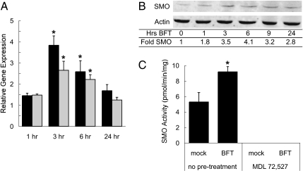
The ability of recombinant, purified BFT (5 nM final concentration) to stimulate SMO expression was evaluated in two human colonic epithelial cell lines, HT29/c1 and T84. As measured by qRT-PCR, BFT rapidly induced SMO gene expression in both cell types, resulting in a two- to fourfold increase after a 3- or 6-h exposure (Fig. 1A). The magnitude and time course of stimulation of SMO transcription by BFT closely parallels that seen in response to H. pylori or TNF-α (21, 22). In addition to induction of SMO at the gene expression level, BFT also similarly increased SMO protein levels (Fig. 1B) and enzyme activity (Fig. 1C), suggesting that increased transcription is the primary mechanism by which BFT up-regulates SMO.
Fig. 1.
BFT induces SMO in human colon epithelial cell lines. T84 and/or HT29/c1 cells were treated with 5 nM purified, recombinant BFT for the indicated time points. (A) SMO gene expression in T84 (black bars) and HT29/c1 (gray bars) cells was quantified by qRT-PCR as described in Materials and Methods and data from three independent experiments analyzed in triplicate are presented (mean + SEM). Statistically significant (*P < 0.05 by t test) increase in SMO expression vs. untreated cells at the indicated time point. (B) HT29/c1 total cellular lysates were analyzed via quantitative fluorescent Western blotting as described in Materials and Methods and SMO protein levels were normalized to β-actin (representative of two independent experiments). (C) HT29/c1 cells were exposed to BFT for 6 h and SMO and APAO enzyme activity (pmol/min/mg protein) were determined (mean of two independent experiments analyzed in triplicate; *P < 0.05 by t test). Pretreatment overnight with MDL 72527 at concentrations of 10, 25, 50, or 100 μM rendered SMO activity undetectable. APAO enzyme activity was not detected in any samples.
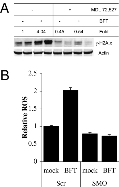
To evaluate the specific cellular responses to BFT that are mediated by polyamine catabolism, HT29/c1 and T84 cells were pretreated with MDL 72527. As expected, MDL 72527 treatment completely inhibited SMO enzyme activity at concentrations >10 μM (Fig. 1C). T84 cells, control or pretreated with 25 μM MDL 72527, were exposed to 5 nM BFT and cell lysates were analyzed by immunoblotting for γ-H2A.x, a phosphorylated histone variant that is recognized as a marker of cellular DNA damage (25). A 6-h exposure of T84 cells to BFT significantly induced γ-H2A.x, a response that could be completely inhibited by MDL 72527 (Fig. 2A).
Fig. 2.
BFT induces SMO-dependent ROS and DNA damage in T84 cells. (A) T84 cells were pretreated with or without 25 μM MDL 72527 before stimulation with 5 nM BFT for 6 h. Total cellular lysates were analyzed by quantitative fluorescent Western blotting and γ-H2A.x levels were normalized to β-actin as described in Materials and Methods. The presented data and fold changes in protein levels include two samples per treatment group representative of multiple independent experiments. (B) T84 cell lines stably expressing nontargeting (Scr) and SMO-specific (SMO) shRNA were stimulated with BFT. Intracellular ROS levels were measured via CM-H2DCFDA fluorescence.
BFT Induces Polyamine Catabolism via SMO Rather than the Spermidine/Spermine-N1-Acetyltransferase–N1-acetylpolyamine oxidase (SSAT–APAO) Pathway.
MDL 72527 is a potent inhibitor of both APAO and SMO (26, 27). To conclusively demonstrate that BFT exposure stimulates SMO-dependent ROS generation, T84 cell lines stably expressing nontargeting or SMO-specific short hairpin RNA (shRNA) were created. Cells were exposed to BFT as described above and intracellular ROS levels were measured using the oxidation-sensitive fluorescent probe CM-H2DCFDA. A twofold induction of ROS was observed in control cells but was eliminated in the SMO knockdown cell line (Fig. 2B). Further, BFT exposure did not increase SSAT or APAO expression in either control or SMO knockdown cells and APAO enzyme activity levels remained undetectable in BFT-treated cells (Fig. 1C and Fig. S1). Finally, mice infected with ETBF exhibited no change in SSAT enzyme activity (Fig. S2). Taken together, these data indicate that, consistent with previous reports (21, 28), SMO is the source of ROS leading to cytotoxic DNA damage in response to BFT.
ETBF Induces SMO in Mouse Models of Colitis.
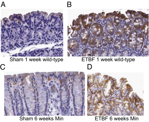
On the basis of the ability of BFT to induce SMO expression in human colon epithelial cell lines, we next evaluated the effect of ETBF infection on SMO expression in colon and cecum tissue of wild-type and Min C57BL/6 mice. Immunohistochemical staining for SMO on Swiss-rolled colon tissue from C57BL/6 mice revealed that colon tissues from sham-inoculated mice were negative for SMO staining with the exception of regions of mild-to-moderate apical staining. In contrast, slides from ETBF-inoculated mice contained regions of intense SMO staining that extended deeper into the colonic crypts (Fig. 3). Cecum nuclear extracts from sham- and ETBF-infected mice were also analyzed by Western blotting to detect differences in SMO expression. In the small number of animals studied, nuclear SMO protein levels were elevated in mice killed 1 or 3 d postinfection compared with controls (Fig. S3).
Fig. 3.
ETBF-infected mice exhibit regions of intense SMO staining. C57BL/6 mice were sham or ETBF inoculated and killed after 1 wk (wild type) or 6 wk (Min). Rolled colon tissues were fixed, embedded, and analyzed by immunohistochemistry using specific antisera against SMO. Representative images are displayed from (A) wild type, sham, 1 wk; (B) wild type, ETBF, 1 wk; (C) Min, sham, 6 wk; (D) Min, ETBF, 6 wk. Sham mice had regions of mild-to-moderate apical SMO staining. ETBF-infected mice contained regions of intense SMO staining that extended deeper into the crypts.
Following sham or ETBF infection for 1 wk, Smo gene expression was measured by qRT-PCR in cecum (Fig. S4A) and colon (Fig. S4B) tissues. Consistent with our in vitro data, ETBF infection (with or without MDL 72527) significantly induced Smo gene expression in both cecum and colon tissues. No significant differences in Smo expression between sham vs. sham+MDL and ETBF vs. ETBF+MDL groups were observed.
MDL 72527 Reduces Chronic Intestinal Inflammation and Proliferation in ETBF-Infected Min Mice.
H&E stained intestinal tissue was evaluated for gross histopathological features as described in Materials and Methods. Scores reflecting relative inflammation (on a 0–4 scale) and proliferation (on a 0–3 scale) were assigned to each slide and median scores were calculated for each group (sham, sham+MDL 72527, ETBF, and ETBF+MDL 72527). The ETBF group exhibited signicantly higher inflammation and proliferation scores compared with the ETBF+MDL 72527 group. In fact, chronic inflammation and intestinal proliferation scores for ETBF+MDL 72527 mice were not statistically greater than the sham or sham+MDL 72527 groups (Fig. S5).
The previous study by Wu and colleagues (16) demonstrated that ETBF infection induces a rapid and persistent Stat3-driven Th17 immune response that is required for colon tumorigenesis. The authors demonstrated that colonic expression of the genes encoding IL-1β, IL-17A, IL-6, and IL-23A, cytokines associated with differentiation, expansion, and effector functions of Th17 cells (29, 30), were up-regulated in ETBF-infected mice. Therefore, we assessed whether decreased chronic inflammation in ETBF+MDL 72527 mice was associated with changes in gene expression levels of these markers of the Th17 immune response. Treatment of ETBF-infected Min mice with MDL 72527 abrogated the chronic induction of both IL-1β (ETBF group 3.1-fold median increase in expression vs. sham, ETBF+MDL group 1.2-fold, P = 0.03) and IL-17A (2.6-fold increase in ETBF group vs. SHAM, no change in ETBF+MDL group, P = 0.01) gene expression measured 6 wk postinoculation. Taken together, these histopathological and gene expression data indicate that pharmacological inhibition of SMO by MDL 72527 reduces ETBF-associated intestinal chronic inflammation and proliferation.
ETBF-Induced Colon Tumorigenesis Is Significantly Inhibited by MDL 72527.
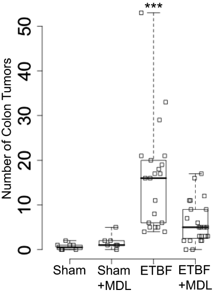
We used a model of ETBF-induced colon tumorigenesis in Min mice (16) to directly test the hypothesis that induction of SMO represents a molecular link between chronic inflammatory stimuli and epithelial carcinogenesis. Mice infected with ETBF developed significant numbers of colon tumors by 6 wk postinoculation (median = 16; Fig. 4). However, 69% fewer tumors were observed in ETBF-inoculated mice treated with MDL 72527 (median = 5). Sham-inoculated animals developed very few colon polyps whether treated with or without MDL 72527. These results indicate that the SMO contributes to ETBF-induced colon tumorigenesis and is therefore a potential target for chemopreventive or chemotherapeutic drug development.
Fig. 4.
MDL 72527 significantly inhibits ETBF-induced colon tumorigenesis in Min mice. Min mice were sham or ETBF inoculated with or without MDL 72527 (20 mg/kg 3 d per wk) for 6 wk and colon polyps were counted as described in Materials and Methods. The number of tumors for each mouse analyzed is plotted as an open square; plot whiskers indicate lower and upper limits of data; boxes are bounded by first and third quartiles of data; and heavy lines denote median tumor number in each treatment group. Sham-inoculated animals developed very few colon polyps whether treated with (median = 1, range = 0–5, n = 8) or without (median = 0.5, range = 0–2, n = 7) MDL 72527. ETBF-induced colon tumorigenesis (median = 16, range = 4–53, n = 21) was significantly reduced by MDL 72527 treatment (median = 5, range = 0–17, n = 23; ***P < 0.001 by Mann-Whitney-Wilcoxon u test for ETBF vs. ETBF+MDL comparison).
Discussion
Chronic inflammation contributes to the genesis of a substantial percentage of epithelial cancers, and it is becoming increasingly clear that in the gastrointestinal tract, commensal and pathogenic microbes are an important link between these processes. A growing number of studies have identified molecular components of the immune response to bacteria that may be involved in either promoting or protecting against intestinal inflammation and carcinogenesis (31–36). The importance of bacterially-induced ROS in inflammation-associated carcinogenesis was demonstrated in mice lacking the H2O2 detoxification enzymes gpx-1 and gpx-2. These animals have a normal phenotype when raised in germ-free conditions, but if colonized with conventional flora, they develop intestinal tumors (18). Although inflammation-associated ROS has been implicated as one of the sources for the mutagens necessary to produce the genetic changes required for the carcinogenic process, the sources of the ROS have not been clearly identified. Whereas immune cells absolutely contribute to infection-associated tumorigenesis, this and other studies have demonsrated that bacteria can stimulate ROS production, cytokine secretion, DNA damage, and cellular proliferation in epithelial cells independent of any immune system contribution (5, 9, 17, 22, 37).
SMO, a polyamine catabolic enzyme, is one such epithelial source of inflammation-induced ROS and DNA damage. Inducers of SMO include proinflammatory cytokines as well as the gastric pathogen H. pylori, a contributor to gastritis, peptic ulcer disease, and gastric adenocarcinoma (21, 22, 38). In addition to H2O2, SMO produces 3-aminopropanal, a byproduct that contributes to the formation of acrolein, a highly reactive aldehyde. Acrolein associated with elevated polyamine catabolism has been strongly implicated in the etiology of diseases including ischemia-reperfusion injuries, renal failure, stroke, and silent brain infarction (39–44). Further, elevated SMO expression has been observed in inflammation-associated human diseases including gastritis (22), ulcerative colitis (23), and prostatic intraepithelial neoplasia (24).
Therefore, the goal of the present study was to use a validated murine carcinogenesis model to determine whether SMO plays a direct role in the development of colon tumors. The data presented above implicate SMO as a molecular link between ETBF-induced chronic inflammation and colon tumorigenesis. BFT rapidly induces SMO expression and leads to SMO-dependent ROS and DNA damage in intestinal epithelial cell lines and mice infected with ETBF exhibit elevated intestinal SMO expression. In addition, inhibition of SMO by MDL 72527 in ETBF-infected mice was shown to (i) reduce chronic intestinal inflammation and proliferation; (ii) decrease the chronic up-regulation of cytokines associated with the Th17 immune response; and, most importantly, (iii) significantly inhibit colon tumorigenesis in Min mice. The results of these studies are particularly intriguing because, in contrast to other published models of intestinal carcinogenesis that rely on chemical irritants, mutagens, and/or immunocompromised mice, the ETBF model used for these experiments incorporates a genetic defect, a bacterial pathogen, and disease physiology that have direct importance in and therefore should be more applicable to human intestinal inflammation and colorectal cancer (12, 13, 16).
MDL 72527, an inhibitor of both SMO and APAO (26, 27), has been demonstrated to increase survival in a model of prostate carcinogenesis (45), in addition to decreasing ETBF-induced colon tumorigenesis. The data presented in this study, combined with additional published and preliminary results, argue strongly that it is SMO that plays an important role in the production of inflammation-associated ROS and DNA damage and therefore represents a potential chemopreventive or chemotherapeutic target (21, 28). Although SMO expression is highly inducible and active isoforms have been documented in both the nucleus and cytosol (46–49), the second polyamine oxidase, APAO, is confined to the peroxisome and therefore its activity seems less likely to yield pathological consequences (50). Further, APAO activity is dependent on acetylated polyamine substrates generated by SSAT, which is not induced by ETBF.
Our data clearly suggest that, whereas MDL 72527 inhibits both SMO and APAO, the possibility that the SSAT/APAO pathway contributes to tumorigenesis is remote. However, this possibility cannot be excluded until specific inhibitors or knockout mice for each polyamine oxidase are developed. Despite this limitation, the current study has validated polyamine catabolism as a therapeutic target for chemoprevention by demonstrating that MDL 72527 significantly decreased intestinal inflammation, proliferation, and tumorigenesis in response to ETBF. Taken together, our body of work suggests that up-regulation of SMO in response to inflammatory stimuli is widespread in human epithelial cells and leads to the production of ROS and DNA damage, two features of the chronic inflammatory microenvironment that have been linked to subsequent carcinogenesis. We have now demonstrated that inhibition of SMO activity by MDL 72527 dramatically reduces colon tumorigenesis in ETBF-infected Min mice. These results merit further research into chemopreventive therapies targeting SMO.
Materials and Methods
ETBF Techniques and Mouse Infections.
The ETBF strain 86-5443-2-2 (51) was propagated and prepared for inoculation as described (12). C57BL/6 mice heterozygous for the MinAPCΔ716 mutation (referred to as Min mice) and wild-type littermates were maintained, treated with antibiotics, sham or ETBF inoculated, and monitored for colonization as described previously (16). Min mice were studied in four independent 6-wk experiments (n = 12–23 each). Wild-type C57BL/6 littermates were used for two independent 1-wk experiments (n = 24–26 each). As described, some groups were administered the polyamine oxidase inhibitor MDL 72527 (52–54). Two doses of MDL 72527 (20 mg/kg in sterile 1× PBS by i.p. injection) were administered before inoculation and then the mice received the drug 3 d per week until being killed.
Tissue Preparation and Colon Polyp Analysis.
Mice were killed by CO2 asphyxiation and cecum and colon tissues were placed in TRIzol (Invitrogen; for RNA purification, see below) or 10% buffered formalin (for histopathology). After fixation, tissues were paraffin-embedded and 5-μm sections were prepared for hematoxylin and eosin (H&E, for histopathological analysis) or SMO immunohistochemical staining. Min mouse colons were prepared and stained as described (16) and polyps were counted for each mouse with a Leica ES2 dissecting microscope by two independent investigators (S.W. and C.L.S.) who were blinded to treatment group assignments. Following these analyses, colons were Swiss-rolled into cryomolds, placed in cassettes with fixed cecum tissue, and processed as described above.
Histopathology and Immunohistochemistry.
Immunohistochemical staining for SMO expression was performed on sham- and ETBF-infected mouse cecum and colon tissues as previously reported (24). H&E stained slides were assigned composite colon and cecum inflammation and proliferation scores by a comparative pathologist (D.L.H.) as previously published (12, 13, 16).
qRT-PCR: Mouse Tissues.
RNA was purified using the Invitrogen PureLink kit, DNase-treated, quantified, and cDNA synthesized (qScript; Quanta Biosciences). Gene expression was measured via TaqMan probe-based qRT-PCR with FAM-labeled gene of interest probe sets, VIC-labeled 18s rRNA control probe set, Gene Expression master mix solution, and the ABI 7500 thermocycler and companion software all obtained from Applied Biosystems.
Cell Culture and Reagents.
The HT29/c1 colon carcinoma cell line was maintained as previously reported (7). The T84 colon carcinoma cell line was maintained in DMEM: F-12K medium supplemented with 5% FBS. The shRNAmir lentiviral system (Open Biosystems) was used to generate stable puromycin-resistant T84 cell lines expressing nontargeting or SMO-specific shRNA.
Recombinant BFT was purified as described (55). For experimental studies, HT29/c1 or T84 cells were rinsed with 1× PBS before addition of BFT for the indicated time points at a concentration of 100 ng/mL (5 nM) in serum-free media supplemented with 10 μg/mL apo-transferrin. When indicated, cells were pretreated with MDL 72527 for 16 h before addition of BFT.
Quantitative Real-Time PCR: Cell Culture Experiments.
RNA was isolated from BFT-treated cells using the PureLink Micro-to-Midi system (Invitrogen), treated with DNase I, and purified over RNEasy spin columns (Qiagen) before cDNA synthesis with Oligo-dT12–18 primers and M-MLV reverse transcriptase (Invitrogen). Gene expression was measured by qRT-PCR (iQ SYBR Green reagents and MyiQ detection system; Bio-Rad) and differences among treatment groups were calculated using the ΔΔCt method (56). A region of cDNA common to all active SMO isoforms was amplified with the primers (5′ GATCCCGGCGGACCATGTGATTGTG) and (5′ CTCAGGCGGGTAGAGGACATCAAA). Glyceraldehyde 3-phosphate dehydrogenase (GapDH; normalization gene) was amplified with the primers (5′ GAAGGTGAAGGTCGGAGTC) and (5′ GAAGATGGTGATGGGATTTC).
Western Blotting and Enzyme Activity Assays.
To obtain total cellular lysates, HT29/c1 or T84 cell pellets were resuspended in 1× PBS with 1% (vol/vol) Nonidet P-40, 0.5% sodium deoxycholate, 0.1% (vol/vol) SDS, 30 μg/mL aprotinin, 100 μM sodium orthovanadate, 10 μg/mL PMSF and 1× protease inhibitor mixture (Roche). Extracts were incubated on ice for 20 min, and insoluble material was pelleted by centrifugation at 13,500 × g and 4 °C for 15 min. Mouse cecum nuclear protein extracts from sham- or ETBF-infected mice were prepared as previously described (16). Quantitative fluorescent Western blotting was performed as previously described (46). To detect SMO, a rabbit polyclonal antibody (21) was used at a 1:1,000 dilution in combination with a mouse monoclonal antibody against β-actin (1:2,000; Sigma-Aldrich) for 1–2 h at room temperature. To detect γ-H2A.x, a mouse monoclonal antibody (1:2,000; Abcam) was used in combination with a rabbit polyclonal antibody against β-actin (1:2,000; Sigma-Aldrich) overnight at 4 °C. Dye-conjugated secondary antirabbit (IRDye800; Rockland Immunochemicals) and antimouse (Alexa Fluor 680; Invitrogen) IgG antibodies were used at a final concentration of 0.1 μg/mL.
Chemiluminescent SMO and APAO enzyme activity assays of HT29/c1 cell pellets and radiographic SSAT enzyme activity assays of mouse tissue were performed as previously reported (28).
Detection of Intracellular ROS.
T84 shRNA cell lines were exposed to BFT as described above. Cells were then washed with 1× PBS and incubated with 10 μM CM-H2DCFDA (Invitrogen) at 37 °C for 90 min in RPMI media without serum or phenol red. Cells were then washed and fluorescence was measured using a Spectra Max M5 plate reader (Molecular Devices).
Statistics.
Differences in mean gene expression, enzyme activity, and ROS levels between in vitro treatment groups among multiple independent experiments were evaluated by Student's t test. Differences in median gene expression, tumor burden, and histopathological inflammation or proliferation scores among in vivo treatment groups across multiple independent experiments were evaluated using the nonparametric Mann–Whitney–Wilcoxon u test. When used, box-and-whisker plots display median (heavy bar), first/third quartiles (boundaries of box), and data range (whiskers). P values <0.05 were considered to be statistically significant.
Supplementary Material
Acknowledgments
The authors gratefully acknowledge Dr. Angelo De Marzo and Jessica Hicks (Department of Pathology, Johns Hopkins University School of Medicine) for assistance with SMO immunohistochemistry. These studies were supported by the Samuel Waxman Cancer Research Foundation (R.A.C.) and National Institutes of Health Grants CA 51085 and CA 98454 (to R.A.C.), and CA 151325, DK 45496, and DK 080817 (to C.L.S.).
Footnotes
The authors declare no conflict of interest.
*This Direct Submission article had a prearranged editor.
This article contains supporting information online at www.pnas.org/lookup/suppl/doi:10.1073/pnas.1010203108/-/DCSupplemental.
References
- 1.Coussens LM, Werb Z. Inflammation and cancer. Nature. 2002;420:860–867. doi: 10.1038/nature01322. [DOI] [PMC free article] [PubMed] [Google Scholar]
- 2.Aggarwal BB, Shishodia S, Sandur SK, Pandey MK, Sethi G. Inflammation and cancer: How hot is the link? Biochem Pharmacol. 2006;72:1605–1621. doi: 10.1016/j.bcp.2006.06.029. [DOI] [PubMed] [Google Scholar]
- 3.Sears CL. Enterotoxigenic Bacteroides fragilis: A rogue among symbiotes. Clin Microbiol Rev. 2009;22:349–369. doi: 10.1128/CMR.00053-08. [DOI] [PMC free article] [PubMed] [Google Scholar]
- 4.Weikel CS, Grieco FD, Reuben J, Myers LL, Sack RB. Human colonic epithelial cells, HT29/C1, treated with crude Bacteroides fragilis enterotoxin dramatically alter their morphology. Infect Immun. 1992;60:321–327. doi: 10.1128/iai.60.2.321-327.1992. [DOI] [PMC free article] [PubMed] [Google Scholar]
- 5.Wu S, Morin PJ, Maouyo D, Sears CL. Bacteroides fragilis enterotoxin induces c-Myc expression and cellular proliferation. Gastroenterology. 2003;124:392–400. doi: 10.1053/gast.2003.50047. [DOI] [PubMed] [Google Scholar]
- 6.Wu S, Lim KC, Huang J, Saidi RF, Sears CL. Bacteroides fragilis enterotoxin cleaves the zonula adherens protein, E-cadherin. Proc Natl Acad Sci USA. 1998;95:14979–14984. doi: 10.1073/pnas.95.25.14979. [DOI] [PMC free article] [PubMed] [Google Scholar]
- 7.Wu S, Rhee KJ, Zhang M, Franco A, Sears CL. Bacteroides fragilis toxin stimulates intestinal epithelial cell shedding and gamma-secretase-dependent E-cadherin cleavage. J Cell Sci. 2007;120:1944–1952. doi: 10.1242/jcs.03455. [DOI] [PMC free article] [PubMed] [Google Scholar]
- 8.Sanfilippo L, et al. Bacteroides fragilis enterotoxin induces the expression of IL-8 and transforming growth factor-beta (TGF-beta) by human colonic epithelial cells. Clin Exp Immunol. 2000;119:456–463. doi: 10.1046/j.1365-2249.2000.01155.x. [DOI] [PMC free article] [PubMed] [Google Scholar]
- 9.Wu S, et al. Bacteroides fragilis enterotoxin induces intestinal epithelial cell secretion of interleukin-8 through mitogen-activated protein kinases and a tyrosine kinase-regulated nuclear factor-kappaB pathway. Infect Immun. 2004;72:5832–5839. doi: 10.1128/IAI.72.10.5832-5839.2004. [DOI] [PMC free article] [PubMed] [Google Scholar]
- 10.Kim JM, et al. Mitogen-activated protein kinase and activator protein-1 dependent signals are essential for Bacteroides fragilis enterotoxin-induced enteritis. Eur J Immunol. 2005;35:2648–2657. doi: 10.1002/eji.200526321. [DOI] [PubMed] [Google Scholar]
- 11.Kim JM, et al. Nuclear factor-kappa B activation pathway in intestinal epithelial cells is a major regulator of chemokine gene expression and neutrophil migration induced by Bacteroides fragilis enterotoxin. Clin Exp Immunol. 2002;130:59–66. doi: 10.1046/j.1365-2249.2002.01921.x. [DOI] [PMC free article] [PubMed] [Google Scholar]
- 12.Rabizadeh S, et al. Enterotoxigenic bacteroides fragilis: A potential instigator of colitis. Inflamm Bowel Dis. 2007;13:1475–1483. doi: 10.1002/ibd.20265. [DOI] [PMC free article] [PubMed] [Google Scholar]
- 13.Rhee KJ, et al. Induction of persistent colitis by a human commensal, enterotoxigenic Bacteroides fragilis, in wild-type C57BL/6 mice. Infect Immun. 2009;77:1708–1718. doi: 10.1128/IAI.00814-08. [DOI] [PMC free article] [PubMed] [Google Scholar]
- 14.Wu S, et al. A human colonic commensal promotes colon tumorigenesis via activation of T helper type 17 T cell responses. Nat Med. 2009;15:1016–1022. doi: 10.1038/nm.2015. [DOI] [PMC free article] [PubMed] [Google Scholar]
- 15.Moser AR, Pitot HC, Dove WF. A dominant mutation that predisposes to multiple intestinal neoplasia in the mouse. Science. 1990;247:322–324. doi: 10.1126/science.2296722. [DOI] [PubMed] [Google Scholar]
- 16.Su LK, et al. Multiple intestinal neoplasia caused by a mutation in the murine homolog of the APC gene. Science. 1992;256:668–670. doi: 10.1126/science.1350108. [DOI] [PubMed] [Google Scholar]
- 17.Huycke MM, Abrams V, Moore DR. Enterococcus faecalis produces extracellular superoxide and hydrogen peroxide that damages colonic epithelial cell DNA. Carcinogenesis. 2002;23:529–536. doi: 10.1093/carcin/23.3.529. [DOI] [PubMed] [Google Scholar]
- 18.Chu FF, et al. Bacteria-induced intestinal cancer in mice with disrupted Gpx1 and Gpx2 genes. Cancer Res. 2004;64:962–968. doi: 10.1158/0008-5472.can-03-2272. [DOI] [PubMed] [Google Scholar]
- 19.Vujcic S, Diegelman P, Bacchi CJ, Kramer DL, Porter CW. Identification and characterization of a novel flavin-containing spermine oxidase of mammalian cell origin. Biochem J. 2002;367:665–675. doi: 10.1042/BJ20020720. [DOI] [PMC free article] [PubMed] [Google Scholar]
- 20.Wang Y, et al. Cloning and characterization of a human polyamine oxidase that is inducible by polyamine analogue exposure. Cancer Res. 2001;61:5370–5373. [PubMed] [Google Scholar]
- 21.Babbar N, Casero RA., Jr Tumor necrosis factor-alpha increases reactive oxygen species by inducing spermine oxidase in human lung epithelial cells: A potential mechanism for inflammation-induced carcinogenesis. Cancer Res. 2006;66:11125–11130. doi: 10.1158/0008-5472.CAN-06-3174. [DOI] [PubMed] [Google Scholar]
- 22.Xu H, et al. Spermine oxidation induced by Helicobacter pylori results in apoptosis and DNA damage: Implications for gastric carcinogenesis. Cancer Res. 2004;64:8521–8525. doi: 10.1158/0008-5472.CAN-04-3511. [DOI] [PubMed] [Google Scholar]
- 23.Hong SK, et al. Increased expression and cellular localization of spermine oxidase in ulcerative colitis and relationship to disease activity. Inflamm Bowel Dis. 2010;16:1557–1566. doi: 10.1002/ibd.21224. [DOI] [PMC free article] [PubMed] [Google Scholar]
- 24.Goodwin AC, et al. Increased spermine oxidase expression in human prostate cancer and prostatic intraepithelial neoplasia tissues. Prostate. 2008;68:766–772. doi: 10.1002/pros.20735. [DOI] [PMC free article] [PubMed] [Google Scholar]
- 25.Bonner WM, et al. GammaH2AX and cancer. Nat Rev Cancer. 2008;8:957–967. doi: 10.1038/nrc2523. [DOI] [PMC free article] [PubMed] [Google Scholar]
- 26.Wang Y, et al. Properties of recombinant human N1-acetylpolyamine oxidase (hPAO): Potential role in determining drug sensitivity. Cancer Chemother Pharmacol. 2005;56:83–90. doi: 10.1007/s00280-004-0936-5. [DOI] [PubMed] [Google Scholar]
- 27.Wang Y, et al. Properties of purified recombinant human polyamine oxidase, PAOh1/SMO. Biochem Biophys Res Commun. 2003;304:605–611. doi: 10.1016/s0006-291x(03)00636-3. [DOI] [PubMed] [Google Scholar]
- 28.Pledgie A, et al. Spermine oxidase SMO(PAOh1), Not N1-acetylpolyamine oxidase PAO, is the primary source of cytotoxic H2O2 in polyamine analogue-treated human breast cancer cell lines. J Biol Chem. 2005;280:39843–39851. doi: 10.1074/jbc.M508177200. [DOI] [PubMed] [Google Scholar]
- 29.Dong C. TH17 cells in development: an updated view of their molecular identity and genetic programming. Nat Rev Immunol. 2008;8:337–348. doi: 10.1038/nri2295. [DOI] [PubMed] [Google Scholar]
- 30.Manel N, Unutmaz D, Littman DR. The differentiation of human T(H)-17 cells requires transforming growth factor-beta and induction of the nuclear receptor RORgammat. Nat Immunol. 2008;9:641–649. doi: 10.1038/ni.1610. [DOI] [PMC free article] [PubMed] [Google Scholar]
- 31.Erdman SE, et al. CD4+ CD25+ regulatory T lymphocytes inhibit microbially induced colon cancer in Rag2-deficient mice. Am J Pathol. 2003;162:691–702. doi: 10.1016/S0002-9440(10)63863-1. [DOI] [PMC free article] [PubMed] [Google Scholar]
- 32.Rao VP, et al. Innate immune inflammatory response against enteric bacteria Helicobacter hepaticus induces mammary adenocarcinoma in mice. Cancer Res. 2006;66:7395–7400. doi: 10.1158/0008-5472.CAN-06-0558. [DOI] [PubMed] [Google Scholar]
- 33.Erdman SE, et al. CD4+CD25+ regulatory lymphocytes induce regression of intestinal tumors in ApcMin/+ mice. Cancer Res. 2005;65:3998–4004. doi: 10.1158/0008-5472.CAN-04-3104. [DOI] [PubMed] [Google Scholar]
- 34.Nagamine CM, et al. Helicobacter hepaticus infection promotes colon tumorigenesis in the BALB/c-Rag2(-/-) Apc(Min/+) mouse. Infect Immun. 2008;76:2758–2766. doi: 10.1128/IAI.01604-07. [DOI] [PMC free article] [PubMed] [Google Scholar]
- 35.Chen GY, Shaw MH, Redondo G, Núñez G. The innate immune receptor Nod1 protects the intestine from inflammation-induced tumorigenesis. Cancer Res. 2008;68:10060–10067. doi: 10.1158/0008-5472.CAN-08-2061. [DOI] [PMC free article] [PubMed] [Google Scholar]
- 36.Rakoff-Nahoum S, Medzhitov R. Regulation of spontaneous intestinal tumorigenesis through the adaptor protein MyD88. Science. 2007;317:124–127. doi: 10.1126/science.1140488. [DOI] [PubMed] [Google Scholar]
- 37.Lofgren JL, et al. Lack of commensal flora in Helicobacter pylori-infected INS-GAS mice reduces gastritis and delays intraepithelial neoplasia. Gastroenterology. 2011;140:210–220. doi: 10.1053/j.gastro.2010.09.048. [DOI] [PMC free article] [PubMed] [Google Scholar]
- 38.Kusters JG, van Vliet AH, Kuipers EJ. Pathogenesis of Helicobacter pylori infection. Clin Microbiol Rev. 2006;19:449–490. doi: 10.1128/CMR.00054-05. [DOI] [PMC free article] [PubMed] [Google Scholar]
- 39.Saiki R, et al. Brain infarction correlates more closely with acrolein than with reactive oxygen species. Biochem Biophys Res Commun. 2010;404:1044–1049. doi: 10.1016/j.bbrc.2010.12.107. [DOI] [PubMed] [Google Scholar]
- 40.Kim GH, et al. The role of polyamine metabolism in neuronal injury following cerebral ischemia. Can J Neurol Sci. 2009;36:14–19. doi: 10.1017/s0317167100006247. [DOI] [PubMed] [Google Scholar]
- 41.Yoshida M, et al. Acrolein, IL-6 and CRP as markers of silent brain infarction. Atherosclerosis. 2009;203:557–562. doi: 10.1016/j.atherosclerosis.2008.07.022. [DOI] [PubMed] [Google Scholar]
- 42.Wood PL, et al. Aldehyde load in ischemia-reperfusion brain injury: Neuroprotection by neutralization of reactive aldehydes with phenelzine. Brain Res. 2006;1122:184–190. doi: 10.1016/j.brainres.2006.09.003. [DOI] [PubMed] [Google Scholar]
- 43.Igarashi K, Ueda S, Yoshida K, Kashiwagi K. Polyamines in renal failure. Amino Acids. 2006;31:477–483. doi: 10.1007/s00726-006-0264-7. [DOI] [PubMed] [Google Scholar]
- 44.Tomitori H, et al. Polyamine oxidase and acrolein as novel biochemical markers for diagnosis of cerebral stroke. Stroke. 2005;36:2609–2613. doi: 10.1161/01.STR.0000190004.36793.2d. [DOI] [PubMed] [Google Scholar]
- 45.Basu HS, et al. A small molecule polyamine oxidase inhibitor blocks androgen-induced oxidative stress and delays prostate cancer progression in the transgenic adenocarcinoma of the mouse prostate model. Cancer Res. 2009;69:7689–7695. doi: 10.1158/0008-5472.CAN-08-2472. [DOI] [PMC free article] [PubMed] [Google Scholar]
- 46.Murray-Stewart T, et al. Nuclear localization of human spermine oxidase isoforms: Possible implications in drug response and disease etiology. FEBS J. 2008;275:2795–2806. doi: 10.1111/j.1742-4658.2008.06419.x. [DOI] [PMC free article] [PubMed] [Google Scholar]
- 47.Murray-Stewart T, Wang Y, Devereux W, Casero RA., Jr Cloning and characterization of multiple human polyamine oxidase splice variants that code for isoenzymes with different biochemical characteristics. Biochem J. 2002;368:673–677. doi: 10.1042/BJ20021587. [DOI] [PMC free article] [PubMed] [Google Scholar]
- 48.Cervelli M, et al. Mouse spermine oxidase gene splice variants. Nuclear subcellular localization of a novel active isoform. Eur J Biochem. 2004;271:760–770. doi: 10.1111/j.1432-1033.2004.03979.x. [DOI] [PubMed] [Google Scholar]
- 49.Bianchi M, Amendola R, Federico R, Polticelli F, Mariottini P. Two short protein domains are responsible for the nuclear localization of the mouse spermine oxidase mu isoform. FEBS J. 2005;272:3052–3059. doi: 10.1111/j.1742-4658.2005.04718.x. [DOI] [PubMed] [Google Scholar]
- 50.Pegg AE. Mammalian polyamine metabolism and function. IUBMB Life. 2009;61:880–894. doi: 10.1002/iub.230. [DOI] [PMC free article] [PubMed] [Google Scholar]
- 51.Myers LL, Shoop DS. Association of enterotoxigenic Bacteroides fragilis with diarrheal disease in young pigs. Am J Vet Res. 1987;48:774–775. [PubMed] [Google Scholar]
- 52.Bey P, Bolkenius FN, Seiler N, Casara P. N-2,3-Butadienyl-1,4-butanediamine derivatives: potent irreversible inactivators of mammalian polyamine oxidase. J Med Chem. 1985;28:1–2. doi: 10.1021/jm00379a001. [DOI] [PubMed] [Google Scholar]
- 53.Seiler N, Duranton B, Raul F. The polyamine oxidase inactivator MDL 72527. Prog Drug Res. 2002;59:1–40. doi: 10.1007/978-3-0348-8171-5_1. [DOI] [PubMed] [Google Scholar]
- 54.Bolkenius FN, Bey P, Seiler N. Specific inhibition of polyamine oxidase in vivo is a method for the elucidation of its physiological role. Biochim Biophys Acta. 1985;838:69–76. doi: 10.1016/0304-4165(85)90251-x. [DOI] [PubMed] [Google Scholar]
- 55.Wu S, Dreyfus LA, Tzianabos AO, Hayashi C, Sears CL. Diversity of the metalloprotease toxin produced by enterotoxigenic Bacteroides fragilis. Infect Immun. 2002;70:2463–2471. doi: 10.1128/IAI.70.5.2463-2471.2002. [DOI] [PMC free article] [PubMed] [Google Scholar]
- 56.Livak KJ, Schmittgen TD. Analysis of relative gene expression data using real-timequantitative PCR and the 2(-Delta Delta C(T)) Method. Methods. 2001;25:402–408. doi: 10.1006/meth.2001.1262. [DOI] [PubMed] [Google Scholar]
Associated Data
This section collects any data citations, data availability statements, or supplementary materials included in this article.


