Abstract
Spatiotemporal brain activation profiles were obtained from 27 middle school students experiencing difficulties in reading comprehension as well as word-level skills (RD) and 23 age- and IQ-matched non-reading impaired students during performance of an oral pseudoword reading task using Magnetoencephalography (MEG). Based on their scores on standardized reading fluency tests 1 year later, students with RD who showed significant improvement were classified as Adequate Responders (AR) whereas those not demonstrating such gains were classified as Inadequate Responders (IR). At baseline, activation profiles of the AR group featured increased activity in the left supramarginal and angular gyri, as well as in the superior and middle temporal gyri, bilaterally compared to IR. The degree of activity in these regions was a significant predictor of the amount of subsequent gains in reading fluency. These results extend previous functional brain imaging findings of beginning readers, suggesting that recruitment of brain areas that typically serve as key components of the brain circuit for reading is an important factor in determining response to intervention in older struggling readers.
Keywords: Phonological decoding, Dyslexia, Educational remediation, Magnetoencephalography, Functional brain imaging, Temporo-parietal cortex
INTRODUCTION
The majority of functional brain imaging studies on students with reading difficulties (RD) concur in demonstrating reduced neurophysiological and hemodynamic activity in posterior temporal and inferior parietal regions of the left hemisphere (e.g., Hoeft, Meyler, et al., 2007; Shaywitz et al., 2002; Simos, Breier, Fletcher, Bergman, & Papanicolaou, 2000; Simos et al., 2002, 2005, 2011; Temple et al., 2001). Failure to engage these regions has been found early during reading development, often despite adequate classroom reading instruction and may serve as a predictor of the capacity of individual students to benefit from instruction (Gabrieli, 2009; Hoeft, Ueno, et al., 2007; Simos et al., 2005).
With few exceptions, imaging studies have used relatively homogeneous community samples of elementary school children selected on the basis of low scores on standardized achievement tests indicating the presence of reading difficulties, primarily at the word level (decoding, word recognition, and fluency). In recent years, however, there is increasing interest on students who struggle with more complex (and educationally significant) areas such as reading comprehension. Such difficulties become more prominent as students enter middle school, when educational history and family environment interact in complex ways to compound any primary cognitive and language deficits. Such diverse cognitive and social factors may limit the potential for functional plasticity in response to remedial instruction at both the cognitive and neurophysiological levels. One of the main research questions addressed in the present study is whether signs of adequate engagement of the neurophysiological processes typically supporting reading automaticity predict individual capacity to benefit from remedial instruction among students who face a variety of interacting socioeconomic, cognitive and linguistic adversities. The study investigated features of reading-related brain function during performance of a speeded phonological decoding task associated with the capacity of individual struggling readers to benefit from small-group remedial instruction. The primary target group consisted of 27 students recruited from a larger Grade 6–8 educational study (Vaughn, Cirino, et al., 2010; Vaughn, Wanzek, et al., 2010) as being at-risk for further academic failure in part because of poor reading comprehension performance, while also scoring below grade level on standardized decoding assessments. These children received remedial instruction focusing on reading fluency, vocabulary, and reading comprehension skills.
Source current density maps of neurophysiological activity were obtained at the beginning of the study on a reading-aloud task involving relatively rapid presentation of three-letter pseudowords. Selection of stimuli maintained decoding demands relatively low to prevent floor effects in individual in-scanner performance, while attempting to simulate the demands for rapid processing of print that appears to be critical for effective text comprehension (e.g., Jenkins, Fuchs, van den Broek, Espin, & Deno, 2003; Kim, Petscher, Schatschneider, & Foorman, 2010; Yovanoff, Duesbery, Alonzo, & Tindal, 2005). The degree of response to educational remediation for each student was determined based on 1-year follow-up reading assessments, and individual differences in neurophysiology were then examined using spatiotemporal activation profiles obtained at baseline (pre-intervention).
In comparison to a group of 23 children comparable in age and IQ who scored in the average range on reading measures, we hypothesized that the activation profiles of students who later demonstrated positive response to intervention would show adequate engagement of temporo-parietal and inferior parietal regions in the left hemisphere, resembling those of typically achieving readers. In contrast, pre-intervention profiles for inadequate responders would closely resemble those typically observed in previous imaging studies in children with RD. In addition, it was predicted that degree of activity in these regions would correlate strongly with the amount of gains made by individual students post-intervention. Given the nature of the task, we predicted that these associations would be stronger for achievement measures which mainly assess phonological decoding automaticity, rather than measures of word reading fluency.
METHOD
Participants
The current sample included 27 adolescent struggling readers who did not pass the school-administered Texas Assessment of Knowledge and Skills (TAKS), a criterion-referenced reading comprehension assessment that represents the state accountability test. These individuals were volunteers, who met the selection criteria specified below, from the treatment groups of an intervention study designed for students in Grades 6–8 (Vaughn, Cirino, et al., 2010; Vaughn, Wanzek, et al., 2010). While all of the ~450 struggling readers who received intervention were invited and actively recruited to participate through letters to parents and follow-up phone calls, response rates for imaging studies in school-identified samples are typically very small (e.g., Meyler, Keller, Cherkassky, Gabrieli, & Just, 2008; Simos, Fletcher, Sarkari, Billingsley-Marshall, et al., 2007). In this study, many parents did not respond to the letter of invitation and did not have accurate contact information at the school. We also required scores below the 25th percentile (standard score of 90) on the Phonetic Decoding (pseudoword) Efficiency subtest of the Test of Word Reading Efficiency (TOWRE; Torgesen, Wagner, & Rashotte, 1999), a norm referenced word reading fluency assessment, because the TAKS cannot be used as a pre-post measure and relatively few of the children in the larger intervention study had reading difficulties restricted to reading comprehension. This measure was adopted given its close similarity to the activation task used in the present study (speeded naming of pseudowords) with respect to the component operations involved.
Individual differences in response to intervention were assessed on a group basis using change in standard score on the TOWRE Phonetic Decoding Efficiency subtest between baseline and 1-year follow-up visit (a 7-point difference cutoff was used). Sixteen children were classified as Adequate Responders (AR group), demonstrating a mean improvement on the TOWRE composite of 15 ± 4 points (range, 7 to 25 points), and 11 as Inadequate Responders (IR group), improving on average by only 0.7 ± 5 points (range, −4 to 6 points). Other studies of elementary school children have demonstrated that the TOWRE reliably detects changes in responder status (Fletcher et al., in press).
At follow-up three students in the AR group still read at or below the 25th percentile on the TOWRE Phonetic Decoding efficiency subtest, but all demonstrated significant changes in pseudoword fluency performance. The majority of AR students also showed strong gains on sight word reading fluency, indicated by difference scores on the corresponding TOWRE subtest averaging 8 ± 6 points. Number of points gained on this subtest ranged between 1 and 18 for this group, with only one student scoring below the 25th percentile at follow-up. All but one student in the IR group maintained scores lower than the 25th percentile post-intervention on both TOWRE subtests. Supporting the responder status grouping, we found significant Session (Baseline, Follow-up) by Group interactions on standard scores from the TOWRE Sight Word Efficiency, F(1,25) = 11.81, p = .002), and TOWRE Phonetic Decoding Efficiency subtests, F(1,25) = 22.60, p = .0001). Pairwise comparisons indicated that improvement on TOWRE scores was restricted to the AR group on the Sight Word subscale, F(1,15) = 37.10, p = .0001, and on the Phonetic Decoding subscale, F(1,15) = 44.96, p = .0001. No comparisons within the IR group approximated the critical level of alpha (all p values > .3). Moreover, although the two groups did not differ significantly on either measure at baseline (p > .2), AR students as a group significantly outperformed IR students at follow-up on both the Sight Word, F(1,25) = 8.15, p = .009), and Phonetic Decoding subtests, F(1,25) = 8.76, p = .007, subtests.
A third group of 23 children who had never experienced difficulties in reading (NI group) served as comparisons, showing standard scores>92 on the TOWRE composite index. Table 1 displays demographic and psychoeducational information for each of the three groups of participants, which were comparable on age and handedness. On average (and as expected), NI students scored higher on the IQ measures than each of the two RD sub-groups, although these differences did not reach statistical significance. There was also a tendency for a higher proportion of boys than girls in the NI group, and a significantly higher proportion of minority students in both RD sub-groups as compared to the NI group. As expected, both RD sub-groups scored significantly lower than the NI group on measures of reading and spelling, but did not differ from each other on these measures.
Table 1.
Participant demographic and psychoeducational data (M ± SD)
| NI (n = 23) | AR (n = 16) | IR (n = 11) | |
|---|---|---|---|
| Gender (b/g)1 | 15/8 | 7/9 | 6/5 |
| Age (mo) | 153 ± 12 | 159 ± 9 | 156 ± 16 |
| Ethnicity (C, H, A-AM2)3 | 12/3/8 | 0/5/11 | 0/9/2 |
| Handedness (R/L)4 | 22/3 | 15/2 | 10/1 |
| VIQ | 102 ± 12 | 98 ± 16 | 93 ± 13 |
| PIQ | 102 ± 11 | 94 ± 20 | 95 ± 13 |
| FSIQ | 102 ± 10 | 96 ± 14 | 94 ± 11 |
| WJ-III RC | 104 ± 8†§ | 84 ± 9† | 83 ± 11§ |
| WJ-IIIWA | 105 ± 10†§ | 85 ± 8† | 85 ± 9§ |
| WJ-III L WID | 102 ± 11†§ | 83 ± 11† | 81 ± 14§ |
| WJ-III Spelling | 107 ± 9†§ | 84 ± 16† | 80 ± 14§ |
| WRE | |||
| Time 1 | 97 ± 7#@ | 85 ± 9# | 84 ± 6@ |
| Time 2 | 102 ± 6#§ | 94 ± 7#¢ | 85 ± 6§¢ |
| PsWRE | |||
| Time 1 | 98 ± 9†§ | 80 ± 7† | 84 ± 7§ |
| Time 2 | 104 ± 7*§ | 95 ± 10*¢ | 84 ± 7§¢ |
Note. Significant pairwise group differences on achievement measures:
p < .0001
p < .0001
p < .01
p < .01
p < .01
p < .05
Abbreviations, Woodcock-Johnson III: WJ-III RC (Reading Comprehension), WJ-III WA (Word Attack), WJ-III LWID (Letter-Word Identification). Test of Word Reading Efficiency: WRE (Sight Word) and PsWRE (Phonetic Decoding) subtests. Time 1 and Time 2 indicate assessments at baseline (concurrent with MEG testing) and at the 1-year follow-up (post intervention), respectively.
Groups were matched for gender (Phi = .22, p = .3).
C: Caucasian; H: Hispanic; A-AM: African American.
There were significant group differences in ethnicity (Phi = .82, p = .001).
Groups were matched for handedness (Phi = .09, p = .75).
Presence of symptoms indicative of ADHD was assessed by means of the Child Behavior Checklist Parent Form (CBCL; Achenbach, 1991) or the Inattention and Hyperactivity-Impulsivity scales of the teacher-completed Strengths and Weaknesses of ADHD symptoms and Normal Behavior (SWAN; Swanson et al., 2005). All participants had T scores<55 on the former scale or a mean score lower than 1.67 on the latter scale, indicating low risk for ADHD (Chen, Faraone, Biederman, & Tsuang, 1994). In addition to not having scores on the CBCL or SWAN in the clinically significant range, none of the participants had a clinical diagnosis of ADHD.
Materials and Procedure
Intervention
Students in the larger intervention study (n ~ 450) were randomly assigned to receive intervention in either large groups of 10–15 students or small groups of approximately 5 students. Since both groups received the same intervention and the results did not indicate an effect of group size (Vaughn, Wanzek, et al., 2010), we collapsed across these interventions to maximize the sample size. For the interventions, the instruction took place for 45–50min per day (regular class period) throughout the school year (September to May). Three phases of instruction composed the yearlong intervention. In Phase I (approximately 7–8 weeks), word study and fluency were emphasized, with additional instruction provided in vocabulary and comprehension. Phase II (approximately 17–18 weeks) emphasized vocabulary and comprehension, with additional instruction and practice in the application of the word study and fluency skills and strategies learned in Phase I. Phase III (8–10 weeks) continued the instructional emphasis on vocabulary and comprehension, with more time spent on independent student application of skills and strategies. The results of the study demonstrated relatively small effects across a range of measures of decoding, fluency, and comprehension compared to a business as usual comparison group (Vaughn, Cirino, et al., 2010; Vaughn, Wanzek, et al., 2010), which is consistent with most large-scale studies of secondary school students (Vaughn & Fletcher, in press). However, many students made reading gains from pre- to post-test, which is the focus of the present study.
Imaging task
The current imaging study was performed in accordance with the Declaration of Helsinki and approved by the Institutional Review Board of the University of Texas Medical School—Houston. All participants provided informed consent and were financially compensated for their time. Each participant was tested on a pseudoword reading task involving three-letter pronounceable non-words (e.g., lan), presented at a relatively high rate of 1 s (duration) with a randomly variable interstimulus interval of 3–4 s. A total of 100 stimuli were presented, randomly arranged in four blocks of 25 items each, through a Sony LCD projector (Model VPL-PX21) on a back-projection screen located approximately 60 cm in front of the participant. Letter strings subtended 2.0° of horizontal visual angle. The task and stimuli were identical to those used in a recent large-scale MEG study on RD (Simos et al., 2011).
Imaging procedures
MEG recordings were obtained with a whole-head neuromagnetometer array (4D Neuroimaging, Magnes WH3600) consisting of 248 first-order axial gradiometer coils. The magnetic flux measurements were digitized at 250 Hz, filtered with a bandpass filter between 0.1 and 20 Hz and subjected to baseline adjustment (using the 150 ms prestimulus recording) and to a noise reduction algorithm that is part of the 4D-Neuroimaging software. The single-trial event-related field segments (ERFs) in response to 60–80 stimulus presentations, were averaged after excluding those containing eye movement or other myogenic or mechanical artifacts.
High-resolution brain MRIs were acquired on a Philips 3T scanner equipped with an 8 channel phase array head coil and SENSE (Sensitivity Encoding) technology. After a conventional scout sequence, a three-dimensional T1-weighted SPGR sequence was performed in the coronal plane to obtain whole brain coverage. Acquisition parameters of the 3D ultrafast gradient echo (turbo factor=2) were as follows: repetition time/echo time=6.5–6.7/3.04–3.14; flip angle=8°; field of view=240×240 mm; matrix=256×256; slice thickness=1.5 mm, in-plane pixel dimensions (x, y)=0.94, 0.94 mm; number of excitations (NEX)=2.
To identify the intracranial origin of ERFs, the magnetic flux distribution at successive points (4 ms apart) was analyzed using a minimum norm model to obtain estimates of the time-varying strength of intracranial currents (MNE Software, v. 2.5; Hämäläinen & Ilmoniemi, 1994). Estimated current sources were anatomically constrained by an MRI-derived surface model of each participant's brain using the automated gyral-based labeling system of Desikan and colleagues (2006) in Freesurfer software (Dale, Fischl, & Sereno, 1999). Minimum norm estimates attempt to reconstruct the intracranial sources of activity by identifying the smallest distribution of dipoles (e.g., minimum norm) that can account for the magnetic flux distribution recorded simultaneously over the entire head surface at successive time points. Minimum norm estimates have been shown to display adequate spatial resolution to distinguish near-simultaneous adjacent cortical sources. Solving the inverse problem using the MNE method requires a cortical surface model which is created using automated extraction techniques. Actual estimation of the activity sources is derived by defining a solution source space, using a grid-spacing of several millimeters, to model each vertex (cortical patch) as a potential current dipole perpendicular to the cortical surface. The inverse solution is subsequently reduced to obtaining an estimate of the scalar distribution of dipole strength across current sources within orientation-specific cortical patches of approximately 3000 vertices covering the entire cortical surface. Co-registration of each MEG dataset with its corresponding MRI dataset was performed using an automated co-registration routine within the MNE Software suite.
Based on previous reports on the cortical areas that constitute major components of the brain mechanism for reading in children (e.g., Démonet, Taylor, & Chaix, 2004; Jobard, Crivello, & Tzourio-Mazoyer, 2003; Maisog, Einbinder, Flowers, Turkeltaub, & Eden, 2008; Schlaggar & McCandliss, 2007; Shaywitz & Shaywitz, 2006), the following regions of interest (ROIs) were examined (in both hemispheres): superior (STG; BA 22) and middle temporal gyri (MTG; BA 21); supramarginal gyrus (SMG; BA 40); angular gyrus (ANG; BA 39); pars opercularis of the inferior frontal gyrus (IFG; BA 44); and fusiform gyrus (BA 37). Preliminary Equivalent Current Dipole modeling of magnetic activity using procedures described in detail elsewhere (Simos, Fletcher, Sarkari, Billingsley, et al., 2007) have indicated that these regions accounted for a vast majority (94%) of activity sources found in these regions (in both hemispheres) during phonological decoding tasks. Furthermore, this selection of ROIs excluded areas with weak evidence of serving as crucial components of the brain mechanism for reading pseudowords (such as BA46, BA47, BA19, and superior temporal sulcus). The anatomical extent of ROIs was determined automatically using the cortical map provided by Freesurfer (Desikan et al., 2006). The program outputs a current estimate value for each voxel and each 4 ms time point. These values were then used to compute the two dependent measures used in the analyses outlined in the following paragraphs, namely: (1) the average current across all voxels defining each of the ROIs listed above and across all of the 4 ms time points comprising 7 successive 100ms time windows (100–200, 200–300ms, etc. up to 800 ms); and (2) the latency (in ms after stimulus onset) when the averaged current in a given ROI reached peak amplitude within the entire recording epoch.
Analytic approach
Statistical analyses addressed three main research questions: (1) Whether group differences existed in the degree of regional activity (with a focus on differences between IR and AR groups) and assess whether such differences were time- and hemisphere- dependent (i.e., more systematic for particular time windows and/or restricted to one hemisphere); (2) Whether group differences were present in peak latency; and (3) Whether activity in certain ROIs and time windows correlated with the degree of change in standardized reading fluency measures obtained at the 1-year follow-up.
To address the first research question, the average current for each 100ms time window and each ROI were initially submitted to an analysis of variance (ANOVA) with Area (6), Hemisphere (2), and Time window (7) as the within subjects variables, and Group (3) as the between subjects variable. Significant four-way interactions involving Group were further evaluated by examining three-way (e.g., Time by Hemisphere by Group for each ROI) or two-way interactions (e.g., Time by Group or Hemisphere by Group for each ROI) which, if significant, were explored by testing Group simple main effects at each time window or hemisphere, respectively. To address the second question, a similar approach to ANOVA was performed, with Area, Hemisphere, and Peak latency (the time point in the ERF epoch when average MNE current reached peak value) as the within subjects variables, and Group as the between subjects variable. All ANOVA results were evaluated using the Huynh-Feldt method as a precaution against inhomogeneity of variance problems.
The third research question was addressed through a series of partial correlations between reading fluency difference scores (the difference between baseline and follow-up on TOWRE Sight Word Reading and TOWRE Decoding standard scores) and average current or peak latency in ROIs associated with significant group differences in previous analyses.
RESULTS
In-scanner Task Performance
Significant Group main effects (controlling for age) were found for performance on the pseudoword reading task, F(2,47)=17.58, p=.0001. Performance was significantly higher for the NI group (M=88.8; SD=8%), as compared to the AR (M=67.5; SD=19%) and IR groups (M=62.3; SD=18%), whereas the latter two groups did not differ from each other (p>.6).
MEG Data
Group differences in the degree of activity
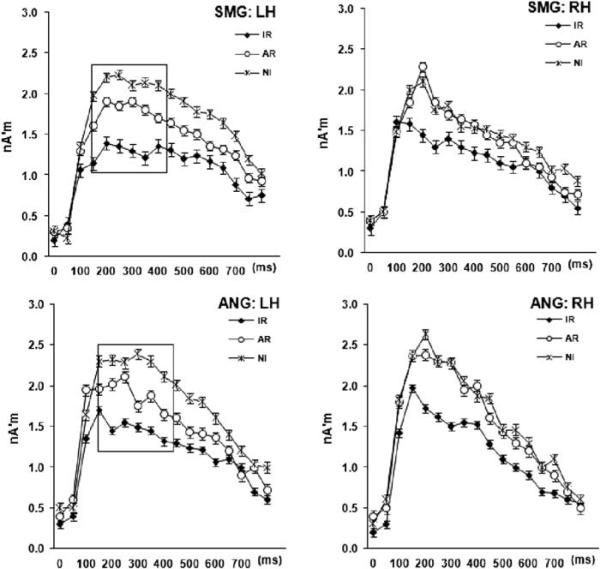
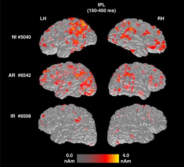
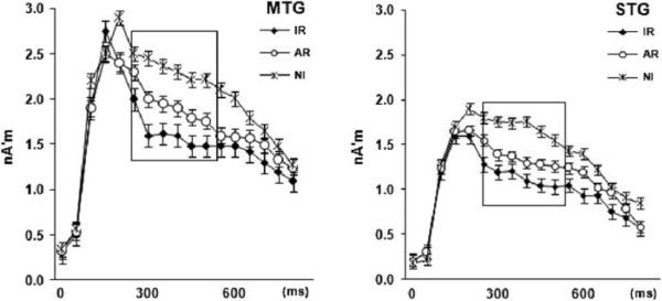
The omnibus ANOVA revealed a significant Time by Hemisphere by ROI by Group interaction, F(60,1410)=1.54, p=.01, which was further explored by performing three-way ANOVAs (Time by Hemisphere by Group) separately for each ROI. As shown in Figures 1 and 2, IR students displayed reduced degree of activity than both AR and NI students in two key areas of the brain circuit for reading: SMG and ANG in the left hemisphere. In addition, significantly reduced activity among IR students was found, bilaterally, in STG and MTG. In the latter ROIs, however, degree of activity did not vary systematically as a function of responder status, being smaller for IR as compared to NI students only (see Figure 3). ANOVA effects which are described in more detail below were assessed at α=05/6=.0083.
Fig. 1.
Time course of neurophysiological activity associated with pseudoword reading estimated between 0 and 800 ms post-stimulus onset in the supramarginal (SMG) and angular (ANG) gyri in the left (LH) and right hemispheres (RH). Significant group differences were only found in the left hemisphere between 200 and 500 ms (indicated by solid rectangles).
Fig. 2.
Baseline brain activation maps from three representative participants: a non-impaired reader (NI; top row), a student who later showed adequate response to intervention (AR; middle row), and a student who did not show adequate response (IR; bottom row). When these recordings were made, this AR student scored 87 and 77 points on the TOWRE Sight Word and Pseudoword reading efficiency subtests, respectively (1-year follow-up scores were 96 and 87 points). Corresponding scores for the IR student shown here, were 80/81 and 83/81 points. The relative intensity of activated voxels is shown at the bottom of the figure. Each set of images presents activity in inferior parietal (IPL) regions, comprised of the angular and supramarginal gyri, where significant group differences were observed, between 200–500 ms, in the left (LH) and right (RH) hemispheres.
Fig. 3.
Time course of estimated neurophysiological activity (in nanoAmpere'meters- nA'm) associated with pseudoword reading in the middle (MTG) and superior (STG) temporal gyri averaged across hemispheres. The latency of significant group differences is indicated by solid rectangles.
Group differences in SMG, F(12,282)=2.82, p=.004, η2=.17, and ANG, F(12,282)=2.71, p=.007, η2=.11, varied between hemispheres as indicated by a Hemisphere by Time by Group interaction. Follow-up ANOVAs revealed a significant Group simple main effect in the left SMG between 200 and 500 ms, F(2,42)=11.40, p=.0001, η2=.22 (this difference approached significance in ANG, F (2,42)=4.30, p=.01, η2=.12). There were no group differences in the right hemisphere at any latency (p>.2, see Figure 1). Pairwise tests showed that students in the NI group had higher degree of activity than IR students (p=.0001) in SMG, who also displayed reduced degree of activity than the AR group (p=.03) for the same region. A similar pattern of group differences was noted in ANG (p=.002 and p=.03, respectively).
Time by Group interactions were found in STG, F(12,282)=2.35, p=.008, η2=.09 and MTG, F(12,282)=2.97, p=.003, η2=.19. Follow-up ANOVAs revealed that significant Group differences, across hemispheres, were restricted to late time windows (300–600 ms) in STG, F(2,42)=9.32, p=.0001, η2=.20, and MTG, F(2,42)=5.57, p=.007, η2=.18. Pairwise group comparisons revealed that NI participants showed greater degree of activity than IR students in each of the two ROIs (p=.001 for STG and p=.008 for MTG; see Figure 3). No other pairwise comparisons came out significant (p>.4 in all cases). Finally, a Group main effect was found for pars opercularis, F(1,47)=6.18, p=.004, and follow-up pairwise tests showed that, throughout the recorded epoch, activity was significantly higher for the NI as compared to the other two groups (p<.02 for both comparisons), which did not differ from each other (p>.9).
Significant hemisphere asymmetries (L>R) in the degree of activity were restricted to SMG, F(1,22)=36.51, p=.0001 between 200 and 500 ms and were noted only for the NI group (p>.3 for the other two groups). A non-significant tendency in the same direction was noted for ANG (p=.01). There were no significant differences in peak latency between the three groups.
Correlations between neurophysiological activity and reading ability
To preserve the temporal information that is inherent in the MNE time series data, Pearson correlation coefficients were computed between estimates of the magnitude of regional neurophysiological activity for each time window in each of the six ROIs and achievement standard difference scores (post-intervention minus pre-intervention TOWRE Word reading efficiency and TOWRE Pseudoword reading efficiency scores). We used difference scores as opposed to post intervention scores to control for individual differences in baseline performance (both within and across subgroups). Initially, these analyses were conducted on the entire sample of students with RD regardless of RTI status (N=27). Table 2 reports significant coefficients (p<.003 to control for family-wise Type I error). Results of the correlational analyses were generally consistent with the findings of group differences in the degree of activity. Specifically, degree of activity in three left hemisphere regions (STG, SMG, and ANG) emerged as significant, positive correlates of subsequent response to intervention.
Table 2.
Pearson correlation coefficients between degree of activity in three left hemisphere ROIs and amount of longitudinal change in TOWRE standard scores for the entire group of poor readers (N = 27)
| Latency window (ms) |
|||||||
|---|---|---|---|---|---|---|---|
| ROI | TOWRE Subscale | 200–300 | 300–400 | 400–500 | 500–600 | 600–700 | 700–800 |
| SMG | WRE | .58 | .57 | ||||
| PsWRE | .72 | .60 | (.53) | ||||
| ANG | WRE | .57 | |||||
| PsWRE | .61 | .57 | (.52) | .62 | .63 | ||
| STG | PsWRE | (.54) | .56 | ||||
Note. Only significant coefficients at a = .003 are shown (r > .56). Coefficients approaching that value are shown in italics. Test of Word Reading Efficiency: WRE (Sight Word) and PsWRE (Pseudoword).
Given that there was substantial non-shared variance between word and pseudoword reading efficiency difference scores (as indicated by a moderate degree of linear association between the two measures, r(27)=.64), differences in the profiles of associations between each measure and MNE variables were possible. Inspection of Table 2 reveals that correlations for degree of activity in SMG and ANG were generally stronger, and covering longer latency windows, when computed with the amount of longitudinal change in TOWRE Decoding than those computed with TOWRE Sight Word reading efficiency difference scores. This finding was expected given the nature of the activation task (reading of pseudowords presented at a relatively rapid rate). Moreover, significant activation-performance associations involving activity in the left STG were restricted to the Decoding TOWRE subtest.
Careful examination of the distribution of individual scores in the univariate regression plots of Figure 4 suggests that most of the systematic covariance between reading fluency and MNE values was present among AR students. This impression was confirmed by the pattern of Pearson correlation coefficients computed separately for each of the two RD subgroups. The linear mapping of individual difference scores onto MNE measures was significant only among AR students. Corresponding coefficients among IR students ranged in the small-positive to moderate-negative range, which was probably due to the restricted range of reading scores in this group. We also computed partial correlations, using each student's baseline reading score as a covariate, as a complementary strategy to control for autoregressive effects on reading measures, leading to the same general pattern of results.
Fig. 4.
Regression plots of TOWRE Pseudoword Reading Efficiency difference scores (representing change in standard scores between baseline and 1-year follow-up) over degree of time-dependent neurophysiological activity in left hemisphere SMG (300–400 ms, upper left panel), ANG (200–300 ms, upper right panel), and STG (500–600 ms, lower left panel). Higher levels of activity at baseline predicted greater improvement in reading efficiency for the entire group of struggling readers, although a stronger association was noted for students who showed adequate response to intervention (open circles).
Neurophysiological predictors of reading outcomes
As shown in Table 2, three MNE variables correlated significantly with TOWRE Sight Word outcomes (pre-post difference scores): Degree of activity in SMG between 200 and 300 ms and also between 700 and 800 ms, and degree of activity in ANG between 200 and 300 ms (all in the left hemisphere). These measures showed only moderate intercorrelations (ranging between r=.45 and .60), and minimally correlated residuals (r<.2; when entered together in a preliminary multiple regression model as predictors of TOWRE Sight Word difference scores). The overall model was significant, Adj. R2=.42, F(2,24)=6.11, p=.003, although individual regression coefficients did not reach significance: SMG 200–300 ms (β=.22; t=1,36; p>.1), SMG 700–800 ms (β=.28; t=1,70; p>.1), ANG 200–300 ms (β=.25; t=1,45; p>.1).
With respect to TOWRE Pseudoword Reading difference scores Table 2 lists six significant correlates, representing degree of activity in three left hemisphere temporo-parietal areas. Inspection of the intercorrelation matrix revealed that three of the six variables [SMG (400–500 ms), ANG (400–500 ms and 600–700 ms)] correlated very strongly with the remaining variables (r>.7). Two of the three variables were, in addition, associated with correlated residuals (r>.4) when all six measures were entered together as independent variables in a preliminary multiple regression model predicting TOWRE pseudoword reading outcomes. The final multiple regression model, after excluding these three variables was significant, Adj R2=.50, F(2,24)=8.34, p=.0001. However, individual regression coefficients failed to reach significance: SMG 300–400 ms (β=.32; t=1.72; p>.1), ANG 200–300 ms (β=.30; t=1.55; p>.1), and STG 500–600 ms (β=.24; t=1.25; p>.1). The relatively small sample size and the relatively high covariances between predictors (.47<r<.66) may account for this result. Thus, although each of the three MNE measures included in the final model showed a rather linear relationship with TOWRE Pseudoword Reading difference scores across RD sub-groups (as shown in the univariate regression plots in Figure 4), results of the multiple regression analyses may only be considered as evidence for the joint contribution of pre-intervention degree of activity in temporo-parietal regions in affecting reading outcomes.
DISCUSSION
The main outcome of the present study was that adequate engagement of left hemisphere temporo-parietal and inferior parietal regions during phonological decoding predicted response to intervention at a 1-year follow-up assessment. This observation is supported by two sets of findings: (1) the degree of pre-intervention activity in these regions (left SMG, STG, and ANG) reliably differentiated students who showed adequate response to intervention from students who did not show notable gains in decoding automaticity; and (2) positive linear associations between regional degree of activity at Time 1 and degree of change in reading fluency scores were restricted to the AR subgroup. Correlations between degree of activity and performance were stronger and more temporally persistent within the recorded epoch for pseudoword than for sight word reading fluency scores, further attesting to the process-specificity of the findings. These results were expected given the nature of the activation task, imposing significant demands for rapid visual/orthographic processing and decoding of meaningless, pronounceable letter-strings, while ensuring relatively high reading accuracy rates even among struggling readers. As with previous brain imaging studies of educational interventions on elementary school children (e.g., Hoeft, Ueno, et al., 2007; Odegard, Ring, Smith, Biggan, & Black, 2008; Shaywitz et al., 2004; Simos et al., 2002, 2005; Simos, Fletcher, Sarkari, Billingsley, et al., 2007; Temple et al., 2003), as well as those conducted with adults (Eden et al., 2004), our findings show that pre-existing neurophysiological activity in left temporo-parietal and inferior parietal cortex underlies successful remedial instruction.
Both groups of RD students in the present study appeared to be comparably engaged in the MEG activation task as indicated by similar levels of in-scanner performance (reading accuracy). Moreover, the two RD sub-groups were comparable on age, minority status, IQ, standardized word-level reading accuracy and fluency tests and on tests of spelling and text comprehension ability. In addition, all students received comparable amounts of remedial instruction, targeting word- and text-level reading skills. Thus, it appears that in children in advanced stages of reading acquisition (at least chronologically), temporoparietal and inferior parietal cortices in the left hemisphere maintain a crucial role in the brain mechanism that supports acquisition of elementary phonological decoding operations.
We also found that, at baseline, brain activation profiles in students who fail to respond to intervention resemble those which generally distinguish children experiencing difficulties in basic reading skills from NI students (Odegard et al., 2008; Simos, Fletcher, Sarkari, Billingsley, et al., 2007). This finding corroborates previous functional imaging reports of marked underactivation of left hemisphere temporo-parietal (STG: Shaywitz et al., 2002; SMG: Cao, Bitan, Chou, Burman, & Booth, 2006; Hoeft, Meyler, et al., 2007; Hoeft, Ueno, et al., 2007; Maisog et al., 2008; Simos, Fletcher, Sarkari, Billingsley, et al., 2007; Simos et al., 2011; van der Mark et al., 2009; and ANG: Eden & Zeffiro, 1998; Temple et al., 2001) during phonological decoding tasks in struggling readers. These earlier results are presently extended to include middle-school, adolescent students who were initially recruited as at risk for academic failure rather than for well-defined core dyslexia deficits. Nevertheless, it is important to consider when interpreting the present findings that the two retrospectively determined subgroups of struggling readers presented with deficits in both basic reading skills and text comprehension. The current results also highlight an important source of individual differences among RD students, namely each student's potential to benefit from remedial instruction. This additional source of individual variability emerged as an important determinant of the magnitude of differences in the degree of regional activation between the RD and NI groups, with the majority of significant differences present between NI students and the subgroup of inadequate responders to intervention.
Although group differences in peak latency of regional activity did not reach significance in the present study, it is worth noting that activity in the left hemisphere SMG and ANG accounting for individual differences in reading ability was restricted to specific latency windows. Moreover, correlational data highlight the importance of engagement of these regions in pseudoword reading starting as early as 200 ms after stimulus onset. Given the nature of the task and converging evidence that these regions host neurophysiological processes involved in converting print-to-sound, this finding may bear relevance within the broader context of aberrant functional connectivity in RD (during tasks emphasizing phonological assembly; Cao, Bitan, & Booth, 2008; Hampson et al., 2006; Horwitz, Rumsey, & Donohue, 1998; Pugh et al., 2000). Thus the present finding is consistent with the notion that early engagement of the left SMG and ANG not only precedes inferior frontal activation, as shown previously using MEG (e.g., Marinković, 2004; Salmelin, Helenius, & Service, 2000), but also provides neuronal input to IFG which is crucial for the overall efficiency of the reading circuit. Whereas this form of functional organization appears to be present in typically developing students, it may also be maintained in students who experience reading difficulties readily amenable to education remediation. It is thus possible that for children who fail to respond to intervention, neurophysiological processes in one or more temporo-parietal areas are so severely deficient that the overall functionality of the brain circuit for reading cannot then become “jump-started” by remedial instruction. Alternatively, a more generalized dysfunction affecting additional circuits for various other cognitive functions may be present in these children, but at present we can only capture the deficit in the circuit responsible for decoding given the nature of the activation task used. This possibility is not very likely given that IR students performed at comparable levels to AR students on a variety of cognitive/language tasks that are part of the IQ tests they were administered.
There are several factors which could limit the impact of remedial instruction on brain function, particularly if the intervention itself does not yield robust results. In this study, the effects of the intervention in the larger educational study were small, a finding commonly encountered in remedial studies of older children with reading disabilities (Vaughn & Fletcher, in press). The sample size in the imaging study was also small, but the students subsequently assigned to the AR group clearly exhibited a greater degree of pre-intervention activity in left temporo-parietal regions, relative to the IR group. The ultimate goal of brain imaging studies, however, is to describe the outline, and the typical and atypical function of the entire network of brain areas that enable reading acquisition. For this purpose it is imperative to examine patterns of structural and functional connections between key brain regions that constitute this network. Whereas some progress has recently been made, mainly through correlational analyses of hemodynamic data (e.g., Cao et al., 2008; Davis et al., 2010; Hampson et al., 2006; Horwitz et al., 1998; Pugh et al., 2000), exploration of the relative timing of regional activation may provide crucial supplementary information. Given the nature of the relevant spatiotemporal data and also the extent of individual differences in demographics, educational history, cognitive abilities and basic patterns of brain activity, this enterprise requires very large samples (e.g., Hoeft, Meyler et al., 2007; Shaywitz et al., 2004; Simos et al., 2011).
Although the present results are promising, at the least, test–retest reliability studies are necessary to assess the stability of pre-intervention estimates of neurophysiological activity in key temporo-parietal areas. Nevertheless, the value of functional neuroimaging as a prognostic adjunct to standard behavioral measures in predicting the outcome of intervention in RD is emphasized by the recent development of objective data analytic techniques with the ability to accurately predict long-term reading gains (e.g., Hoeft et al., 2011). Similarly, such methodological advances in the analysis of group-wise functional brain data have proven to be of value in differentiating between various diagnostic groups (e.g., Pollonini et al., 2010; Tsiaras et al., 2011). This line of evidence further highlights the implications of brain-based research for the development of educational practices.
ACKNOWLEDGMENTS
This research was supported in part by grant P50 HD052117 from the Eunice Kennedy Shriver National Institute of Child Health and Human Development (NICHD). The content is solely the responsibility of the authors and does not necessarily represent the official views of the NICHD or the National Institutes of Health.
Footnotes
The authors declare no conflict of interest.
REFERENCES
- Achenbach TM. Manual for the child behavior checklist/4–18 & 1991 profile. University of Vermont, Department of Psychiatry; Burlington, VT: 1991. [Google Scholar]
- Cao F, Bitan T, Booth JR. Effective brain connectivity in children with reading difficulties during phonological processing. Brain and Language. 2008;107:91–101. doi: 10.1016/j.bandl.2007.12.009. [DOI] [PMC free article] [PubMed] [Google Scholar]
- Cao F, Bitan T, Chou TL, Burman DD, Booth JR. Deficient orthographic and phonological representations in children with dyslexia revealed by brain activation patterns. Journal of Child Psychology and Psychiatry. 2006;47:1041–1050. doi: 10.1111/j.1469-7610.2006.01684.x. [DOI] [PMC free article] [PubMed] [Google Scholar]
- Chen WJ, Faraone SV, Biederman J, Tsuang MT. Diagnostic accuracy of the Child Behavior Checklist scales for attention-deficit hyperactivity disorder: A receiver-operating characteristic analysis. Journal of Consulting and Clinical Psychology. 1994;62:1017–1025. doi: 10.1037/0022-006X.62.5.1017. [DOI] [PubMed] [Google Scholar]
- Dale AM, Fischl B, Sereno MI. Cortical surface-based analysis. I. Segmentation and surface reconstruction. Neuroimage. 1999;9:179–194. doi: 10.1006/nimg.1998.0395. [DOI] [PubMed] [Google Scholar]
- Davis N, Fan Q, Compton DL, Fuchs D, Fuchs LS, Cutting LE, Anderson AW. Influences of neural pathway integrity on children's response to reading instruction. Frontiers in Systems Neuroscience. 2010;4:150. doi: 10.3389/fnsys.2010.00150. [DOI] [PMC free article] [PubMed] [Google Scholar]
- Démonet JF, Taylor MJ, Chaix Y. Developmental dyslexia. Lancet. 2004;363:1451–1460. doi: 10.1016/S0140-6736(04)16106-0. [DOI] [PubMed] [Google Scholar]
- Desikan RS, Ségonne F, Fischl B, Quinn BT, Dickerson BC, Blacker D, Killiany RJ. An automated labeling system for subdividing the human cerebral cortex on MRI scans into gyral based regions of interest. Neuroimage. 2006;31:968–980. doi: 10.1016/j.neuroimage.2006.01.021. [DOI] [PubMed] [Google Scholar]
- Eden GF, Jones KM, Cappell K, Gareau L, Wood FB, Zeffiro TA, Flowers DL. Neural changes following remediation in adult developmental dyslexia. Neuron. 2004;44:411–422. doi: 10.1016/j.neuron.2004.10.019. [DOI] [PubMed] [Google Scholar]
- Eden GF, Zeffiro TA. Neural systems affected in developmental dyslexia revealed by functional neuroimaging. Neuron. 1998;21:279–282. doi: 10.1016/s0896-6273(00)80537-1. [DOI] [PubMed] [Google Scholar]
- Fletcher JM, Stuebing KK, Barth AE, Denton CA, Cirino PT, Francis DJ, Vaughn SR. Cognitive correlates of inadequate response to intervention. School Psychology Review. in press. [PMC free article] [PubMed] [Google Scholar]
- Gabrieli JD. Dyslexia: A new synergy between education and cognitive neuroscience. Science. 2009;325:280–283. doi: 10.1126/science.1171999. [DOI] [PubMed] [Google Scholar]
- Hämäläinen MS, Ilmoniemi RJ. Interpreting magnetic fields of the brain: Minimum norm estimates. Medical & Biological Engineering & Computing. 1994;32:35–42. doi: 10.1007/BF02512476. [DOI] [PubMed] [Google Scholar]
- Hampson M, Tokoglu F, Sun Z, Schafer RJ, Skudlarski P, Gore JC, Constable RT. Connectivity-behavior analysis reveals that functional connectivity between left BA39 and Broca's area varies with reading ability. Neuroimage. 2006;31:513–519. doi: 10.1016/j.neuroimage.2005.12.040. [DOI] [PubMed] [Google Scholar]
- Hoeft F, McCandliss BD, Black JM, Gantman A, Zakerani N, Hulme C, Gabrieli JD. Neural systems predicting long-term outcome in dyslexia. Proceedings of the National Academy of Sciences (U S A) 2011;108:361–366. doi: 10.1073/pnas.1008950108. [DOI] [PMC free article] [PubMed] [Google Scholar]
- Hoeft F, Meyler A, Hernandez A, Juel C, Taylor-Hill H, Martindale JL, Gabrieli JD. Functional and morphometric brain dissociation between dyslexia and reading ability. Proceedings of the National Academy of Sciences of the United States of America. 2007;104:4234–4239. doi: 10.1073/pnas.0609399104. [DOI] [PMC free article] [PubMed] [Google Scholar]
- Hoeft F, Ueno T, Reiss AL, Meyler A, Whitfield-Gabrieli S, Glover GH, Gabrieli JD. Prediction of children's reading skills using behavioral, functional, and structural neuroimaging measures. Behavioral Neuroscience. 2007;121:602–613. doi: 10.1037/0735-7044.121.3.602. [DOI] [PubMed] [Google Scholar]
- Horwitz B, Rumsey JM, Donohue BC. Functional connectivity of the angular gyrus in normal reading and dyslexia. Proceedings of the National Academy of Sciences of the United States of America. 1998;95:8939–8944. doi: 10.1073/pnas.95.15.8939. [DOI] [PMC free article] [PubMed] [Google Scholar]
- Jenkins R, Fuchs LS, van den Broek P, Espin C, Deno SL. Sources of individual differences in reading comprehension and reading fluency. Educational Psychology. 2003;95:719–729. [Google Scholar]
- Jobard G, Crivello F, Tzourio-Mazoyer N. Evaluation of the dual route theory of reading: A metanalysis of 35 neuroimaging studies. Neuroimage. 2003;20:693–712. doi: 10.1016/S1053-8119(03)00343-4. [DOI] [PubMed] [Google Scholar]
- Kim YS, Petscher Y, Schatschneider C, Foorman B. Does growth rate in oral reading fluency matter in predicting reading comprehension achievement? Journal of Educational Psychology. 2010;102:652–667. [Google Scholar]
- Maisog JM, Einbinder ER, Flowers DL, Turkeltaub PE, Eden GF. A meta-analysis of functional neuroimaging studies of dyslexia. Annals of the New York Academy of Sciences. 2008;1145:237–259. doi: 10.1196/annals.1416.024. [DOI] [PubMed] [Google Scholar]
- Marinković K. Spatiotemporal dynamics of word processing in the human cortex. Neuroscientist. 2004;10:142–152. doi: 10.1177/1073858403261018. [DOI] [PMC free article] [PubMed] [Google Scholar]
- Meyler A, Keller TA, Cherkassky VL, Gabrieli JD, Just MA. Modifying the brain activation of poor readers during sentence comprehension with extended remedial instruction: A longitudinal study of neuroplasticity. Neuropsychologia. 2008;46:2580–2592. doi: 10.1016/j.neuropsychologia.2008.03.012. [DOI] [PMC free article] [PubMed] [Google Scholar]
- Odegard TN, Ring J, Smith S, Biggan J, Black J. Differentiating the neural response to intervention in children with developmental dyslexia. Annals of Dyslexia. 2008;58:1–14. doi: 10.1007/s11881-008-0014-5. [DOI] [PubMed] [Google Scholar]
- Pollonini L, Paditar U, Situ N, Rezaie R, Papanicolaou AC, Zouridakis G. Functional connectivity networks in the autistic and healthy brain assessed using granger causality. IEEE Engineering in Medicine and Biology. 2010;2010:1730–1733. doi: 10.1109/IEMBS.2010.5626702. [DOI] [PubMed] [Google Scholar]
- Pugh KR, Mencl WE, Shaywitz BA, Shaywitz SE, Fulbright RK, Constable RT, Gore JC. The angular gyrus in developmental dyslexia: Task-specific differences in functional connectivity within posterior cortex. Psychological Science. 2000;11:51–56. doi: 10.1111/1467-9280.00214. [DOI] [PubMed] [Google Scholar]
- Salmelin R, Helenius P, Service E. Neurophysiology of fluent and impaired reading: A magnetoencephalographic approach. Journal of Clinical Neurophysiology. 2000;17:163–174. doi: 10.1097/00004691-200003000-00005. [DOI] [PubMed] [Google Scholar]
- Schlaggar BL, McCandliss BD. Development of neural systems for reading. Annual Review of Neuroscience. 2007;30:475–503. doi: 10.1146/annurev.neuro.28.061604.135645. [DOI] [PubMed] [Google Scholar]
- Shaywitz BA, Shaywitz SE, Blachman BA, Pugh KR, Fulbright RK, Skudlarski P, Gore JC. Development of left occipitotemporal systems for skilled reading in children after a phonologically-based intervention. Biological Psychiatry. 2004;55:926–933. doi: 10.1016/j.biopsych.2003.12.019. [DOI] [PubMed] [Google Scholar]
- Shaywitz BA, Shaywitz SE, Pugh KR, Mencl WE, Fulbright RK, Skudlarski P, Gore JC. Disruption of posterior brain systems for reading in children with developmental dyslexia. Biological Psychiatry. 2002;52:101–110. doi: 10.1016/s0006-3223(02)01365-3. [DOI] [PubMed] [Google Scholar]
- Shaywitz SE, Shaywitz BA. Dyslexia (specific reading disability) Biological Psychiatry. 2006;57:1301–1309. doi: 10.1016/j.biopsych.2005.01.043. [DOI] [PubMed] [Google Scholar]
- Simos PG, Breier JI, Fletcher JM, Bergman E, Papanicolaou AC. Cerebral mechanisms involved in word reading in dyslexic children: A magnetic source imaging approach. Cerebral Cortex. 2000;10:809–816. doi: 10.1093/cercor/10.8.809. [DOI] [PubMed] [Google Scholar]
- Simos PG, Fletcher JM, Bergman E, Breier JI, Foorman BR, Castillo EM, Papanicolaou AC. Dyslexiaspecific brain activation profile become normal following successful remedial training. Neurology. 2002;58:1203–1213. doi: 10.1212/wnl.58.8.1203. [DOI] [PubMed] [Google Scholar]
- Simos PG, Fletcher JM, Sarkari S, Billingsley RL, Denton C, Papanicolaou AC. Altering the brain circuits for reading through intervention: A magnetic source imaging study. Neuropsychology. 2007;21:485–496. doi: 10.1037/0894-4105.21.4.485. [DOI] [PubMed] [Google Scholar]
- Simos PG, Fletcher JM, Sarkari S, Billingsley RL, Francis DJ, Castillo EM, Papanicolaou AC. Early development of neurophysiological processes involved in normal reading and reading disability: A magnetic source imaging study. Neuropsychology. 2005;19:787–798. doi: 10.1037/0894-4105.19.6.787. [DOI] [PubMed] [Google Scholar]
- Simos PG, Fletcher JM, Sarkari S, Billingsley-Marshall R, Denton CA, Papanicolaou AC. Intensive instruction affects brain magnetic activity associated with oral word reading in children with persistent reading disabilities. Journal of Learning Disabilities. 2007;40:37–48. doi: 10.1177/00222194070400010301. [DOI] [PubMed] [Google Scholar]
- Simos PG, Rezaie R, Fletcher JM, Juranek J, Passaro AP, Li Z, Papanicolaou AC. Functional disruption of the brain mechanism for reading: Effects of comorbidity and task difficulty among children with developmental learning problems. Neuropsychology. 2011 doi: 10.1037/a0022550. Epub ahead of print. [DOI] [PMC free article] [PubMed] [Google Scholar]
- Swanson J, Schuck S, Mann M, Carlson C, Hartman K, Sergeant J, McCleary R. Categorical and dimensional definitions and evaluations of symptoms of ADHD: The SNAP and the SWAN Ratings Scales. 2005 Retrieved from http://www.adhd.net. [PMC free article] [PubMed]
- Temple E, Deutsch GK, Poldrack RA, Miller SL, Tallal P, Merzenich MM, Gabrieli JD. Neural deficits in children with dyslexia ameliorated by behavioral remediation: Evidence from functional MRI. Proceedings of the National Academy of Sciences of the United States of America. 2003;100:2860–2865. doi: 10.1073/pnas.0030098100. [DOI] [PMC free article] [PubMed] [Google Scholar]
- Temple E, Poldrack RA, Salidis J, Deutsch GK, Tallal P, Merzenich MM, Gabrieli JD. Disrupted neural responses to phonological and orthographic processing in dyslexic children: An fMRI study. Neuroreport. 2001;12:299–307. doi: 10.1097/00001756-200102120-00024. [DOI] [PubMed] [Google Scholar]
- Torgesen JK, Wagner R, Rashotte C. Test of word reading efficiency. Pro-Ed; Austin, TX: 1999. [Google Scholar]
- Tsiaras V, Simos P, Rezaie R, Sheth B, Garyfallidis E, Martinez Castillo E, Papanicolaou AC. Extracting biomarkers of autism from MEG based resting state functional connectivity networks. Computers in Biology and Medicine. 2011 doi: 10.1016/j.compbiomed.2011.04.004. Epub ahead of print. [DOI] [PubMed] [Google Scholar]
- van der Mark S, Bucher K, Maurer U, Schulz E, Brem S, Buckelmuller J, Brandeis D. Children with dyslexia lack multiple specializations along the visual word-form (VWF) system. Neuroimage. 2009;47:1940–1949. doi: 10.1016/j.neuroimage.2009.05.021. [DOI] [PubMed] [Google Scholar]
- Vaughn S, Cirino PT, Wanzek J, Wexler J, Fletcher JM, Denton CA, Francis DJ. Response to intervention for middle school students with reading difficulties: Effects of a primary and secondary intervention. School Psychology Review. 2010;39:3–21. [PMC free article] [PubMed] [Google Scholar]
- Vaughn S, Fletcher JM. RTI in secondary schools. Journal of Learning Disabilities. in press. [Google Scholar]
- Vaughn S, Wanzek J, Wexler J, Barth A, Cirino PT, Fletcher J, Francis D. The relative effects of group size on reading progress of older students with reading difficulties. Reading and Writing. 2010;23:931–956. doi: 10.1007/s11145-009-9183-9. [DOI] [PMC free article] [PubMed] [Google Scholar]
- Yovanoff P, Duesbery L, Alonzo J, Tindal G. Grade-level invariance of a theoretical causal structure predicting reading comprehension with vocabulary and oral reading fluency. Educational Measurement: Issues and Practice. 2005;24:4–12. [Google Scholar]


