Abstract
Previous studies have demonstrated that Polarization Sensitive Optical Coherence Tomography (PS-OCT) can be used to image the remineralization of early artificial caries lesion on smooth enamel surfaces of human and bovine teeth. However, most new dental decay is found in the pits and fissures of the occlusal surfaces of posterior dentition and it is in these high risk areas where the performance of new caries imaging devices need to be investigated. The purpose of this study was to demonstrate that PS-OCT can be used to measure the subsequent remineralization of artificial lesions produced in the pits and fissures of extracted 3rd molars. A PS-OCT system operating at 1310-nm was used to acquire polarization resolved images of occlusal surfaces exposed to a demineralizing solution at pH-4.5 followed by a fluoride containing remineralizing solution at pH-7.0 containing 2-ppm fluoride. The integrated reflectivity was calculated to a depth of 200-μm in the entire lesion area using an automated image processing algorithm. Although a well-defined surface zone was clearly resolved in only a few of the samples that underwent remineralization, the PS-OCT measurements indicated a significant (p<0.05) reduction in the integrated reflectivity between the severity of the lesions that were exposed to the remineralization solution and those that were not. The lesion depth and mineral loss were also measured with polarized light microscopy and transverse microradiography after sectioning the teeth. These results show that PS-OCT can be used to non-destructively monitor the remineralization potential of anti-caries agents in the important pits and fissures of the occlusal surface.
Keywords: Enamel, caries, PS-OCT, image processing algorithms, early demineralization, edge detection
1. INTRODUCTION
Previous studies in our laboratory have demonstrated that PS-OCT can be used successfully to measure changes in the reflectivity of demineralized areas on smooth enamel surfaces after exposure to a remineralizing solution 1–4.
If carious lesions are detected early enough, it is likely that they can be arrested/reversed by non-surgical means through fluoride therapy, anti-bacterial therapy, dietary changes, or by low intensity laser irradiation5,6. Therefore, one cannot overstate the importance of detecting the decay in the early stage of development at which point non-invasive preventive measures can be taken to halt further decay and new technologies for the assessment of surface demineralization for clinical trials need to be validated for the most prevalent lesion types, such as pre-cavitated lesions on proximal and occlusal surfaces, root caries and secondary caries around restorations.
Accurate determination of the degree of lesion activity and severity is of paramount importance for the effective employment of the treatment strategies mentioned above. Since optical diagnostic tools exploit changes in the light scattering of the lesion they have great potential for the diagnosis of the current “state of the lesion”, i.e., whether or not the caries lesion is active and expanding or whether the lesion has been arrested and is undergoing remineralization. Therefore, new technologies must be able to determine whether active caries lesions have been arrested and/or have undergone remineralization. PS-OCT is uniquely capable of this task since it provides a measure of the reflectivity from each layer of the lesion and is able to show the formation of a zone of increased mineral density and reduced light scattering due to remineralization. This is a very important distinction between PS-OCT and other optical caries detection methods, such as the Diagnodent and QLF, that only provide a single measure of lesion severity representing fluorescence loss or gain from the lesion and are not capable of resolving the internal lesion structure. Although, recent efforts in QLF development have focused on using other means to show the presence of a surface zone which involve hydration/dehydration of the lesion during image acquisition, it is more advantageous to be able to acquire in depth images of the lesion structure 7. Such data is also valuable for caries management by risk assessment in the patient and for determining the appropriate form of intervention. A non-destructive, quantitative method of monitoring demineralization and remineralization “in vivo” with high sensitivity would be invaluable for use in short term clinical trials for various anti-caries agents such as fluoride dentifrices and antimicrobials.
In previous studies we investigated the remineralization of smooth enamel surfaces employing two caries models. The first model involved pH cycling to produce lesions with a well-defined surface zone of intact enamel1 while the second model used a different demineralization model to produce a surface softened lesion3. Both models showed markedly different outcomes after exposure to the remineralization solution. Studies have shown that remineralization requires the presence of residual partially dissolved crystals to serve as a template for growth 8. Furthermore, remineralization has been observed to proceed from the outside of the lesion towards the lesion body, therefore as the remineralization takes place in the surface zone of the lesion the diffusion pathways to the lesion body are blocked thus preventing further remineralization of the lesion body. However, the lesion does become arrested since further dissolution in the lesion body is also blocked. This is typically how lesions are arrested naturally. We observed that the surface softened lesion model yields the greatest change in mineral content upon remineralization since it does not contain a well-defined surface layer that inhibits diffusion 8.
The purpose of this study was to demonstrate that PS-OCT can be used to measure the subsequent remineralization of artificial lesions produced in the pits and fissures of extracted 3rd molars. Most new dental decay is found in the pits and fissures of the occlusal surfaces of posterior dentition and it is in these high risk areas where the performance of new caries imaging need to be investigated. Measurement in these important areas is difficult due to the challenging topography and because the enamel solubility is highly variable in these areas and it is not possible to produce lesions of a consistent severity.
2. MATERIALS AND METHODS
2.1 Artificial Occlusal Surface Caries
Extracted teeth from patients in the San Francisco Bay area were collected with CHR approval, cleaned, sterilized with gamma radiation, and stored in a moist environment to preserve tissue hydration with 0.1% thymol added to prevent bacterial growth. Sound human 3rd molar teeth (n=20) were mounted on acrylic blocks after root resection. As shown in Fig. 1, small incisions were made using a CO2 laser to demarcate the area of interest in the OCT images. An acid resistant varnish, red nail polish, Revlon(New York, NY), was applied on all areas if the teeth outside the 2×2 mm2 occlusal surface region, which was scanned using the PS-OCT system. Artificial lesions were formed by exposing the teeth for 5 days to a 50 mL aliquot of an acetate buffer solution containing 2.0 mmol/L calcium, 2.0 mmol/L phosphate and 0.075 mol/L acetate maintained at pH 4.5 and a temperature of 37°C. After the lesions were created, the samples were exposed for 21 days in a 50 mL remineralizing solution of 1.5 mmol/L calcium, 0.9 mmol/L phosphate, 150 mmol/L KCl, 20 mmol/L cacodylate buffer maintained at pH 7.0 and 37°C. 2 ppm F- in the form of NaF was added to the solution to enhance the remineralization effect. This is a “surface softened” model, which produces subsurface dissolution without erosion and a well defined surface zone after exposure to a remineralization solution that was visible using PS-OCT 3.
Fig. 1.
Microscope image after demineralization (A) and after remineralization (B). Yellow dotted lines mark approximate location of OCT scan.
2.2 Polarization Sensitive Optical Coherence Tomographic Imaging (PS-OCT)
A single-mode fiber autocorrelator-based Optical Coherence Domain Reflectometry (OCDR) system with polarization switching probe, high efficiency piezoelectric fiber-stretchers and two balanced InGaAs receivers that was designed and fabricated by Optiphase, Inc. (Van Nuys, CA) was integrated with a broadband high power superluminescent diode (SLD), Denselight (Jessup, MD) with an output power of 45-mW and a bandwidth of 35 nm and a high-speed XY-scanning system, ESP 300 controller & 850-HS stages, Newport (Irvine, CA) and used for in vitro optical tomography. The system was configured to provide an axial resolution at 22-μm in air and 14-μm in enamel and a lateral resolution of approximately 50-μm over the depth of foucs of 10 mm. The all-fiber OCDR system has been previously described in greater detail9,10. The PS-OCT system was completely controlled using LabVIEW™ software, National Instruments (Austin TX). The samples were scanned with PS-OCT following 5 days of demineralization and once more following 21 days of remineralization. A total of 30 to 40 b-scans were acquired for each tooth at 25-μm intervals for the 3-D tomographic image, which encompassed a 2 × 2 mm2 area of the exposed tooth surface.
2.3 Polarized Light Microscopy (PLM) and Digital Transverse Microradiography (TMR)
After sample imaging was completed, approximately 200 μm thick serial sections were acquired using an Isomet 5000 saw (Buehler, IL), for polarized light microscopy (PLM) and digital transverse microradiography (TMR) in order to confirm remineralization. PLM was carried out using a Meiji Techno RZT microscope (Meiji Techno Co., LTD, Saitama, Japan) with an integrated digital camera, Canon EOS Digital Rebel XT (Canon Inc., Tokyo, Japan). The sample sections were imbibed in water and examined in the brightfield mode with crossed polarizers and a red I plate with 500 nm retardation. A custom-built digital transverse microradiography (TMR) system was used to measure mineral loss in the different partitions of the sample. A high-speed motion control system with Newport UTM150 and 850G stages and an ESP300 controller coupled to a video microscopy and laser targeting system was used for precise positioning of the tooth samples in the field of view of the imaging system. The volume percent mineral for each sample thin section was determined by comparison with a calibration curve of x-ray intensity versus sample thickness created using sound enamel sections of 86.3 ± 1.9% volume percent mineral varying from 50 to 300 μm in thickness. The calibration curve was validated via comparison with cross sectional microhardness measurements. The volume percent mineral determined using microradiography for section thickness ranging from 50 to 300 μm highly correlated with the volume percent mineral determined using microhardness r2 = 0.99 (See paper #7162–33 in this proceedings).
2.4 PS-OCT Image Processing and Assessment of Lesion Severity
Images obtained from the PS-OCT orthogonal polarization scans (⊥-axis) were filtered using a 3×3 median filter in order to reduce the speckle noise prior to processing using MATLAB.. Vertical line profiles were taken from the orthogonal polarization PS-OCT scans to a real depth of 200-μm, yielding the integrated reflectivity, ΔR, of the regions in units of (dB·μm). This process was automated by using a program written in Java (see other paper #7162–33 in this proceedings). A 3×3 median filter and an automatic edge detection scheme were also applied prior to obtaining the integrated reflectivity for the entire 3-D tomographic image. An average integrated reflectivity was calculated by averaging the integrated reflectivity values within the entire 2 × 2 mm2 area.
3. RESULTS AND DISCUSSION
PS-OCT orthogonal polarization scans that were taken along the dotted line in Fig. 1, are shown in Fig. 2. Figure 2A shows the demineralized occlusal surface, and the signal intensity in the remineralized occlusal surface shown in Figure 2B was noticeably reduced compared to images from the demineralized lesion. This was anticipated since exposure to a fluoride containing remineralization solution is expected to increase the mineral content of the lesion by redeposition of fluorapatite and hydroxyapatite. If the mineral content in the lesion area is increased to a sufficient level than the reflectivity should manifest a decrease in reflectivity (scattering) 1,3,11 We were only able to resolve a distinct remineralization layer on a few of the samples using PS-OCT, PLM and TMR. Figure 3 shows one of the samples for which a distinct remineralization. Reflected light from the tooth surface is often not visible in the orthogonal polarization image (⊥-axis) and it is necessary to examine the images from both polarizations to identify the remineralization layer 3,4. This layer was determined by overlaying the two images to identify the position of the tooth surface, since the parallel polarization (||-axis) scans manifest high reflectivity at the tooth surface.
Fig. 2.
PS-OCT orthogonal polarization scans after demineralization (A) and after remineralization (B). Remineralized lesion shows decrease in overall reflectivity and a surface zone of lower reflectivity after treatment.
Fig. 3.
PS-OCT, TMR, and PLM images of one of the samples after remineralization. The orange layer in the (⊥-axis) PS-OCT scan (A) represents the remineralization layer. The remineralization layer can also be distinguished in the TMR (B) and PLM images (C).
The signal in orthogonal polarization is minimal near the sound enamel surface and high at the demineralized regions due to depolarization of the incident polarized light and backscattering. Remineralized lesions typically have a layer of enhanced mineral content tens of microns thick at the lesion surface. PS-OCT is ideally suited for measuring the thickness of this surface zone3,4. Remineralization layer thickness is the distance between the surface of the sample and the edge of demineralization. PS-OCT can be used to measure the distance between the edge of the parallel polarization scan and the edge of the orthogonal polarization scan in order to determine remineralization layer. The remineralization layer derived from this method using PS-OCT is shown as the orange color layer in Fig. 3A, and it is consistent with TMR (Fig. 3B).
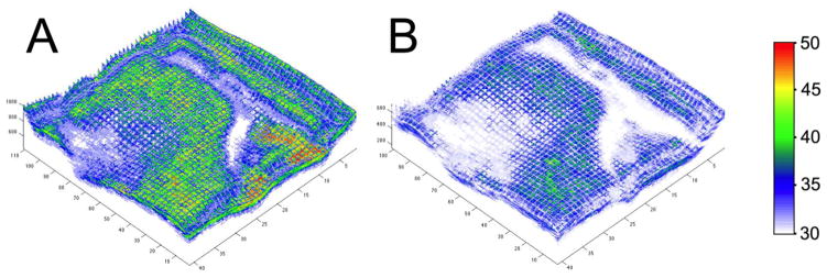
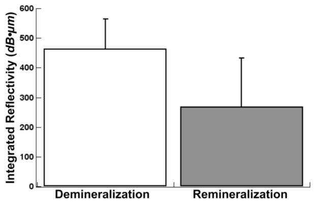
Multiple b-scans were assembled to yield a full 3-D tomographic image of the entire 2×2 mm2 region of interest (ROI) as shown in Fig. 4. The integrated reflectivity was calculated for the entire ROI before and after remineralization to a depth of 200-μm from the surface. A paired t-test was used to show that the average integrated reflectivity was significantly lower, p<0.05, after remineralization, Fig. 5. The average integrated reflectivity values, ΔR, were 465±99 vs. 268±160 before and after remineralization, respectively. 3-D images show the severity and the location of the areas of demineralization.
Fig. 4.
Tomographic PS-OCT images (⊥-axis) after demineralization (A) and after remineralization (B). Remineralized areas show decrease in overall reflectivity with the existence of a surface zone or area of lower reflectivity after treatment.
Fig. 5.
Mean ± s.d. average integrated reflectivity to a depth of 200-μm of the 2×2 mm2 occlusal surface measured with PS-OCT. Bars not sharing any common color were significantly different, p<0.05 (n=20).
The purpose of this study was to determine whether PS-OCT could be used to nondestructively assess demineralization and remineralization on occlusal surfaces and achieve improved discrimation of lesion severity by using an automatic integration routine that identified the position of the tooth surface and integrated the reflectivity to a depth of 200-μm so that changes over a large region of interest could be rapidly calculated.
Remineralization occurs throughout the body of the lesion but is typically most dramatic near the lesion surface. There was a distinct surface layer of low reflectivity/higher mineral content in only a few of the samples after exposure to the remineralization solution, but this layer was visible in those samples with all three imaging modalities PS-OCT, PLM and TMR. PS-OCT was successful in measuring a significant difference in the integrated reflectivity after exposure to the remineralization solution and the images are consistent with the PLM and TMR images acquired after sectioning of the teeth. Most new dental decay is found in the pits and fissures of the occlusal surfaces, therefore it is more appropriate to evaluate the efficacy of anti-caries agents in these high risk areas as opposed to smooth surfaces and this study has demonstrated that nondestructive assessment is feasible using PS-OCT in such areas.
The edge-detection algorithms were successful in processing the full 3D PS-OCT tomographic images of the region of interest. Such 3-D images are valuable for visualization and analysis of the entire lesion. Integration over the entire region of interest instead of integrating the lesion reflectivity a single a-scan or b-scan allows improved discrimination of lesion severity for the non-destructive assessment of the performance of anti-caries agents. The automated approach employed in this study is advantageous for exploiting the high speed FD-OCT systems currently being developed which are capable of acquiring entire tomographic images of large areas of the tooth such as the 2×2 mm2 ROI in this study at video rates (>30Hz) producing an enormous volume of data that can only be analyzed effectively in a realistic time frame using an automated processing algorithms.
Acknowledgments
The authors acknowledge the support of NIH grants R01-DE017869 and R01-DE14698.
References
- 1.Jones RS, Darling CL, Featherstone JDB, Fried D. Remineralization of in vitro Dental Caries Assessed with Polarization Sensitive Optical Coherence Tomography. J of Biomed Opt. 2006;11:014016, 014011–014019. doi: 10.1117/1.2161192. [DOI] [PubMed] [Google Scholar]
- 2.Jones RS, Fried D. Quantifying the remineralization of artificial caries lesions using PS-OCT. 2006;6137:613701–613708. [Google Scholar]
- 3.Jones RS, Fried D. Remineralization of Enamel Caries Can Decrease Optical Reflectivity. J Dent Res. 2006;85:804–808. doi: 10.1177/154405910608500905. [DOI] [PMC free article] [PubMed] [Google Scholar]
- 4.Can AM, Darling CL, Fried D. Lasers in Dentistry XIV. Vol. 6843. San Jose, CA, USA: 2008. High-resolution PS-OCT of enamel remineralization; pp. 68430T–68437. [DOI] [PMC free article] [PubMed] [Google Scholar]
- 5.NIH. Report No 18. 2001. [Google Scholar]
- 6.Featherstone JDB. Prevention and reversal of dental caries:role of low level fluoride. Community Dent Oral Epidemiol. 1999;27:31–40. doi: 10.1111/j.1600-0528.1999.tb01989.x. [DOI] [PubMed] [Google Scholar]
- 7.van der Veen MH, de Josselin de Jong E, Al-Kateeb S. Caries Activity Detection by Dehydration with Qualitative Light Fluorescence. Early detection of Dental caries II. 1999;4:251–260. [Google Scholar]
- 8.ten Cate JM, Arends J. Remineralization of artificial enamel lesions in vitro. Caries Res. 1977;11:277–286. doi: 10.1159/000260279. [DOI] [PubMed] [Google Scholar]
- 9.Bush J, Davis P, Marcus MA. All-Fiber Optic Coherence Domain Interferometric Techniques. Fiber Optic Sensor Technology II. 2000;4204:71–80. [Google Scholar]
- 10.Ngaotheppitak P, Darling CL, Fried D, Bush J, Bell S. PS-OCT of occlusal and interproximal caries lesions viewed from occlusal surfaces. Lasers in Dentistry X. 2006;6137:61370L. [Google Scholar]
- 11.Darling CL, Huynh GD, Fried D. Light Scattering Properties of Natural and Artificially Demineralized Dental Enamel at 1310-nm. J Biomed Optics. 2006;11:034023, 034021-034011. doi: 10.1117/1.2204603. [DOI] [PubMed] [Google Scholar]


