Abstract
Dopamine (DA) D2 receptors expressed in DA neurons (D2 autoreceptors) exert a negative feedback regulation that reduces DA neuron firing, DA synthesis and DA release. As D2 receptors are mostly expressed in postsynaptic neurons, pharmacological and genetic approaches have been unable to definitively address the in vivo contribution of D2 autoreceptors to DA-mediated behaviors. We found that midbrain DA neurons from mice deficient in D2 autoreceptors (Drd2loxP/loxP; Dat+/IRES-cre, referred to as autoDrd2KO mice) lacked DA-mediated somatodendritic synaptic responses and inhibition of DA release. AutoDrd2KO mice displayed elevated DA synthesis and release, hyperlocomotion and supersensitivity to the psychomotor effects of cocaine. The mice also exhibited increased place preference for cocaine and enhanced motivation for food reward. Our results highlight the importance of D2 autoreceptors in the regulation of DA neurotransmission and demonstrate that D2 autoreceptors are important for normal motor function, food-seeking behavior, and sensitivity to the locomotor and rewarding properties of cocaine.
DA neurotransmission participates in complex brain functions, including the initiation and planning of motor activity, the identification of salient stimuli that predict reward, and the spatio-temporal organization of goal-oriented behaviors1. The importance of central dopaminergic systems has been appreciated for decades as a result of the motor, cognitive, emotional and social deficits that constitute the hallmarks of frequent human disorders, such as Parkinson’s disease, schizophrenia, attention-deficit and hyperactivity disorder, and compulsive drug abuse2,3. Natural rewards, such as food or sex, exert their reinforcing properties by eliciting a fast increase in extracellular DA in the brain4. Drugs of abuse take advantage of this system by increasing extracellular DA to levels that greatly exceed those triggered by any natural reward3. Addictive drugs use various mechanisms to raise extracellular DA levels. For example, nicotine and opiates increase DA neuron firing, cocaine blocks DA reuptake, and amphetamine and other phenylethylamines release DA from DA terminals5. Research in humans3, monkeys6 and rodents7 has shown that increased vulnerability to drug addiction correlates with reduced availability of striatal D2 receptors, and healthy non-abusing volunteers expressing low levels of D2 receptors report more pleasant experiences when taking drugs of abuse8,9. These results appear to conflict with studies performed in mutant mice that lack D2 receptors, which showed reduced or absent reinforcing properties for drugs of abuse, such as cocaine, morphine and ethanol10–14. This apparent contradiction may result from the different roles that D2 receptors have in distinct neuronal types.
The majority of D2 receptors are located on postsynaptic non-dopaminergic neurons that integrate several brain circuits. In addition, D2 autoreceptors present in the somas, dendrites and terminals of DA neurons exert an ultra-short negative feedback regulation of DA transmission15–19. Although somatodendritic D2 autoreceptors modulate firing rate15,17,18, those located on nerve terminals regulate DA synthesis and release16,19. Locally released DA inhibits the activity of DA neurons, and therefore inhibits the subsequent release of DA in terminal regions17,20. In addition, activation of autoreceptors present on dopaminergic terminals diminishes terminal excitability and the probability of further DA release20. Because DA autoreceptors produce feedback inhibition of DA transmission, impaired autoreceptor function would likely lead to increased DA neuron excitability and augmented DA release, posing a risk factor for impulsive behavior, hyperactivity, drug addiction and vulnerability to relapse. In fact, a recent study found that highly impulsive individuals are characterized by diminished midbrain autoreceptor availability21.
In vivo blockade or stimulation of D2 autoreceptors has been hampered by the fact that receptor-targeted compounds also interact with postsynaptic D2 receptors, which are more abundantly expressed in all DA target areas. In addition, the pharmacological properties of D2 receptors are very similar to those of D3 and D4 receptors (all members of the D2-like subfamily), and, as a result, stimulation or inhibition of the individual D2-like receptor subtypes in vivo is impractical22. Genetic approaches undertaken to study the functional role of D2 autoreceptors have also failed to settle these issues. Although mutant mice lacking D2 receptors have revealed some of the ex vivo properties of D2 autoreceptors23, Drd2−/− mice have not been used to specifically study D2 autoreceptor function because the simultaneous loss of all pre- and postsynaptic D2 receptors elicits a number of diverse overlapping phenotypes (for example, Drd2−/− mice are dwarfs owing to deficits in the growth hormone axis24). Although impaired DA autoreceptor function may substantially modify motor performance, motivational states and subjective values of reinforcers, the in vivo contribution of this inhibitory system to DA-mediated behaviors remains unknown. To circumvent this difficulty, we generated conditional mutant mice by deleting the D2 receptor gene (Drd2) in dopaminergic neurons (autoDrd2KO mice). AutoDrd2KO mice lacked D2 autoreceptors and expressed postsynaptic D2 receptors normally on non-dopaminergic neurons in all of the brain and peripheral regions that we examined. This mouse model allowed us to directly evaluate the importance of D2 autoreceptor inhibitory control in dopaminergic neurotransmission, DA-mediated locomotor activity and the rewarding properties of food and cocaine.
RESULTS
Lack of autoinhibition in DA neurons from autoDrd2KO mice
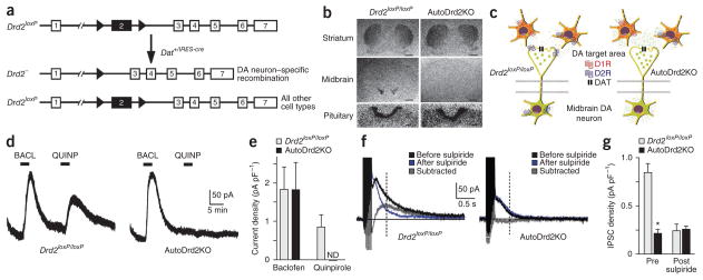
We generated autoDrd2KO mice by consecutively breeding Drd2loxP/loxP and Dat+/IRES-cre mice (Fig. 1a and Supplementary Fig. 1). These two C57BL/6J congenic (≥10) parental strains were overtly normal in all of the tested parameters (Supplementary Fig. 2 and ref. 25). [3H]Nemonapride-binding autoradiography revealed normal D2 receptor levels in forebrain and pituitary sections of autoDrd2KO mice (Fig. 1b), but no signal was detected in midbrain sections. Identical results were obtained by in situ hybridization using a Drd2 exon 2 antisense riboprobe (Supplementary Fig. 3a). Thus, autoDrd2KO mice are authentic D2 autoreceptor knockout mice (Fig. 1c). AutoDrd2KO mice are viable and a broad physical and anatomical inspection did not reveal any overt phenotypic differences from their Drd2loxP/loxP littermates (Supplementary Fig. 3b).
Figure 1.
Selective ablation of DA D2 autoreceptors prevents somatodendritic D2 like–mediated inhibition of midbrain DA neurons. (a) Schematic of conditional mutagenesis in the mouse D2 receptor gene Drd2. Exon 2 (black) is flanked by loxP sites (black triangles). Drd2 exon 2 is excised by Cre in dopaminergic neurons in Drd2loxP/loxP; Dat+/IRES-cre, mice. (b) [3H]Nemonapride-binding autoradiography. Scale bars represent 1 mm for brain sections and 100 μm for pituitary sections. (c) Schematic comparing midbrain DA neurons of Drd2loxP/loxP and autoDrd2KO mice. The absence of D2 autoreceptors predicts enhanced DA synthesis and release. (d) Whole-cell voltage-clamp recordings (Vh = −55 mV) from midbrain DA neurons. Baclofen (BACL, 5 μM) and quinpirole (QUINP, 200 nM) were applied as indicated by horizontal black bars. (e) The current density induced by each agonist was plotted for neurons obtained from Drd2loxP/loxP and autoDrd2KO mice (n = 6–7). ND, not detected. (f) The averages of five traces showing IPSCs evoked by electrical stimulation before (black) and after (blue) sulpiride application, as well as the sulpiride-sensitive component (gray), are plotted. The dashed vertical lines indicate the average time to peak of the sulpiride-sensitive component of the IPSC (0.43 ± 0.10 s, n = 7) in Drd2loxP/loxP neurons. (g) IPSC densities measured at the average time to peak before and after sulpiride are shown for Drd2loxP/loxP and autoDrd2KO mice (n = 6–8). *P < 0.005. Error bars represent s.e.m.
The soma and dendrites of midbrain DA neurons express autoreceptors that modulate firing rates by inducing inhibitory currents15,17. Voltage-clamp recordings (−55 mV) from midbrain dopaminergic neurons revealed that the D2-like (includes D2, D3 and D4 receptors) agonist quinpirole (0.2 μM) induced a slow hyperpolarizing current in neurons obtained from Drd2loxP/loxP mice, but not from autoDrd2KO mice (Fig. 1d,e), whereas dopaminergic neurons recorded from Drd2loxP/loxP and autoDrd2KO midbrain slices responded equally to the GABAB agonist baclofen (5 μM). A train of electrical stimulation, applied in the presence of AMPA, NMDA, GABAA and α-adrenergic blockers evoked inhibitory postsynaptic currents (IPSCs) that were reduced by the D2-like receptor antagonist sulpiride (150 nM) in Drd2loxP/loxP mouse neurons, but not in those from autoDrd2KO mice (Fig. 1f,g). Thus, endogenous DA release acting on somatodendritic D2 autoreceptors constitutes a major component of the total G protein– coupled receptor–mediated inhibitory response in control mice (Fig. 1g). The lack of an effect of quinpirole and sulpiride on midbrain dopaminergic neurons of autoDrd2KO mice strongly suggests that D2 is the predominant D2-like autoreceptor responsible for feedback inhibition of midbrain dopaminergic neuronal activity via somatodendritic actions. Notably, the IPSC density that we recorded from autoDrd2KO mouse neurons was similar to what we observed in neurons from control mice treated with sulpiride (Fig. 1g), indicating that no other G protein–coupled receptor–mediated inhibitory mechanism compensated for the lack of D2 autoreceptors. Together, these results suggest that the endogenous DA-mediated inhibitory regulation of DA neuron firing is severely impaired in autoDrd2KO mice.
Tight control of DA release and synthesis by D2 autoreceptors
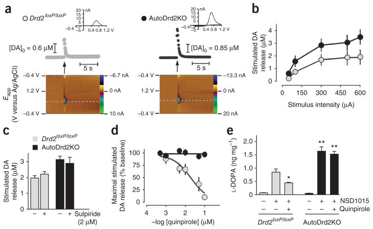
We used fast-scan cyclic voltammetry (FSCV) in dorsal striatal slices to investigate how D2-like autoreceptors present on dopaminergic terminals regulate DA release at a subsecond resolution. The amount of DA released by a single 300-μA pulse was significantly higher (~60%, P < 0.001) in autoDrd2KO mice than in controls (Fig. 2a). Greater DA release was observed at all stimulus intensities (Fig. 2b). In this single-pulse procedure, sulpiride (2 μM) did not affect DA release in striatal slices of either genotype (Fig. 2c), as was previously reported26,27.
Figure 2.
Increased DA release and DA synthesis in autoDrd2KO mice. (a) DA release in the dorsal striatum evoked by a single stimulus pulse (300–600 μA, 0.6 ms per phase, biphasic; arrows). Top, time course of DA concentration changes. Insets represent the background-subtracted cyclic voltammograms indicative of DA. Bottom, two-dimensional representations of the voltammetric data. The voltammetric current is plotted against the applied potential (Eapp) and the acquisition time. (b) Input-output relationship of DA release elicited by single-pulse stimulation across a range of stimulus intensities in the dorsal striatum of Drd2loxP/loxP (n = 4) and autoDrd2KO mice (n = 5) (F1,29 = 10.27, P < 0.001). (c) Stimulated DA release in autoDrd2KO (n = 16) and control mice (n = 11) does not change in the presence of 2 μM sulpiride (Drd2loxP/loxP mice, n = 8; autoDrd2KO mice, n = 7). (d) Effect of quinpirole on electrically stimulated DA release (F5,34 = 17.94, P < 0.001). (e) Tyrosine hydroxylase activity assessed by L-DOPA accumulation in striata of Drd2loxP/loxP and autoDrd2KO mice receiving saline or 100 mg per kg, intraperitoneal, of NSD1015. Quinpirole (0.5 mg per kg, intraperitoneal) was given 30 min before NSD1015 (two-way ANOVA genotype × treatment interaction: F2,17 = 8.58, P < 0.005; treatment: F2,17 = 48.15, *P < 0.05 between NSD1015 treated mice receiving or not receiving quinpirole; genotype: F1,17 = 34.84, **P < 0.001, post hoc Fisher analysis). Error bars represent s.e.m.
The D2-like agonist quinpirole strongly inhibited electrically stimulated DA release in dorsal striatum of Drd2loxP/loxP mice in a concentration-dependent manner with a half maximal inhibitory concentration of 19 ± 0.3 nM (Fig. 2d). We found that DA transients in autoDrd2KO mice were insensitive to quinpirole, indicating that D2 autoreceptors were absent from DA terminals (Fig. 2d). We tested whether the increased DA release could result from a larger releasable pool of DA generated by the lack of DA synthesis inhibition in DA terminals28. Tyrosine hydroxylase activity, assessed by L-3,4-dihydroxyphenylalanine (L-DOPA) accumulation, in striata from autoDrd2KO mice was twice that observed in Drd2loxP/loxP littermates (Fig. 2e). Notably, quinpirole (0.5 mg per kg of body weight) decreased tyrosine hydroxylase activity in Drd2loxP/loxP, but not in autoDrd2KO mice (Fig. 2e), suggesting that the D2 receptor is the only D2-like receptor to participate in the feedback inhibition of DA synthesis. Striatal DA content was similar in mice of both genotypes (Drd2loxP/loxP, 68.2 ± 10.3 pmol per mg of tissue; autoDrd2KO, 64.1 ± 7.6 pmol per mg of tissue).
AutoDrd2KO mice display locomotor hyperactivity
AutoDrd2KO mice showed increased locomotor activity and normal habituation in a novel open field (Fig. 3a). Hyperactivity resulted from higher frequency of movement initiations (Drd2loxP/loxP, 325.0 ± 39.3 initiations; autoDrd2KO, 531.7 ± 29.7 initiations; one-way ANOVA genotype: F1,26 = 18.788, P < 0.001) rather than from increased velocity (Drd2loxP/loxP, 33.7 ± 4.1 cm s−1; autoDrd2KO, 35.0 ± 2.4 cm s−1; one-way ANOVA genotype: F1,26 = 0.092, P = 0.763). AutoDrd2KO mice avoided the central area of the arena, as did their Drd2loxP/loxP littermates (Fig. 3b), which is different from other hyperactive mouse models29–31. On subsequent days, when the open field constituted a somewhat familiar environment, locomotor scores diminished in both genotypes, but always remained between 40 to 60% higher in autoDrd2KO mice (Fig. 3c).
Figure 3.
Spontaneous locomotor hyperactivity in autoDrd2KO mice. (a) Locomotor activity in a novel open field for 60 min (repeated-measures ANOVA genotype: F1,21 = 6.32, P < 0.05). (b) AutoDrd2KO mice avoided the center of the open field, similar to control mice (one-way ANOVA: F1,27 = 0.17, P = 0.68). (c) Locomotor activity along three consecutive days (repeated-measures ANOVA time: F2,50 = 28.22, *P < 0.01 compared to day 1; repeated-measures ANOVA genotype: F1,25 = 15.60, **P < 0.001 compared to Drd2loxP/loxP mice). Both genotypes habituate similarly (time × genotype interaction: F2,50 = 2.97, P = 0.06). (d) Locomotor activity during 30 min after quinpirole (two-way ANOVA treatment: F1,45 = 15.18, #P < 0.001; genotype: F1,67 = 17.00, ##P < 0.001). Error bars represent s.e.m.
We further studied autoDrd2KO mice in other approach/avoidance conflicts. Both autoDrd2KO and Drd2loxP/loxP mice avoided entering the open arms of an elevated plus maze and the lit compartment of a light/dark preference arena to a similar extent (Supplementary Fig. 4a–d). In a novel object test, mice of both genotypes showed similar reaction times (Supplementary Fig. 4e). Finally, autoDrd2KO mice were as adept as their control littermates in performance on a rotarod at a fixed speed and in an accelerating protocol (Supplementary Fig. 4f). Altogether, autoDrd2KO mice were hyperactive, but showed normal risk assessment and motor coordination and no signs of anxiety-like behavior or inattention. Low doses of quinpirole decreased the locomotor activity of Drd2loxP/loxP mice, but not of autoDrd2KO mice (Fig. 3d), indicating that stimulation of D2 autoreceptors diminishes locomotor activity probably by reducing DA neuron firing and DA release.
AutoDrd2KO mice display increased sensitivity to cocaine
DA transporters (DATs) and D2 autoreceptors work together to limit extracellular DA levels via rapid DA reuptake and inhibition of neuronal firing and DA release, respectively26,32,33. To examine the kinetics of DAT-mediated DA uptake in the absence of D2 autoreceptors, we used single-pulse stimulation FSCV in dorsal striatal slices from auto-Drd2KO mice and their Drd2loxP/loxP littermates. The DAT inhibitor cocaine (10 μM) increased stimulation-induced DA levels in mice of both genotypes (Fig. 4a) and similar effects were observed with the DAT blocker methylphenidate (data not shown). DA clearance from the extracellular space followed the same kinetics in autoDrd2KO and Drd2loxP/loxP mice (Fig. 4b). Similarly, both DAT inhibitors decreased DA clearance (increasedτ values; Fig. 4b) to similar extents in slices from both genotypes. Thus, DAT-mediated reuptake remains normal in autoDrd2KO mice despite the complete loss of D2 autoreceptors.
Figure 4.
Normal DA reuptake and supersensitivity for cocaine in autoDrd2KO mice. (a) Representative electrically evoked (one pulse, arrows) DA signals before and after cocaine application. (b) Decay time constants (τ) of DA signal in the absence or presence of DAT blockers cocaine (COC) or methylphenidate (MPH) measured in autoDrd2KO (n = 14) and Drd2loxP/loxP mice (n = 11) (*P < 0.01). Error bars represent s.e.m. (c) Differential locomotor response to cocaine over 30 min (two-way ANOVA treatment: F2,39 = 88.91, #P < 0.001; genotype: F1,39 = 34.23, **P < 0.001; genotype × treatment interaction: F2,39 = 7.22, P < 0.05, post hoc Fisher analysis). (d) Mean s min−1 + s.e.m. spent on the drug-paired floor before and after 4 d of place preference conditioning using 5 mg per kg cocaine in Drd2loxP/loxP (n = 4) and autoDrd2KO mice (n = 4) (repeated-measures ANOVA conditioning: F1,6 = 93.05, ##P < 0.001; repeated-measures ANOVA genotype: F1,6 = 0.76, P = 0.42). (e) A tenfold lower dose of cocaine (0.5 mg per kg) induced place preference in autoDrd2KO mice (n = 6), but not in Drd2loxP/loxP mice (n = 6) (repeated-measures ANOVA genotype: F1,10 = 13.18, ***P < 0.05). Dashed lines indicate 50% of the test time (30 s). Error bars represent s.e.m.
We next sought to assess the in vivo effects of cocaine. AutoDrd2KO mice were supersensitive to the locomotor stimulant effects of cocaine. At 5 mg per kg (intraperitoneal), cocaine increased the locomotor activity of auto-Drd2KO mice 1.4-fold and had no effect on Drd2loxP/loxP mice (Fig. 4c). At 15 mg per kg, cocaine increased locomotor activity of Drd2loxP/loxP and autoDrd2KO mice by 2.8- and 3.8-fold, respectively (Fig. 4c). The rewarding effect of cocaine was evaluated using a conditioned place preference procedure. At 5 mg per kg (intraperitoneal), cocaine induced robust conditioned responses, increasing the time spent on the drug-paired floor in mice of both genotypes (Fig. 4d). However, only autoDrd2KO mice showed conditioned responses when 0.5 mg per kg cocaine doses were used (Fig. 4e). These results indicate that reward sensitivity for cocaine is increased in the absence of D2 autoreceptors.
Supramaximal DA release in autoDrd2KO mice
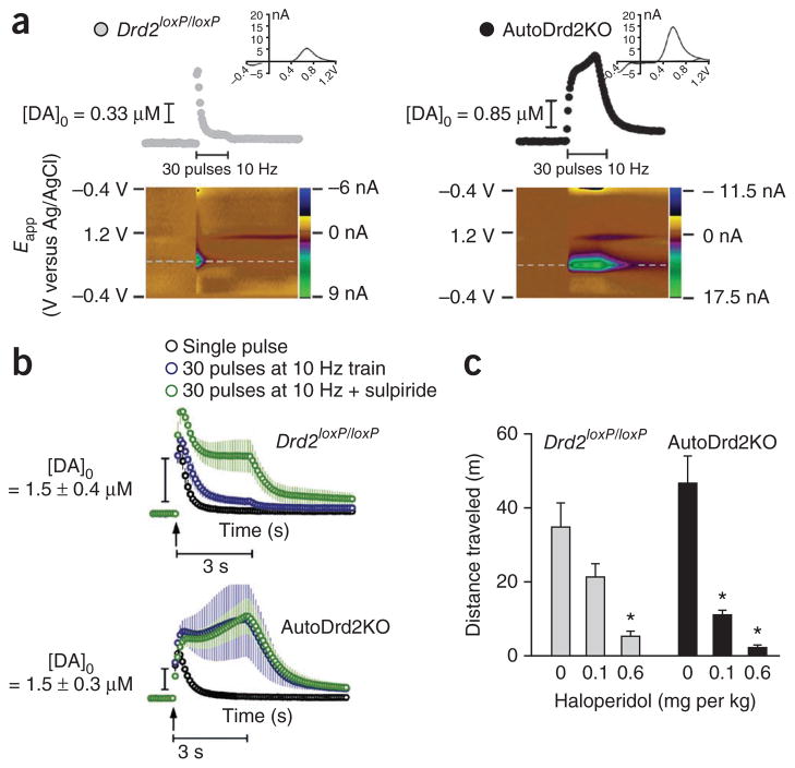
To investigate the functional consequences of the lack of D2 autoreceptors in the regulation of DA release during sustained activity, 30-pulse stimulus trains at 10 Hz (10-min intertrain intervals) were delivered and DA levels were measured with FSCV in dorsal striatal slices (Fig. 5a; note the different temporal profile of DA concentration changes compared with single pulse in Fig. 2a). In Drd2loxP/loxP slices, stimulus trains evoked a characteristic peak and steady-state DA profile that shifted to a sustained high DA level profile on sulpiride application. In autoDrd2KO mice, train-evoked DA levels were not only sustained, but were also increased during stimulation to supramaximal levels that were insensitive to sulpiride (Fig. 5b). Thus, D2 autoreceptors exert an inhibitory control of phasic DA release via rapid feedback inhibition of release combined with inhibition of DA synthesis. Acute D2 receptor blockade only prevents the rapid release inhibition, whereas the full extent of D2 autoreceptor actions can be seen in autoDrd2KO mice.
Figure 5.
AutoDrd2KO mice displayed supramaximal DA release during train stimulation. (a) DA release in dorsal striatum evoked by trains of 30 pulses delivered at 10 Hz and 10-min intervals (pulse duration of 0.6 ms, biphasic, amplitude of 600 μA). Top, time course of DA concentration changes with insets and color plots as described in Figure 2a. (b) Effect of sulpiride on train-evoked DA release. Each figure represents average concentration-time plots for eight Drd2loxP/loxP and nine autoDrd2KO mice. (c) Horizontal locomotor activity recorded over 30 min in mice receiving saline, 0.1 or 0.6 mg per kg (intraperitoneal) of haloperidol (two-way ANOVA drug: F2,17 = 21.71, *P < 0.001 compared with saline). Error bars represent s.e.m.
The loss of D2 autoreceptor modulation observed in autoDrd2KO mice also appears to have noticeable in vivo effects. AutoDrd2KO mice were supersensitive to the inhibitory effects of the D2 receptor antagonist haloperidol. At 0.1 mg per kg, haloperidol reduced locomotor activity of Drd2loxP/loxP and autoDrd2KO mice by 38% and 76%, respectively (Fig. 5c). At a higher dose of haloperidol (0.6 mg per kg), locomotor activity was further reduced by 85% in Drd2loxP/loxP mice, whereas autoDrd2KO mice were rendered mostly akinetic. These findings are consistent with the idea that haloperidol blockade of postsynaptic D2 receptors competes with a concurrent rise in extracellular DA elicited by D2 autoreceptor blockade in normal mice. Given that this latter effect does not occur when D2 autoreceptors are absent, autoDrd2KO mice are supersensitive to the effect of this drug.
Increased motivation to work for food in autoDrd2KO mice
Given that DA participates in reward-guided behavior1,34,35, we sought to examine the effects of D2 autoreceptor loss in motivation to perform rewarded operant behaviors. Food-restricted mice of both genotypes were subjected to a fixed ratio schedule (FR3) that escalated every 3 d to FR10, FR30 and FR100. Up to FR30, all mice worked to obtain approximately 120 food pellets of 20 mg each and showed no differences in satiety and motivation to self-administer food (Fig. 6a). At FR100, however, Drd2loxP/loxP mice decreased the number of pellets obtained (abandoned after pressing for 2.8 ± 0.3 h), whereas autoDrd2KO mice continued pressing the reward-paired lever for more than 4.4 ± 0.8 h and obtained a number of pellets that was even higher than those obtained under the lower fixed ratio value regimes (Fig. 6a).
Figure 6.
AutoDrd2KO mice displayed increased motivation to work for a natural reward. (a) Mice (n = 7 per genotype) were subjected to an escalating fixed ratio schedule (pressing 3, 10, 30 and 100 times) (repeated-measures ANOVA genotype: F1,11 = 4.92, *P < 0.05; interaction: F3,33 = 4.35, P < 0.05). (b) Progressive ratio (2n) schedule. Left, number of presses (one-way ANOVA presses: F1,14 = 6.37, **P < 0.01). Right, maximum number of pellets obtained (break point; one-way ANOVA: F1,14 = 8.94, *P < 0.05). (c) Two day extinction protocol for 60 min (no food delivered) (repeated-measures ANOVA, post hoc Fisher analysis genotype: F1,14 = 11.58, *P < 0.05). Error bars represent s.e.m.
A progressive ratio (PR2n) procedure indicated that these differences were due to greater motivation in autoDrd2KO mice. AutoDrd2KO mice outperformed their control siblings by pressing many more times on the lever (Fig. 6b) and obtaining more pellets (Fig. 6b). Mice of both genotypes exhibited a similar profile of loss of responding following cessation of reward delivery (Fig. 6c) indicating that the increased operant responding in autoDrd2−/− mice is not due to hyperactivity or differences in extinction, but rather to higher motivation to work for food.
DISCUSSION
Our data indicate that targeted inactivation of Drd2 specifically in Dat (also known as Slc6a3)-expressing neurons induces a total loss of Drd2 expression in midbrain DA neurons and, consequently, prevents DA-mediated IPSCs in these neurons and DA-mediated autoinhibition of DA release in striatal DA terminals. Thus, although five subtypes of DA receptors orchestrate all DA postsynaptic responses, DA-mediated autoinhibition of DA neuron activity, DA release and DA synthesis appear to be mainly conveyed by the D2 receptor subtype. There has been a controversy in the literature, with reports suggesting the participation of D3 receptors acting as autoreceptors that control DA neurotranmission21,36,37 and others presenting evidence against this possibility22. Our results settle this debate by demonstrating that no other member of the D2-like subfamily is able to compensate for the loss of D2 autoreceptor function. Another controversy that autoDrd2KO mice help to clarify is whether D2 autoreceptors regulate DA uptake, as has been suggested32,38,39. Our analysis of DA transient decay and the effects of the DAT inhibitors cocaine and methylphenidate shows that DAT-mediated DA reuptake is not altered in the absence of D2 autoreceptors. Moreover, the exaggerated level of extracellular DA detected during sustained afferent activation that mimics phasic DA release revealed the tight regulatory control that D2 autoreceptors normally exert on tuning DA release. Despite normal DAT function, autoDrd2KO mice were supersensitive to cocaine, perhaps owing to the combined additive effects of DAT blockade and absence of presynaptic inhibition that further elevates extracellular DA and maximizes stimulation of postsynaptic DA receptors.
In contrast with full D2 receptor knockout mice, which lack D2 receptors in all cell types and have reduced locomotor activity40,41 and impaired reward responses for cocaine10,13 and other drugs of abuse11,12,14, autoDrd2KO mice displayed hyperactivity in novel and familiar environments and enhanced motivation to seek reward. The phenotypic differences observed between Drd2−/− and autoDrd2KO mice highlight the value of cell-specific conditional mutant mouse models for determining the importance of the same gene product in different neurons. It is conceivable that the phenotypes observed in each mutant mouse model are the results of the primary absence of D2 receptors combined with secondary compensatory mechanisms that are likely to develop with chronic receptor loss, and, in the case of autoDrd2KO mice, chronic hyperdopaminergia.
It has been proposed that lower D2 receptor levels in humans3, monkeys6 and rodents7 predispose them to compulsive drug self-administration. In this context, it has been hypothesized that repetitive drug use compensates for the otherwise decreased activation of postsynaptic D2 receptors participating in reward circuits3,8. However, it is not clear from those studies whether lower D2 receptor availability is a result of reduced D2 receptor density or increased DA release competing with the labeled ligand. In addition, downregulation of postsynaptic D2 receptors may result as a compensatory mechanism for excessive dopaminergic transmission. We found that autoDrd2KO mice are supersensitive to the rewarding properties of cocaine and have excessive dopaminergic transmission, suggesting that low levels of presynaptic D2 autoreceptors can also predict enhanced susceptibility to drug-seeking and drug abuse. In fact, a recent human study found that low levels of D2 receptors in the midbrain correlated with higher impulsivity and stronger subjective desire for amphetamine21. We found increased locomotor responses and conditioned place preference for cocaine in autoDrd2KO mice, indicating that reduced D2 autoreceptor–mediated inhibition of DA cell firing and DA release enhance the postsynaptic effects elicited by drugs that increase extracellular DA concentration.
AutoDrd2KO mice also showed enhanced motivation to work for food. In the escalated fixed ratio experiment in which the mice had to press a lever 100 times to receive one pellet, autoDrd2KO mice not only outperformed their control littermates, but also developed compulsive bar-pressing behavior, as they worked to obtain many more pellets than those required for their daily food intake needs (Fig. 6a). Thus, autoDrd2KO mice may constitute a valuable model for connecting the neurobiology of motivation with that of compulsive behavior. Altogether, our results highlight the critical regulatory role that D2 autoreceptors have in DA neurotransmission and suggest that transcriptional regulation of Drd2 in DA neurons may contribute to the individual behavioral reactions toward natural rewards and drugs of abuse.
METHODS
Methods and any associated references are available in the online version of the paper at http://www.nature.com/natureneuroscience/.
Supplementary Material
Acknowledgments
We thank J. Sztein, G. Levin, C. Bäckman, V. Rodríguez, R. Lorenzo, S. Nemirovsky, M. Peper, S. Merani, C. Carbone, F. Maschi and M. Baetscher for their scientific and technical assistance. This work was supported in part by an International Research Scholar Grant of the Howard Hughes Medical Institute (M.R.), Universidad de Buenos Aires (M.R.) and Agencia Nacional de Promoción Científica y Tecnológica (M.R.), National Science Foundation grant INT-9901278 (M.J.L. and M.R.) and US National Institutes of Health grant R01-MH61326 (M.J.L.). E.P.B., D.N. and D.M.G. received doctoral fellowships from the Consejo Nacional de Investigaciones Científicas y Técnicas, Argentina. Y.M., J.H.S., V.A.A. and D.M.L. were supported by the Division of Intramural Clinical and Biological Research of the National Institute on Alcohol Abuse and Alcoholism. J.H.S. and V.A.A. received support from the Intramural Program of the National Institute of Neurological Disorders and Stroke.
Footnotes
AUTHOR CONTRIBUTIONS
D.M.G. and M.R. generated the conditional mutant mice. D.N. and E.P.B. characterized, raised and maintained mouse colonies and performed backcrossing. E.P.B. and D.N. conducted neurochemical, histological and behavioral experiments. Y.M. and J.H.S. conducted electrochemical and electrophysiological experiments. E.P.B., Y.M., J.H.S., D.N. and M.R. prepared the figures. E.P.B., Y.M. and M.R. wrote the manuscript. All of the authors designed experiments, analyzed data and edited the manuscript.
COMPETING FINANCIAL INTERESTS
The authors declare no competing financial interests.
Reprints and permissions information is available online at http://www.nature.com/reprints/index.html.
Supplementary information is available on the Nature Neuroscience website.
References
- 1.Schultz W. Dopamine signals for reward value and risk: basic and recent data. Behav Brain Funct. 2010;6:24. doi: 10.1186/1744-9081-6-24. [DOI] [PMC free article] [PubMed] [Google Scholar]
- 2.Goto Y, Otani S, Grace AA. The Yin and Yang of dopamine release: a new perspective. Neuropharmacology. 2007;53:583–587. doi: 10.1016/j.neuropharm.2007.07.007. [DOI] [PMC free article] [PubMed] [Google Scholar]
- 3.Volkow ND, Fowler JS, Wang GJ, Baler R, Telang F. Imaging dopamine’s role in drug abuse and addiction. Neuropharmacology. 2009;56 (suppl 1):3–8. doi: 10.1016/j.neuropharm.2008.05.022. [DOI] [PMC free article] [PubMed] [Google Scholar]
- 4.Kenny PJ. Reward mechanisms in obesity: new insights and future directions. Neuron. 2011;69:664–679. doi: 10.1016/j.neuron.2011.02.016. [DOI] [PMC free article] [PubMed] [Google Scholar]
- 5.Sulzer D. How addictive drugs disrupt presynaptic dopamine neurotransmission. Neuron. 2011;69:628–649. doi: 10.1016/j.neuron.2011.02.010. [DOI] [PMC free article] [PubMed] [Google Scholar]
- 6.Morgan D, et al. Social dominance in monkeys: dopamine D2 receptors and cocaine self-administration. Nat Neurosci. 2002;5:169–174. doi: 10.1038/nn798. [DOI] [PubMed] [Google Scholar]
- 7.Thanos PK, et al. Overexpression of dopamine D2 receptors reduces alcohol self-administration. J Neurochem. 2001;78:1094–1103. doi: 10.1046/j.1471-4159.2001.00492.x. [DOI] [PubMed] [Google Scholar]
- 8.Volkow ND, et al. Prediction of reinforcing responses to psychostimulants in humans by brain dopamine D2 receptor levels. Am J Psychiatry. 1999;156:1440–1443. doi: 10.1176/ajp.156.9.1440. [DOI] [PubMed] [Google Scholar]
- 9.Volkow ND, et al. Brain DA D2 receptors predict reinforcing effects of stimulants in humans: replication study. Synapse. 2002;46:79–82. doi: 10.1002/syn.10137. [DOI] [PubMed] [Google Scholar]
- 10.Chausmer AL, et al. Cocaine-induced locomotor activity and cocaine discrimination in dopamine D2 receptor mutant mice. Psychopharmacology (Berl) 2002;163:54–61. doi: 10.1007/s00213-002-1142-y. [DOI] [PubMed] [Google Scholar]
- 11.Maldonado R, et al. Absence of opiate rewarding effects in mice lacking dopamine D2 receptors. Nature. 1997;388:586–589. doi: 10.1038/41567. [DOI] [PubMed] [Google Scholar]
- 12.Phillips TJ, et al. Alcohol preference and sensitivity are markedly reduced in mice lacking dopamine D2 receptors. Nat Neurosci. 1998;1:610–615. doi: 10.1038/2843. [DOI] [PubMed] [Google Scholar]
- 13.Welter M, et al. Absence of dopamine D2 receptors unmasks an inhibitory control over the brain circuitries activated by cocaine. Proc Natl Acad Sci USA. 2007;104:6840–6845. doi: 10.1073/pnas.0610790104. [DOI] [PMC free article] [PubMed] [Google Scholar]
- 14.Cunningham CL, et al. Ethanol-conditioned place preference is reduced in dopamine D2 receptor–deficient mice. Pharmacol Biochem Behav. 2000;67:693–699. doi: 10.1016/s0091-3057(00)00414-7. [DOI] [PubMed] [Google Scholar]
- 15.Bunney BS, Aghajanian GK, Roth RH. Comparison of effects of L-DOPA, amphetamine and apomorphine on firing rate of rat dopaminergic neurones. Nat New Biol. 1973;245:123–125. doi: 10.1038/newbio245123a0. [DOI] [PubMed] [Google Scholar]
- 16.Cubeddu LX, Hoffmann IS. Operational characteristics of the inhibitory feedback mechanism for regulation of dopamine release via presynaptic receptors. J Pharmacol Exp Ther. 1982;223:497–501. [PubMed] [Google Scholar]
- 17.Ford CP, Gantz SC, Phillips PE, Williams JT. Control of extracellular dopamine at dendrite and axon terminals. J Neurosci. 2010;30:6975–6983. doi: 10.1523/JNEUROSCI.1020-10.2010. [DOI] [PMC free article] [PubMed] [Google Scholar]
- 18.Paladini CA, Robinson S, Morikawa H, Williams JT, Palmiter RD. Dopamine controls the firing pattern of dopamine neurons via a network feedback mechanism. Proc Natl Acad Sci USA. 2003;100:2866–2871. doi: 10.1073/pnas.0138018100. [DOI] [PMC free article] [PubMed] [Google Scholar]
- 19.Wolf ME, Roth RH. Autoreceptor regulation of dopamine synthesis. Ann NY Acad Sci. 1990;604:323–343. doi: 10.1111/j.1749-6632.1990.tb32003.x. [DOI] [PubMed] [Google Scholar]
- 20.Schmitz Y, Benoit-Marand M, Gonon F, Sulzer D. Presynaptic regulation of dopaminergic neurotransmission. J Neurochem. 2003;87:273–289. doi: 10.1046/j.1471-4159.2003.02050.x. [DOI] [PubMed] [Google Scholar]
- 21.Buckholtz JW, et al. Dopaminergic network differences in human impulsivity. Science. 2010;329:532. doi: 10.1126/science.1185778. [DOI] [PMC free article] [PubMed] [Google Scholar]
- 22.Koeltzow TE, et al. Alterations in dopamine release but not dopamine autoreceptor function in dopamine D3 receptor mutant mice. J Neurosci. 1998;18:2231–2238. doi: 10.1523/JNEUROSCI.18-06-02231.1998. [DOI] [PMC free article] [PubMed] [Google Scholar]
- 23.Mercuri NB, et al. Loss of autoreceptor function in dopaminergic neurons from dopamine D2 receptor deficient mice. Neuroscience. 1997;79:323–327. doi: 10.1016/s0306-4522(97)00135-8. [DOI] [PubMed] [Google Scholar]
- 24.Díaz-Torga G, et al. Disruption of the D2 dopamine receptor alters GH and IGF-I secretion and causes dwarfism in male mice. Endocrinology. 2002;143:1270–1279. doi: 10.1210/endo.143.4.8750. [DOI] [PubMed] [Google Scholar]
- 25.Bäckman CM, et al. Characterization of a mouse strain expressing Cre recombinase from the 3′ untranslated region of the dopamine transporter locus. Genesis. 2006;44:383–390. doi: 10.1002/dvg.20228. [DOI] [PubMed] [Google Scholar]
- 26.Kennedy RT, Jones SR, Wightman RM. Dynamic observation of dopamine autoreceptor effects in rat striatal slices. J Neurochem. 1992;59:449–455. doi: 10.1111/j.1471-4159.1992.tb09391.x. [DOI] [PubMed] [Google Scholar]
- 27.Phillips PE, Hancock PJ, Stamford JA. Time window of autoreceptor-mediated inhibition of limbic and striatal dopamine release. Synapse. 2002;44:15–22. doi: 10.1002/syn.10049. [DOI] [PubMed] [Google Scholar]
- 28.Kehr W, Carlsson A, Lindqvist M, Magnusson T, Atack C. Evidence for a receptor-mediated feedback control of striatal tyrosine hydroxylase activity. J Pharm Pharmacol. 1972;24:744–747. doi: 10.1111/j.2042-7158.1972.tb09104.x. [DOI] [PubMed] [Google Scholar]
- 29.Avale ME, et al. The dopamine D4 receptor is essential for hyperactivity and impaired behavioral inhibition in a mouse model of attention deficit/hyperactivity disorder. Mol Psychiatry. 2004;9:718–726. doi: 10.1038/sj.mp.4001474. [DOI] [PubMed] [Google Scholar]
- 30.Giros B, Jaber M, Jones SR, Wightman RM, Caron MG. Hyperlocomotion and indifference to cocaine and amphetamine in mice lacking the dopamine transporter. Nature. 1996;379:606–612. doi: 10.1038/379606a0. [DOI] [PubMed] [Google Scholar]
- 31.Zhuang X, et al. Hyperactivity and impaired response habituation in hyperdopaminergic mice. Proc Natl Acad Sci USA. 2001;98:1982–1987. doi: 10.1073/pnas.98.4.1982. [DOI] [PMC free article] [PubMed] [Google Scholar]
- 32.Dickinson SD, et al. Dopamine D2 receptor-deficient mice exhibit decreased dopamine transporter function but no changes in dopamine release in dorsal striatum. J Neurochem. 1999;72:148–156. doi: 10.1046/j.1471-4159.1999.0720148.x. [DOI] [PubMed] [Google Scholar]
- 33.Jones SR, et al. Loss of autoreceptor functions in mice lacking the dopamine transporter. Nat Neurosci. 1999;2:649–655. doi: 10.1038/10204. [DOI] [PubMed] [Google Scholar]
- 34.Berridge KC. The debate over dopamine’s role in reward: the case for incentive salience. Psychopharmacology (Berl) 2007;191:391–431. doi: 10.1007/s00213-006-0578-x. [DOI] [PubMed] [Google Scholar]
- 35.Salamone JD, Correa M. Motivational views of reinforcement: implications for understanding the behavioral functions of nucleus accumbens dopamine. Behav Brain Res. 2002;137:3–25. doi: 10.1016/s0166-4328(02)00282-6. [DOI] [PubMed] [Google Scholar]
- 36.Joseph JD, et al. Dopamine autoreceptor regulation of release and uptake in mouse brain slices in the absence of D(3) receptors. Neuroscience. 2002;112:39–49. doi: 10.1016/s0306-4522(02)00067-2. [DOI] [PubMed] [Google Scholar]
- 37.Maina FK, Mathews TA. A functional fast scan cyclic voltammetry assay to characterize dopamine D2 and D3 autoreceptors in the mouse striatum. ACS Chem Neurosci. 2010;1:450–462. doi: 10.1021/cn100003u. [DOI] [PMC free article] [PubMed] [Google Scholar]
- 38.Benoit-Marand M, Borrelli E, Gonon F. Inhibition of dopamine release via presynaptic D2 receptors: time course and functional characteristics in vivo. J Neurosci. 2001;21:9134–9141. doi: 10.1523/JNEUROSCI.21-23-09134.2001. [DOI] [PMC free article] [PubMed] [Google Scholar]
- 39.Schmitz Y, Schmauss C, Sulzer D. Altered dopamine release and uptake kinetics in mice lacking D2 receptors. J Neurosci. 2002;22:8002–8009. doi: 10.1523/JNEUROSCI.22-18-08002.2002. [DOI] [PMC free article] [PubMed] [Google Scholar]
- 40.Baik JH, et al. Parkinsonian-like locomotor impairment in mice lacking dopamine D2 receptors. Nature. 1995;377:424–428. doi: 10.1038/377424a0. [DOI] [PubMed] [Google Scholar]
- 41.Kelly MA, et al. Locomotor activity in D2 dopamine receptor–deficient mice is determined by gene dosage, genetic background, and developmental adaptations. J Neurosci. 1998;18:3470–3479. doi: 10.1523/JNEUROSCI.18-09-03470.1998. [DOI] [PMC free article] [PubMed] [Google Scholar]
Associated Data
This section collects any data citations, data availability statements, or supplementary materials included in this article.


