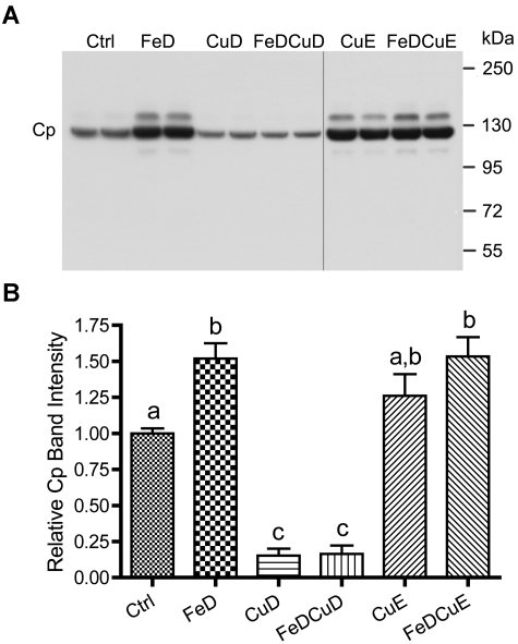
Figure 3.
Cp protein expression in rat serum. A representative Western blot is shown (top panel), representing 2 rats per dietary treatment group. Quantitative data from all rats is also given (n = 8-12 rats/group; bottom panel). Bars with different letters atop error bars are statistically different (P < .05) from each other. Data are expressed as means ± SD. The image is of one blot from a single x-ray film; the vertical black line indicates where a blank lane with nonchemiluminescent molecular weight markers was removed from the image.

