Abstract
 Injuries of the spine in pregnant women are rare. Unstable fractures, incomplete neurological deficits and failed conservative treatment are indications for operative stabilization. So far, only posterior stabilization techniques performed during pregnancy have been published in case reports. This Grand Round case presentation describes a 24-year-old woman in the 19th week of gestation who was involved in a motorcycle accident as a pillion rider. Radiological examination revealed a complete burst fracture (type AO A3.3) of T8 with a slight, yet clinical unapparent narrowing of the spinal canal and a stable T5 fracture (type AO A1.2). Despite analgesia with morphine, conservative treatment failed and it was not possible to mobilize the patient. Hence, an anterior thoracoscopic-assisted reduction and stabilization in left lateral position with single lung ventilation was performed as the therapy of choice. Intraoperatively, the body of T8 was removed and plate was used to stabilize and reduce the fracture. Finally, a tricortical iliac bone graft was implanted into the bony defect. Intraoperative fluoroscopy was merely used to verify the positioning of the implants. Postoperative examination of the foetus revealed normal findings. The patient was discharged with paracetamol as residual pain medication. The degree of kyphosis of the T8 fracture was successfully reduced from 20° to 13° (segmental standard value 12°). Further clinical and radiological course of the patient was uneventful. If suitable implants are available and good bone structure exists, solely anterior thoracoscopic-assisted reduction and stabilization of an unstable thoracic burst fracture can be performed safely. In the present case, it was possible to avoid intraoperative prone positioning of the pregnant patient as well as reaching a minimum of radiation exposure.
Keywords: Pregnancy, Burst fracture, Thoracolumbar spine, Anterior surgery, Thoracoscopic spine surgery
Case presentation
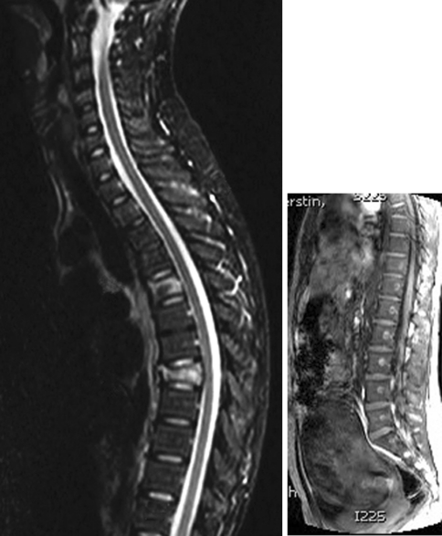
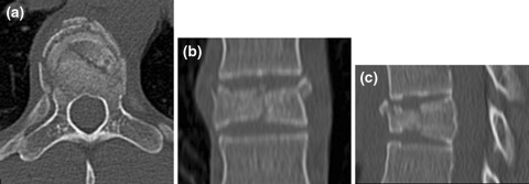
A 24-year-old woman in the 19th week of pregnancy was involved in a motorcycle accident as a pillion rider. On admission she complained about pain in the region of the thoracic spine. No neurological deficit could be detected. Plain radiographs of the thoracic spine as well as an MRI-scan were performed and revealed compression type vertebral fractures, which affected T5 and T8. MRI suggested likely involvement of the posterior wall and the pedicles of T8 (Fig. 1). Consequently, a CT-scan of the T8 vertebral body was performed, which revealed a posterior wall fracture and a slight narrowing of the spinal canal (Fig. 2). No signs of involvement of the posterior ligamentous complex could be detected. Therefore, the fracture of T8 was classified as a complete burst fracture (AO A3.3, according to Magerls classification) [1]. The fracture concerning T5 was a wedged compression fracture (AO A1.2). Local segmental kyphosis of T8 was 20°.
Fig. 1.
Sagittal MRI showing compression fractures of T5 and T8 (a) and foetus (b)
Fig. 2.
Axial (a), coronal (b) and sagittal (c) CT-scan showing burst fracture of T8
The patient remained in bed due to severe pain. She could not be mobilized despite the administration of Fentanyl Transdermal Therapeutic System (TTS) 25 μg/h plus 800 mg Ibuprofen and 1 g Paracetamol on demand. Ultrasound examinations of the foetus were without pathological findings. Despite the administered analgesia, the pain suffered by the patient had not decreased in intensity after 5 days of medical observance. Hence, the patient requested operative treatment.
Rationale for treatment and review of the literature
Traumatic fractures of the thoracolumbar spine in pregnant women are rare. Operative treatment is only recommended in unstable fractures and in cases of incomplete neurological deficit [1]. If possible, surgery can be postponed until delivery of the child [2]. These topics have merely been addressed in case reviews in the current literature.
Surgery and anaesthesia carry a potential risk for both, mother and foetus [3, 4]. To date, only few papers have described treatment modalities for unstable thoracolumbar fractures [2]. Most fractures, however, could be treated conservatively. If surgery was necessary, the access to the thoracolumbar spine has always been from the posterior approach [2]. Presently, no standardized therapeutic strategy can be recommended and treatment should be considered individually, according to the given situation of the pregnant patient.
Thoracoscopic or thoracoscopic-assisted surgery is well established for reconstruction of the anterior spinal column in the thoracolumbar spine [5–7]. Typically, fractures from T4 to L2 can be treated with this technique. Advantages of anterior surgery are direct visualization of the fracture as well as better spinal canal decompression. If instrumented stabilization is necessary, plates and cages can be placed with possibly less radiation exposure compared with posterior pedicle screw fixation. Disadvantages of anterior surgery include a limited extent of fracture reduction compared with posterior surgery as well as more demanding operative technique [6, 8].
To the authors’ best knowledge, this Grand Rounds case presentation describes for the first time an anterior thoracoscopic-assisted reduction, stabilization and fusion of an unstable T8 burst fracture in a pregnant woman using tricortical iliac bone graft and an expandable plate.
In the present case, unbearable pain as a result of a complete burst fracture of the vertebral body of T8 was the indication for surgery. The alternative would have been a prolonged conservative treatment attempt. According to common experience pain caused by unstable fractures resolves after 4–6 weeks, if bony healing occurs. However, immobilization of pregnant women with spinal fractures carries the risks of deep venous thrombosis and pneumonia. The ongoing administration of morphine may harm the foetus. Therefore, the risks of conservative against operative treatment must be weighed.
To stabilize the fracture we could have used either an anterior or a posterior approach to the spine. The drawback of the posterior approach would have been (1) prone positioning of the patient and subsequent increase of the abdominal pressure (2) possibly higher exposure to radiation, whilst placing the pedicle screws (3) damaging of the back muscles.
By approaching the spine from anterior we avoided these hazards entirely. Intraoperatively, we merely used an image intensifier to control the positioning of the plate and screws. Apart from this, the surgical procedure did not differ from our standard technique. Thoracotomies and thoracoscopic surgery have been performed successfully in pregnant females, mostly to treat spontaneous pneumothorax. No surgical complications have been reported in these cases and operative procedures were well tolerated [9–11].
However, maternal and foetal mortality and morbidity due to surgery and anaesthesia remain as major issues [4, 12–14]. In the present case, anaesthesia was performed as a rapid sequence induction to avoid aspiration. The correct position of the tube was reassessed bronchoscopically before single lung ventilation was commenced. During the entire operation, peripheral oxygen saturation was normal and arterial blood gases showed normocapnia. By using Sevofluran and Fenoterol relaxation of the uterus was maintained during surgery. Overall, these precautions might have contributed to the successful anaesthesia of the patient.
Anterior stabilization exclusively for unstable thoracolumbar fractures has been described in the literature [8, 15, 16]. To achieve good fracture reduction, good bone structure and stable implants are prerequisites. Only distraction can be applied for anterior thoracolumbar fracture stabilization with standard plates. Correction of the kyphosis was achieved indirectly through patient positioning and distraction. In our case we used a novel type of anterior-only reduction plate (ArcoFix, Synthes, Oberdorf, Switzerland), which offers the possibility of applying lordotic forces through swivel heads at both ends of the plate. After fixation of the plate in the vertebral bodies, the angle of lordosis or kyphosis can be corrected by changing the angulation of the implant’s cranial and caudal swivel heads. The height can be corrected with the implant’s central sliding mechanism. Both, swivel heads and sliding mechanism are individually locked or unlocked to perform different types of movements. In our pregnant patient we were able to reduce the local segmental kyphosis at T8 from 20° down to a physiologically normal value of 13° [17].
The presented anterior transthoracic technique is suitable to treat fractures from T4 to L2. To reach levels below T12, an incision of the diaphragm is generally necessary [9]. Incision of the diaphragm, however, should be avoided in pregnant women, especially during the last trimester. The increasing size of the uterus and the subsequent ascendency of the diaphragm make it difficult to reach fractures below T11 without incision of the diaphragm. This issue must be addressed during the surgical planning. Furthermore, surgery in the last trimester of pregnancy carries a high risk of abortion.
Procedure
Surgery was performed under general anaesthesia and patient in the left lateral position. As a perioperative antibiotic prophylaxis Cefuroxime was given once shortly before surgery. To prevent vomiting during intubation, Dimenhydrinate was administered orally before surgery. Anaesthesiologists performed rapid sequence induction using Propofol, Rocuronium Bromide and Sufentanil. The use of a double-lumen tube facilitated the one-lung ventilation. The correct position of the tube was reassured by fiberoptic bronchoscopy after patient positioning on the left side. Sevoflurane served to maintain anaesthesia and Sufentanil was given to maintain analgesia. Rocuronium Bromide was used for muscle relaxation. Intraoperative, Fenoterol was infused to maintain relaxation of the uterus.
A right thoracic approach to the spine with a 7-cm incision (minithoracotomy) directly above T8 and two additional ports, one for the thoracoscopic optic, the other for the lung retractor, was performed. Under direct vision, the fractured body of vertebra T8 including the adjacent discs were removed (partial corpectomy) and an anterior titanium plate (ArcoFix, Synthes, Oberdorf, Switzerland) was implanted to bridge the fractured vertebral body from T7 to T9. After verifying the correct positioning of the plate and the screws using an image intensifier, the fracture was primarily reduced by applying lordotic forces, followed by distraction through the ArcoFix plate. The length of the bony defect was measured and a tricortical bone graft was harvested from the right iliac crest. The bone graft was then transplanted to the defect in a pressfit manner. Finally, before closing the wound, two chest tubes were placed through the ports as well as an intrathoracic pain catheter containing Ropivacaine.
Outcome
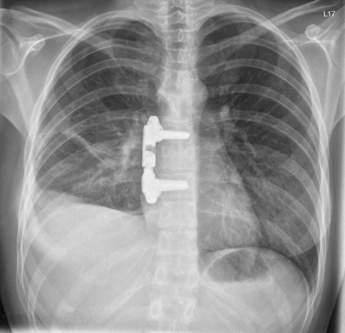
Ultrasound examinations of the foetus on the first postoperative day showed normal movement and values. The patient remained in ICU for three days and was mobilized on the second postoperative day. The postoperative course was uneventful. The pain catheter and the chest tubes were removed on the second postoperative day and pain medication was decreased gradually. Postoperatively, a chest X-ray was done to verify correct implant positioning and to rule out pneumothorax or pleura effusion (Fig. 3). Local segmental kyphosis had been decreased to 13° which is a normal value on this segmental level [17]. The patient was discharged on the eleventh day after the operation.
Fig. 3.
Postoperative chest X-ray with ArcoFix plate
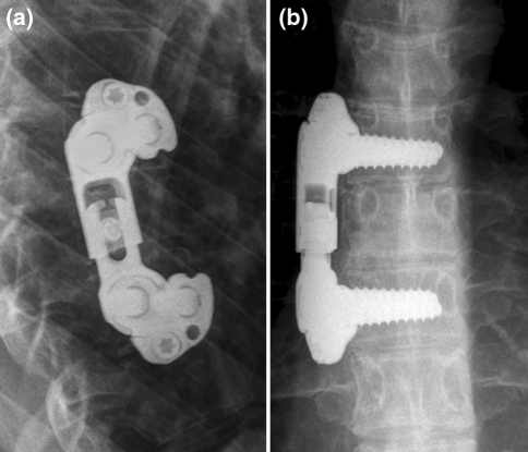
The further course was uneventful. After delivery of a healthy baby, radiological follow-up examinations of the thoracic spine were performed, which displayed a stable implant with solid fusion and no loss of reduction at 12-month follow-up (Fig. 4).
Fig. 4.
Plain X-rays in ap (a) and lateral (b) view 12 months postoperative
Conclusion
Anterior, thoracoscopic-assisted stabilization of unstable thoracolumbar burst fractures in pregnant women can be recommended, if conservative treatment is not possible and the fractured body itself can be reached without incision of the diaphragm. The transthoracic approach proved to be well tolerated. By using an anterior-only reduction plate physiological segmental kyphosis can be restored. The anterior technique reduces exposure to radiation. Furthermore, the anterior technique avoids the increase of abdominal pressure as well as injuries in the back muscles.
Acknowledgments
The authors thank Mrs. Kirsten Stangenberg-Gliss and Ms. Shafreena Zainal-Abidin for assistance in preparation of the manuscript.
References
- 1.Magerl F, Aebi M, Gertzbein SD, Harms J, Nazarian S. A comprehensive classification of thoracic and lumbar injuries. Eur Spine J. 1994;3:184–201. doi: 10.1007/BF02221591. [DOI] [PubMed] [Google Scholar]
- 2.Tanchev P, Dikov D, Novkov H. Thoracolumbar distraction fractures in advanced pregnancy: a contribution of two case reports. Eur Spine J. 2000;9:167–170. doi: 10.1007/s005860050229. [DOI] [PMC free article] [PubMed] [Google Scholar]
- 3.Prokop A, Swol-Ben J, Helling HJ, Neuhaus W, Rehm KE. Trauma im letzten Trimester der Gravidität. Unfallchirurg. 1996;99:450–453. [PubMed] [Google Scholar]
- 4.Nunn CR, Bass JG, Eddy VA. Management of the pregnant patient with acute spinal cord injury. Tenn Med. 1996;89:335–337. [PubMed] [Google Scholar]
- 5.Beisse R. Endoscopic surgery on the thoracolumbar junction of the spine. Eur Spine J. 2006;15:687–704. doi: 10.1007/s00586-005-0994-3. [DOI] [PMC free article] [PubMed] [Google Scholar]
- 6.Khoo LT, Beisse R, Potulski M. Thoracoscopic-assisted treatment of thoracic and lumbar fractures: a series of 371 consecutive cases. Neurosurgery. 2002;51(5 Suppl):S104–S117. [PubMed] [Google Scholar]
- 7.Schnake KJ, Kandziora F, Hoffmann R. Thoracolumbar fractures. Minimally invasive anterior stabilization. Trauma Berufskrankh. 2009;11:87–93. doi: 10.1007/s10039-009-1490-5. [DOI] [Google Scholar]
- 8.Dai LY, Jiang LS, Jiang SD. Anterior-only stabilization using plating with bone structural autograft versus titanium mesh cages for two- or three-column thoracolumbar burst fractures: a prospective randomized study. Spine. 2009;34:1429–1435. doi: 10.1097/BRS.0b013e3181a4e667. [DOI] [PubMed] [Google Scholar]
- 9.VanWinter JT, Nichols FC, III, Pairolero PC, Ney JA, Ogburn PL., Jr Management of spontaneous pneumothorax during pregnancy: case report and review of the literature. Mayo Clin Proc. 1996;71:249–252. doi: 10.4065/71.3.249. [DOI] [PubMed] [Google Scholar]
- 10.Reid CJ, Burgin GA. Video-assisted thoracoscopic surgical pleurodesis for persistent spontaneous pneumothorax in late pregnancy. Anaesth Intensive Care. 2000;28:208–210. doi: 10.1177/0310057X0002800217. [DOI] [PubMed] [Google Scholar]
- 11.Wong MD, Leung WC, Wang JK, Lao TT, Ip MS, Wam WK, Ho JC. Recurrent pneumothorax in pregnancy: what should we do after placing an intercostals drain. Hong Kong Med J. 2006;12:375–380. [PubMed] [Google Scholar]
- 12.Popov I, Ngambu F, Mantel G, Rout C, Moodley J. Acute spinal cord injury in pregnancy: an illustrative case and literature review. J Obstet Gynaecol. 2003;23:596–598. doi: 10.1080/01443610310001604321. [DOI] [PubMed] [Google Scholar]
- 13.Kuczkowski KM, Fouhsy SA, Greenberg M, Benumof JL. Trauma in pregnancy: anaesthetic management of the pregnant trauma victim with unstable cervical spine. Anaesthesia. 2003;58:822. doi: 10.1046/j.1365-2044.2003.03295_28.x. [DOI] [PubMed] [Google Scholar]
- 14.Eason JR, Swaine CN, Jones PI, Gronow MJ, Beaumont A. Unstable cervical fracture. Anaesthetic management for an urgent caesarean section. Anaesthesia. 1987;42:745–749. doi: 10.1111/j.1365-2044.1987.tb05320.x. [DOI] [PubMed] [Google Scholar]
- 15.Kirkpatrick JS. Thoracolumbar fracture management: anterior approach. J Am Acad Orthop Surg. 2003;11:355–363. doi: 10.5435/00124635-200309000-00008. [DOI] [PubMed] [Google Scholar]
- 16.Zdeblick TA, Sasso RC, Vaccaro AR, Chapman JR, Harris MB. Surgical treatment of thoracolumbar fractures. Instr Course Lect. 2009;58:639–644. [PubMed] [Google Scholar]
- 17.Weber W, Wimmer B. Die klinische und röntgenologische Begutachtung von Wirbelsäulenverletzungen nach dem Segmentprinzip. Unfallchirurg. 1991;17:200–207. doi: 10.1007/BF02588687. [DOI] [PubMed] [Google Scholar]


