Abstract
Introduction
The surgical strategies to treat idiopathic scoliosis on adolescents and young adults need a basic reliable classification. King’s and Lenke’s classification are inappropriate because they fail to take shoulders and pelvis into account.
Methods
We propose the answer for the following three questions:
Why are we challenging King’s and Lenke’s systems of classification?
How many frontal and possibly sagittal curves do we need to be able to develop a strategy which is applicable to almost all cases?
How should scoliotic curves be classified?
Results
In double thoracic and lumbar (thoracic predominant) scoliosis, the concepts of “pelvis included” and “pelvis excluded” are not simply based on a semantic distinction, but correspond to different physiopathological entities and require different surgical strategies. In double thoracic curves the concepts of “real double thoracic” and “potential double thoracic” curves are keys to obtain post operative shoulder balance. In lumbar scoliosis the concepts of “real lumbar” and “lumbosacral” curves are necessary to compare results of posterior or anterior approach in surgical strategies. The system proposed in this work involves ten basic curves.
Conclusion
The surgical strategies used to treat idiopathic scoliosis in adolescents and young adults depend on the school of thought as to whether the anterior or posterior approach is preferable and the extent of the vertebral instrumentation. A consensus system of classification of scoliotic curves is required to compare the results obtained using various methods. This has been done in the improved version of King’s system proposed here and should provide an efficient tool for use in comparative studies on surgical methods.
Keywords: Adolescent and adult idiopathic scoliosis, Scoliosis classification, Double thoracic curves, Lumbar scoliosis, Lumbosacral scoliosis, King’s and Lenke’s classification, Pelvis included and pelvis excluded concepts, Spinal balance
Introduction
The aim of surgical treatment for idiopathic scoliosis in adolescents and young adults is to correct the distorted spinal region after performing an instrumented arthrodesis, while at the same time working on the upper trunk to make the shoulders horizontal on the frontal plane, thus bringing C7 straight up above the mid-pelvis and leaving the discs located immediately below the arthrodesis in a horizontal position in the frontal plane and in a lordotic position in the sagittal plane. Achieving this result requires analyzing the patient’s curves, taking the shoulders and pelvis into account and not just applying arbitrary geometric landmarks indiscriminately to all the curves.
We propose the answer for the following three questions:
Why are we challenging King’s [16, 17] and Lenke’s [18] systems of classification?
How many frontal and possibly sagittal curves do we need to be able to develop a strategy, which is applicable to almost all cases?
How should scoliotic curves be classified?
Why we object to the systems of classification proposed by King and Lenke
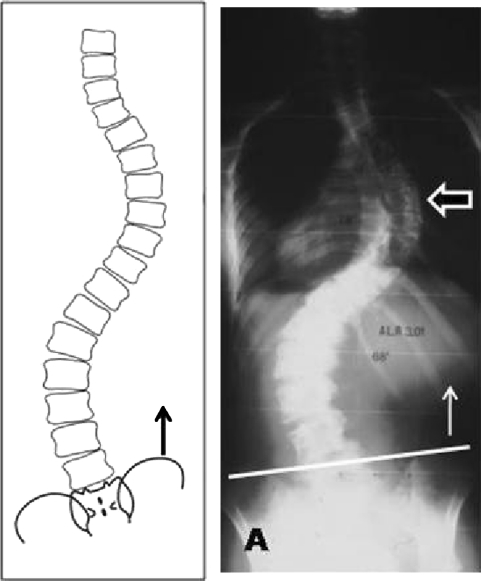
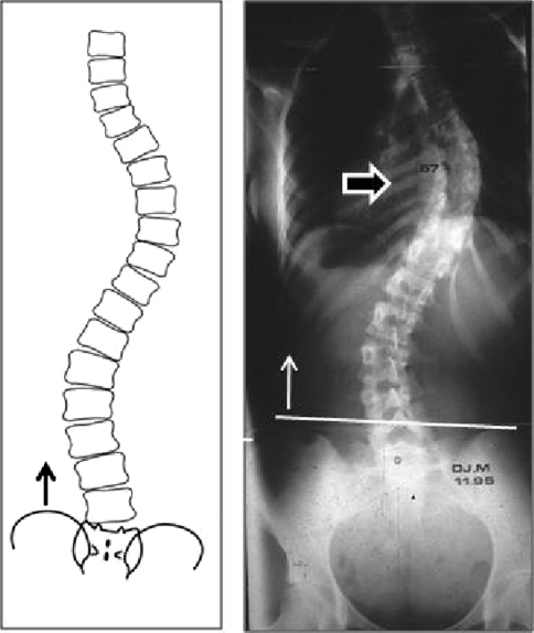
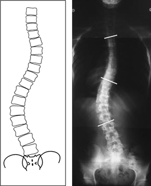
The basis of these two authors’ systems of classifications is not sound. They consistently assume the pelvis to be horizontal. The two reference lines they have adopted, regardless of the position of the pelvis, are a horizontal line based on the sacral plate and a vertical line starting in the middle of the sacrum. These are obviously not natural lines. If one has to use a line at all, the only obvious one is the plumb line from C7 down to the pelvis in the frontal and sagittal planes, which can be used to characterize individual patients’ pre- and postoperative balance. This line is a useful tool for the classification purposes, but it is not sufficient to describe spinal curves. The pelvis of patients with idiopathic scoliosis is sometimes horizontal, but this is often not the case, especially in patients with double thoracic and lumbar (thoracic predominant) curves. Either the iliac crest tilts toward the convex side of the thoracic curve, giving what Salanova [27] has called “pelvis included” (the pelvis is directly involved in the lumbar curve) (Fig. 1), or the iliac crest tilts towards the concave side of the thoracic curve (while the pelvis can remain horizontal), which Salanova has called “pelvis excluded” (the pelvis is not involved in the lumbar curve) (Fig. 2).
Fig. 1.
Double thoracic and lumbar curve scoliosis (thoracic predominant) pelvisincluded the pelvis tilt to convexity of thoracic curve
Fig. 2.
Double thoracic and lumbar curve scoliosis (thoracic predominant) pelvisexcluded the pelvis tilt to concavity of thoracic curve
In double thoracic and lumbar (thoracic predominant) scoliosis, the concepts of pelvis included and excluded are not simply based on a semantic distinction, but correspond to different physiopathological entities and require different surgical strategies [2].
In “pelvis included” scoliosis, it is not possible to decapitate the lumbar curve (the discs cannot be spared) and the arthrodesis has to extend down to L4 (unless the disorder is left to run its natural course, with the potential risks this entails). If the “reverse effect” strategy is used, the underlying discs will remain oblique, or this will soon be the case.
In “pelvis excluded” scoliosis, the lumbar curve can be decapitated, the “reverse effect” strategy will be effective, and the underlying discs will remain horizontal.
With King’s and Lenke’s reference lines, it is not possible to carry out this analysis. For example, the King II type actually includes both “pelvis included” and “pelvis excluded” curves: when the two curves are both of the same grade, the pelvis is usually included. Some authors have used bending lumbar tests to help them decide which vertebrae should be instrumented but, since the lumbar curve is always more flexible than the thoracic one for anatomical reasons, the bending test can mask the contribution of the pelvis and result in strategic errors [12, 19]. One might even say that when dealing with double thoracic and lumbar (thoracic predominant) “pelvis included” curves, the use of the bending lumbar test is the best possible way of getting it wrong. King’s and Lenke’s methods of classification are therefore not appropriate [34].
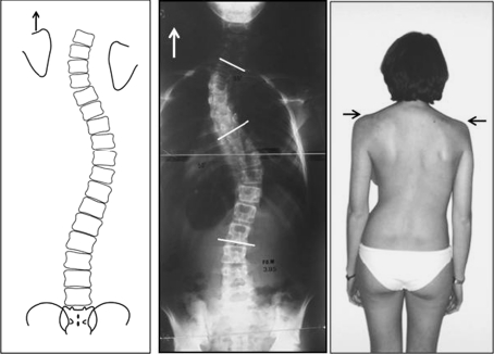
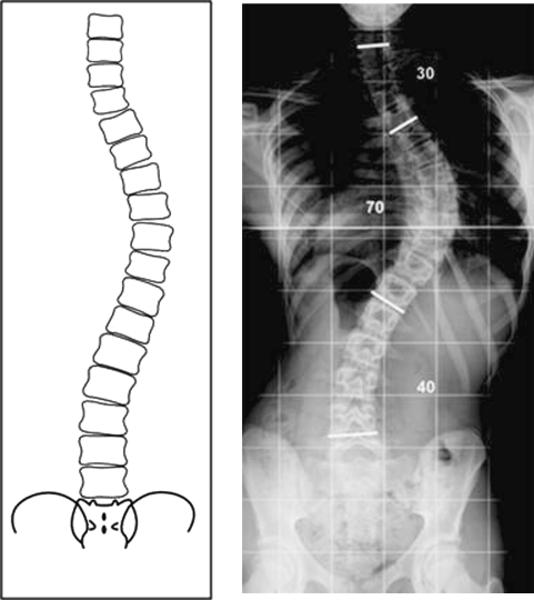
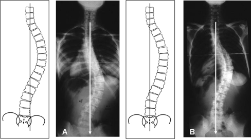
As concerns the above landmarks (spine and shoulders), the King V pattern corresponds to the picture of double thoracic scoliosis described 35 years ago by Moe: a higher left shoulder, a left cervicothoracic curve and a right lower thoracic curve (Fig. 3). But King’s classification does not include “potential” double thoracic scoliosis, i.e., that where the left structural cervicothoracic curve is masked by the lower left shoulder (Fig. 4). Likewise, Lenke pools together in the same class (Lenke I A) a simple thoracic curve (topped with a hemi-curve) and the potential double thoracic curve we characterized in 1997 [4]. However, these two cases should not be instrumented in the same way. It was only quite recently that Ilharreborde [14] defined the difference between real and potential double thoracic curves more clearly. Therefore, there exist two different King V curves: using the bending test on the left side helps to determine the actual pattern of the upper thoracic curve [13, 23, 29].
Fig. 3.
Real double thoracic curve scoliosis (left shoulder is higher)
Fig. 4.
Potential double thoracic curve scoliosis (left shoulder is lower)
In King’s system of classification, two curves have been well defined and do not require any further comment: the King III type, as long as it is restricted to simple thoracic curves (a single thoracic curve with a hemi-curve above and below it) (Fig. 5) and the King I type, which corresponds to a double thoracic and lumbar (lumbar predominant) curve (Fig. 6). The King V type, however, is problematic because it corresponds to a special entity, namely imbalanced thoracolumbar scoliosis, but the author overlooks the existence of balanced and imbalanced thoracolumbar curves [11] (Fig. 7). One might say, as suggested by Perdriolle [21], that a balanced thoracolumbar curve may be a simple thoracic curve extending downward, whereas an imbalanced thoracolumbar curve may be a simple lumbar curve extending upward. This brings us to the various types of lumbar scoliosis, which were poorly defined in Lenke’s system of classification, and were deliberately neglected by King in his system, in which he dealt only with thoracic scoliosis.
Fig. 5.
Simple thoracic curve scoliosis (one thoracic curve with a fractional sus and underlying curve)
Fig. 6.
Double thoracic and lumbar curve scoliosis (lumbar predominant)
Fig. 7.
Thoracolumbar curve scoliosis. a Unbalanced thoracolumbar scoliosis, b Balanced thoracolumbar scoliosis
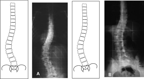
At lumbar level, curves of two kinds can occur: real lumbar scoliosis, which usually involves T11–L3, but can range between T10 and L4, has never been properly defined although it corresponds to a radiological, clinical and progressive entity in adulthood [3]. The second type of lumbar curve is lumbosacral scoliosis, a lumbar curve descending to the sacrum without any counter curves at any level. This is a borderline case of idiopathic scoliosis, because the lumbosacral area shows some small defects, which are typical of malformative scoliosis. A breakdown of lumbosacral scoliosis into various classes was recently presented by Tallet [30], (Fig. 8).
Fig. 8.
Lumbar scoliosis. a Real lumbar scoliosis (T11–L3), b Lumbosacral scoliosis
How many frontal and possibly sagittal curves do we need to be able to define an appropriate strategy for dealing with most of the situations encountered?
The following answers were based on the literature and on the facts mentioned above.
In the frontal plane, ten types of curves are required:
- Double curve scoliosis:
- Triple or quadruple curve scoliosis:
- These curves are actually combinations of the above curves: for example, some cases of scoliosis involve a left cervicothoracic as well as a right thoracic and a left lumbar (pelvis included or excluded) curve.
On the sagittal plane, like other authors, we have not been able to find any connections between frontal and sagittal abnormalities, which might have made it possible to combine both planes in a single system of classification. We can only say, in line with Cotrel [8], that a given frontal curve can be associated with various sagittal abnormalities: a double thoracic scoliosis can be associated, for example, with either two lordotic curves or a junctional kyphosis: the common double thoracic and lumbar curve can be associated with either a flat back or a short kyphosis at the thoracolumbar junction between the two curves. Preoperative sagittal analysis can help to determine the most appropriate strategy (we are talking here about scoliosis in adolescents and young adults of course): this analysis should be performed early enough, giving priority to the frontal X-ray findings (failure to perform frontal analysis is responsible for the most strategic surgical errors), [15, 26, 31].
How should scoliotic curves therefore be classified?
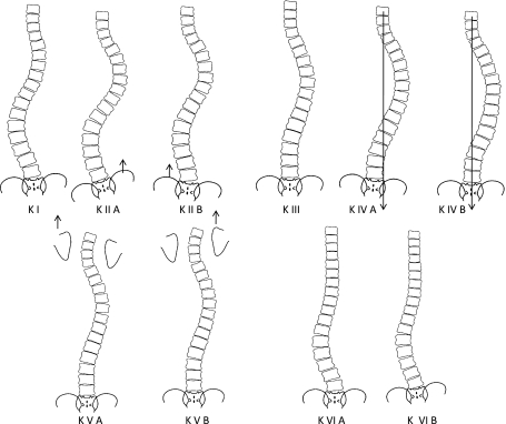
Here we have to choose between two possible alternatives: the first possibility consists in proposing a new system of classification based on the ten curves listed above, which might seem to give the most comprehensive and accurate system. However, this approach would have several disadvantages. It is always difficult to reach a consensus on such controversial topics, and Salanova might have taken to heart the following saying by a French politician called Edgard Faure: “to be right too soon is a serious error”. The English-speaking world is very attached to King’s system of classification and has not really accepted Lenke’s system due to its shortcomings as well as its complexity. In our opinion, the best solution would be to adopt a slightly modified version of King’s system, using the letter K as the reference term.
- King 1
would remain unchanged: double thoracic and lumbar (lumbar predominant) scoliosis, which would be denoted K I
- King 2
could be subdivided into K II A, a double thoracic and lumbar (thoracic predominant) “pelvis included” curve; and K II B, a double thoracic and lumbar (thoracic predominant) “pelvis excluded” curve
- King 3
could be restricted to single thoracic curves, denoted K III
- King 4
could be subdivided into K IV A, an imbalanced thoracolumbar curve; and K IV B, a balanced thoracolumbar curve
- King 5
could be subdivided into K V A, a real double thoracic curve; and K V B, a potential double thoracic curve
It would also be necessary to add definitions for lumbar scoliosis, and therefore to include the following two types:
K VI A a real lumbar curve, and K VI B a lumbosacral curve (Fig. 9).
Fig. 9.
Ten curves of proposed classification
This system of classification can always be completed.
Discussion
Will it be sufficient to use the system of classification proposed above to be able to make valid strategic decisions about managing adolescent and young adult idiopathic scoliosis? The answer is no, because surgical strategies depend on the school of thought. For example, the vertebral instrumentation indicated in our opinion for dealing with the curves we propose to denote K I, K IV A and K VI A is anterior instrumentation, which involves a completely different choice of instrumented vertebrae and a different number of instrumented vertebrae from those involved when it is decided to perform posterior instrumentation [20, 32, 33]. Since surgeons are perfectly free to make their own decisions, this means that comparisons between the outcomes obtained on specific curves have not been possible so far without a general system of classification. Comparisons of treatment strategies are impossible as long as we have not the same language of classifying them. This can be widely seen from the literature. Papers describing therapeutic results are on the decrease, while those reviewing or criticizing the existing systems of classification or the choice of instrumented vertebrae are legion [1, 5–7, 9, 10, 22, 24, 25, 28]. However, comparisons between outcomes will now be possible in the following three cases.
First, in the case of a K III curve, the results obtained using an anterior or posterior surgical approach can be compared if we are dealing with a K III curve as defined above, but not a K V B curve, with which the K III curve is frequently confused. Second, in the case of a K II A curve, a double thoracic and lumbar (thoracic predominant) “pelvis included” curve can be instrumented using either hooks, pedicle screws or hybrid constructs—as long as this type of scoliosis is not confused with the similar “pelvis excluded” K II B curve, the physiopathology of which is quite different.
The third example again involves a K III curve, on which it is proposed to use a posterior approach: the choice of lowest vertebra to be instrumented here is between what King has called the “stable vertebra” or what Salanova has called “la vertèbre d’éléction” (the most obvious choice according to him, i.e., the vertebra just above the most widely open disc). However, surgeons using an anterior approach may choose the neutral lowest vertebra. Comparisons between outcomes will be possible in this case if the authors take care to specify exactly what type of curve was involved.
Despite the differences existing between spinal approaches (which depend on the school of thought and do not come within the scope of this paper), comparisons between of surgical strategies will certainly be much more satisfactory once the serious inaccuracies about the shoulders and pelvis have been eliminated.
Conclusion
King’s and Lenke’s systems of classification do not lend themselves to accurately defining idiopathic scoliotic curves in adolescents and young adults because these systems do not take the shoulders and pelvis into account. This shortcoming has led to the problems reported in many recent studies as to how the most appropriate fusion levels should be chosen. If the simple modified version of King’s system of classification presented here was adopted; it would make it possible to perform comparative studies on the surgical results obtained using various methods on adolescents and young adults with idiopathic scoliosis.
Acknowledgments
The original illustrations have been prepared by the authors with the technical assistance of Amandine Lefebvre.
Conflict of interest None.
References
- 1.Badra M, Felmdan D, Hart R. Thoracic adolescent idiopathic scoliosis. Selection of fusion level. J Pediatr Orthop. 2010;19:465–472. doi: 10.1097/BPB.0b013e32833cb72d. [DOI] [PubMed] [Google Scholar]
- 2.Bergoin M, Hornung H, Gennari JM (1990) Double major curves and CD. In: 7th Proceeding of international Congress on Cotrel-Dubousset instrumentation 1990. Sauramps medical, Montpellier, pp 47–50
- 3.Bergoin M (1996) Histoire naturelle des scolioses lombaires de l’adolescent. Conférence d’enseignement, 1996 Expansion Scientifique Française, Paris, pp 195–210
- 4.Bergoin M, Gennari JM (1997) Formes topographiques et classification des courbures scoliotiques idiopathiques. 1997 Monographie du GEOP. Scoliose idiopathique. Sauramps Médical, Montpellier, pp 107–114
- 5.Behensky H, Cole AA, Freeman BJ, Grevitt MP, Mehdian HS, Webb JK. Fixed lumbar apical vertebral rotation predicts spinal decompensation in Lenke type 3C adolescent idiopathic scoliosis after selective posterior thoracic correction and fusion? Eur Spine J. 2007;16(10):1570–1578. doi: 10.1007/s00586-007-0397-8. [DOI] [PMC free article] [PubMed] [Google Scholar]
- 6.Berryman F, Pynsent P, Fairbank J, Disney S. A new system for measuring three-dimensional back shape in scoliosis. Eur Spine J. 2008;17(5):663–672. doi: 10.1007/s00586-007-0581-x. [DOI] [PMC free article] [PubMed] [Google Scholar]
- 7.Bridwel KH. Selection of instrumentation and fusion levels for scoliosis where to start and where to stop. J Neurosurg Spine. 2004;1:1–8. doi: 10.3171/spi.2004.1.1.0001. [DOI] [PubMed] [Google Scholar]
- 8.Cotrel Y, Dubousset J (1991) L’instrumentation CD en chirurgie rachidienne: principes, techniques, erreurs et pièges. Sauramps Médical 1991, édition française 1992. (English edition)
- 9.Duong L, Cheriet H, Labelle H, et al. Inter observer and intra observer variability in the identification lumbar modifier in adolescent idiopathic scoliosis. J Spinal Disord Tech. 2009;22:448–455. doi: 10.1097/BSD.0b013e3181831ef7. [DOI] [PubMed] [Google Scholar]
- 10.Fischer CR, Kim Y (2011) Selective fusion for adolescent idiopathic scoliosis: a review of current operative strategy. Eur Spine J (Epub ahead of print) [DOI] [PMC free article] [PubMed]
- 11.Gennari JM, Bergoin M (2007) De la nécessité d’une classification des courbures scoliotiques idiopathiques. La gazette de la Société Française d’Orthopédie Pédiatrique (SOFOP) No. 20, Mars Avril, pp 13–15
- 12.Hay D, Izzatt M, Adam C, et al. The use of fulcrum bending radiographs in anterior thoracic scoliosis correction a consecutive surveys of 90 patients. Spine. 2008;33:999–1005. doi: 10.1097/BRS.0b013e31816c8343. [DOI] [PubMed] [Google Scholar]
- 13.Hollenbeck S, Glatttes R, Asher M, et al. The prevalence of increased proximal junctional flexion following posterior instrumentation and arthrodesis for adolescent scoliosis. Spine. 2008;33:1675–1681. doi: 10.1097/BRS.0b013e31817b5bea. [DOI] [PubMed] [Google Scholar]
- 14.Ilharreborde B, Even J, Lefevre Y, et al. How to determine the upper level of instrumentation in Lenke type 1 and 2 adolescent idiopathic scoliosis: a prospective study of 132 patients. J Pediatric Orthop. 2008;28:733–739. doi: 10.1097/BPO.0b013e318185a36b. [DOI] [PubMed] [Google Scholar]
- 15.Jansen RC, Rhijn LW, Duinkerke E, Ooij A. Predictability of the spontaneous lumbar curve correction after selective thoracic fusion in idiopathic scoliosis. Eur Spine J. 2007;16(9):1335–1342. doi: 10.1007/s00586-007-0320-3. [DOI] [PMC free article] [PubMed] [Google Scholar]
- 16.King HA, Moe JH, Bradford DS, Winter RB. The selection of fusion level in thoracic idiopathic scoliosis. J Bone Joint Surg Am. 1983;65:1302–1313. [PubMed] [Google Scholar]
- 17.King HA. Selection of fusion levels for posterior instrumentation and fusion in idiopathic scoliosis. Orthop Clin North Am. 1988;19:247–255. [PubMed] [Google Scholar]
- 18.Lenke LG, Bets RR, Harms J, Bridwell KH, et al. Adolescent idiopathic scoliosis: a new classification to determine the extent of spinal arthrodesis. J Bone Joint Surg Am. 2001;83:1169–1181. [PubMed] [Google Scholar]
- 19.Luk K, Don A, Chong C, et al. Selection of fusion levels adolescent idiopathic scoliosis using fulcrum bending prediction: prospective study. Spine. 2008;33:2192–2198. doi: 10.1097/BRS.0b013e31817bd86a. [DOI] [PubMed] [Google Scholar]
- 20.Min K, Hahn F, Ziebarth K. Short anterior correction of the thoracolumbar/lumbar curve in King 1 idiopathic scoliosis: behaviour of the instrumented and non-instrumented curves and trunk balance? Eur Spine J. 2007;16(1):65–72. doi: 10.1007/s00586-006-0075-2. [DOI] [PMC free article] [PubMed] [Google Scholar]
- 21.Perdriolle R. La scoliose son étude tridimensionnelle. Paris: Maloine Sa; 1979. [Google Scholar]
- 22.Qiu X, Zhang J, Wang Y, et al. A new operative classification of idiopathic scoliosis: a Peking Union Medical College method. Spine. 2005;30:1419–1426. doi: 10.1097/01.brs.0000166531.52232.0c. [DOI] [PubMed] [Google Scholar]
- 23.Qiu XS, Ma WW, Li WG, Wang B, Yu Y, Zhu ZZ, Qian BP, et al. Discrepancy between radiographic shoulder balance and cosmetic shoulder balance in adolescent idiopathic scoliosis patients with double thoracic curve. Eur Spine J. 2009;18(1):45–51. doi: 10.1007/s00586-008-0833-4. [DOI] [PMC free article] [PubMed] [Google Scholar]
- 24.Qui G, Li Q, Wang Y, et al. Comparison of reliability between the PUC and Lenke classification systems of classifying adolescent idiopathic scoliosis. Spine. 2008;33:836–842. doi: 10.1097/BRS.0b013e318187bb10. [DOI] [PubMed] [Google Scholar]
- 25.Rhijn LW, Plasmans CM, Veraart BE. No relationship exists between the correction of the thoracic and the lumbar curves after selective thoracic fusion for adolescent idiopathic scoliosis King type II. Eur Spine J. 2002;11(16):550–555. doi: 10.1007/s00586-002-0414-x. [DOI] [PubMed] [Google Scholar]
- 26.Ritzman T, Upasani V, Bastrom J, et al. Comparison of compensatory curve spontaneous derotation after selective thoracic or lumbar fusions in adolescent idiopathic scoliosis surgery. Spine. 2008;33:2643–2647. doi: 10.1097/BRS.0b013e3181891806. [DOI] [PubMed] [Google Scholar]
- 27.Salanova C (2000) De la fusion simple à l’instrumentation segmentaire. Résonnances Européennes du Rachis RER 3(26):1043–1061
- 28.Smith S, Shaffrey C, Kunts C. Classification systems for adolescent and adult scoliosis. Neuro Surgery. 2008;63:16–24. doi: 10.1227/01.NEU.0000320447.61835.EA. [DOI] [PubMed] [Google Scholar]
- 29.Smyrnis PN, Sekouris N, Papadopoulos G. Surgical assessment of the proximal thoracic curve in adolescent idiopathic scoliosis. Eur Spine J. 2009;18(4):52. doi: 10.1007/s00586-009-0902-3. [DOI] [PMC free article] [PubMed] [Google Scholar]
- 30.Tallet JM (2010) Lumbosacral scoliosis: congrès de la société française de chirurgie rachidienne, 4 et 5 juin 2010: personal communication
- 31.Tanguay F, Mac-Thiong JM, Guise JA, Labelle H. Relation between the sagittal pelvic and lumbar spine geometries following surgical correction of adolescent idiopathic scoliosis. Eur Spine J. 2007;16(4):531–536. doi: 10.1007/s00586-006-0238-1. [DOI] [PMC free article] [PubMed] [Google Scholar]
- 32.Wang T, Zeng B, Xu J, Chen H, Zhang T, Zhou W, et al. Radiographic evaluation of selective anterior thoracolumbar or lumbar fusion for adolescent idiopathic scoliosis. Eur Spine J. 2008;17(8):1012–1018. doi: 10.1007/s00586-007-0510-z. [DOI] [PMC free article] [PubMed] [Google Scholar]
- 33.Wang Y, Fei Q, Qiu G, et al. Anterior spinal fusion versus posterior spinal fusion for moderate lumbar/thoracolumbar adolescent idiopathic scoliosis a prospective study. Spine. 2008;33:2166–2172. doi: 10.1097/BRS.0b013e318185798d. [DOI] [PubMed] [Google Scholar]
- 34.Ward W, Timothy R, Rihn J, Solic J (2008) A comparison of the Lenke and King classification systems in the surgical treatment of idiopathic thoracic scoliosis. Spine 33 [DOI] [PubMed]


