Abstract
Introduction
The main objective of all the sagittal compensating mechanisms is to allow a subject to stand and keep an erect position.
Materials and methods
The cascade of compensating mechanisms appears progressively with the increasing amount of imbalance of the spine until compensation is no longer possible. The loss of lumbar lordosis can be considered as the initiating event of sagittal imbalance. This loss of the normal lordosis pushes the C7 plumb line forward.
Results
The assessment of sagittal balance has to include to be complete: a parameter measuring the global balance of the trunk, either C7 plumb line and sacral plateau, the position of the pelvis rotation by the pelvic tilt, and a description of the position of the lower limbs. Those three parameters have been taken into account by the newly described method called full balance integrated (FBI). This evaluation is easily done on a sagittal full spine standing X-ray from C2 to the pelvis, including the first 10 cm of the femur.
Conclusion
Three questions to answer: What is the value of the pelvis incidence? Is the patient balanced? Are there compensatory mechanisms?
Keywords: Sagittal balance, Compensation mechanism, Imbalance, Calculation method, Full balance integrated, Algorythm
Introduction
The main objective of all the sagittal compensating mechanisms is to allow a subject to stand and keep an erect position. In other words, the gravity line has to fall between the two feet. The cascade of compensating mechanisms appears progressively with the increasing amount of imbalance of the spine until compensation is no longer possible and the individual is no longer able to keep the standing position. If we consider that the objective of these compensating mechanisms is to be able to stand, the balance of the trunk and the position of the lower limbs are inevitably related.
Itoi [8] made one of the first descriptions of the compensating mechanisms involved in progressive kyphosis of the spine seen in osteoporotic patients in 1991. Since then publications dealing with sagittal balance of the spine are numerous reflecting the complexity and importance of the subject [2, 5, 9, 14]. Due to ageing process [9, 15] or disc trauma, the loss of disc height is often the first phenomenon of local loss of balance. The loss of lumbar lordosis can be considered as the initiating event of sagittal imbalance. This loss of the normal lordosis pushes the C7 plumb line forward [11].
Sagittal imbalance cascade
The consequence of the disc height loss leads to the first compensation mechanism: the pelvis tilts backwards to compensate for this imbalance [1, 2, 3, 12]. This movement takes place over the femoral heads and it is called pelvic retroversion. It is a posterior rotation of the pelvis to bring the global balance of the trunk (C7 plumb line) backwards. The consequence of this movement of backward rotation of the pelvis is the extension of the hips. Knowing that pelvic incidence = pelvic tilt + sacral slope and that the pelvic incidence remains constant for a given individual [4], the sacral slope could be zero with respect to the equation of the pelvic incidence. At this point it is important to explain the role of the extension reserve of the hip [7]. It is the difference between the normal position of the hip in the standing position and the maximum extension at this joint. The extension reserve will limit the amount of pelvic retroversion possible. Of course we are not taking into consideration all the other reasons that could limit the extension of the hip such as contractures of the flexor muscles or osteoarthritis. Taking this into consideration, the amount of compensation possible by pelvic retroversion will depend on the pelvic incidence with the addition of this hip extension reserve which is around 5°–6° [7].
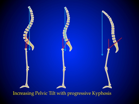
The consequence is that the pelvic backward rotation can continue for some degree so that the pelvic tilt increases putting the femoral heads forward and the sacrum and the spine backwards. This allows the C7 plumb line to stay behind the vertical line passing through the centre between the femoral heads and the gravity line to fall between the two feet (Fig. 1). The full body is balanced but it is a compensated balance, which is less economical. At the same time the posterior spine muscles can act as a posterior tension band to try to restore some lumbar lordosis, but this is an energy consuming process that becomes rapidly painful explaining for some amount the low back pain in combination with facet constraints and overstress.
Fig. 1.
Normal balance spine (left side), progressive loss of lordosis due to lumbar disc collapse leads to anterior moving of the C7 plumb line with a progressive retroversion of the pelvis to compensate (compensated balance) (central drawing), pelvis retroversion had a limit and the progression of the kyphosis imposed a flexion of the knees but it is not enough and C7 plumb line falled in the front of femoral heads (decompensated balance : imbalance) (right side)
If the loss of lordosis continues to progress, the pelvic rotation has a limit dictated by the pelvis anatomy. Pelvis with high incidence angle has more capacity to compensate [16]. When pelvis backward rotation overpasses, the only solution to stand with horizontal eyes axes is to bend the knees in order to keep the gravity line between the two feet. This process needs good psoas and quadriceps muscles activity, which is energy consuming again and not an economical situation. The use of crutches is often the only way to keep the balance.
With progressive imbalance the muscular energy necessary to keep a balance position explains the discomfort and pain suffered by these patients [2].
Clinical assessment and relevance
The clinical importance and recognition of these mechanisms are critical because they give important elements to take into consideration when indicating treatment [12]. The assessment of sagittal balance has to include to be complete: a parameter measuring the global balance of the trunk, either C7 plumb line and sacral plateau, the position of the pelvis rotation by the pelvic tilt, and a description of the position of the lower limbs. Those three parameters have been taken into account by Le Huec [13] in his evaluation method called full balance integrated (FBI).
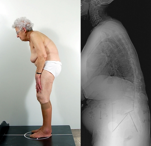
In the most severely imbalanced cases,the patients will present with all signs previously described. Trunk tilted forward, retroversion of the pelvis, apparent flexion but in reality extension of the hips and flexion of the knees (Fig. 2).
Fig. 2.
External aspect of a decompensated balance, excessive posterior muscle strength to maintain the standing position (left : photography), full standing lateral X rays showing the retroverted pelvis and the C7 plumb line in front of femoral heads (right : radiography)
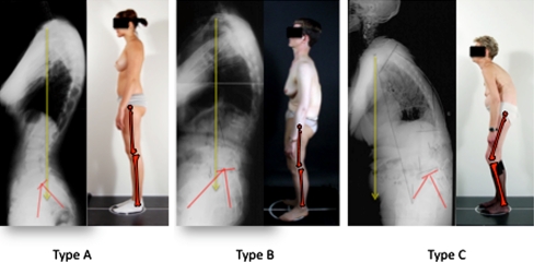
But for more simple cases of spine pathology due to degenerative process and often complaining of radicular claudication for instance, it is important to recognize intermediate cases that present with retroversion of the pelvis consequently extension of the hips but without knee flexion. These patients may appear balanced but are actually compensated balance, which is not economical because they spend muscular energy to keep this position. The issue is to be able to determine when a patient is effectively in pelvic retroversion, in other words, how do we know if the pelvic tilt for a given patient is normal. Several normal population studies provide formulae to calculate the “normal” pelvic tilt for a given pelvic incidence value [4, 16, 18]. If the value obtained for a given pelvic incidence is considered the “normal pelvic tilt” [1], any value above “normal” means a pelvic retroversion. Using these parameters a relatively simple classification of sagittal balance can be described and used before all decision of lumbar spine fusion (Fig. 3). This needs to obtain an X-ray sagittal view of the full spine including the first 10 cm of the femur to integrate all the parameters of the FBI method.
Type A or normal: global balance of the trunk measured with the C7 plumb line falls within 3 cm of the posterior corner of S1 and the pelvic tilt is normal, or in other words, it matches the pelvic tilt calculated for the patient’s pelvic incidence. Lower limbs are completely extended in the standing position. There is no reason to restore balance if surgery is requested.
Type B or compensated balance: global balance of the trunk is still normal (C7 plumb line falling behind the femoral head and within 3 cm of the posterior corner of S1) [11] but the pelvis is retroverted. The pelvic tilt is higher than the “normal” one for the patient’s incidence value [1]. The lower limbs show extension of the hips (femur straight) and the knees are in full extension. This is a very common situation in degenerative spine, mainly for patients complaining of radicular pain or claudication. During the canal decompression it is important to know that the balance is a compensated one, so that if a PLIF or TLIF procedure is requested it is important to restore the appropriate lordosis to fit with the pelvic parameters [12]: high incidence high lordosis and low incidence low lordosis according to Roussouly’s classification [16].
Type C or decompensated balance: global balance of the trunk shows a positive C7 value that falls generally in front of the femoral heads and the pelvis shows a pelvic tilt in retroversion. In the lower limbs, extension of the hips (pelvic retroversion) and flexion of the knees.
Fig. 3.
Three situations according to the balance Type A Well balance economical, Type B Compensated balance, pelvis is retroverted, PT is to important for the incidence angle, Type C Imbalanced: knee flexion, maximum pelvis retroversion, and C7 plumb line in front of femoral head
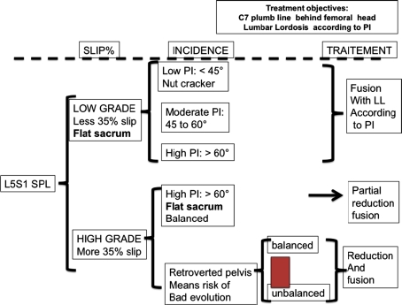
There is a special group of patient requesting a more detailed analysis; it is the patients with spondylolisthesis and -lysis. The clinical relevance is that clinicians need to keep in mind when planning treatment that subjects with L5-S1 spondylolisthesis with are a heterogeneous group with various adaptations of their posture. The common question about high-grade deformities that should or should not be reduced is always debated but the balance analysis suggests that reduction techniques should preferably be used in subjects with evidence of abnormal posture, in order to restore global spino-pelvic balance and improve the biomechanical environment for fusion (Fig. 4).
Fig. 4.
Treatment algorithm for sponylolisthesis with lysis
In conclusion we propose three main steps to the achieve analysis of sagittal balance and determine the presence of compensatory mechanisms allowing to determine the amount of correction requested to restore an ideal economical balance using the FBI method:
What is the value of the pelvis incidence? The knowledge of the pelvis incidence permits to determine expected theoretical values for spino-pelvic positional parameters (Table 1).
Is the patient balanced? Global sagittal alignment is evaluated by analysing the positioning of C7 using measurement of SSA [17] or C7PL/SFD ratio [11].
- Are there compensatory mechanisms?
- In spine level: analysis of this zone consists of measurement of lumbar lordosis and thoracic kyphosis and looking for the presence of compensatory discopathy(ies) and retrolisthesis [10].
- In lower limbs area: are the knees flexed? One must take care of this point considering that knee flessum minimizes the importance of sagittal imbalance on full spine radiographs [13].
Table 1.
Classes of pelvic incidence and corresponding values of spino-pelvic positional parameters from a group control of 154 subjects [1]
| n | PI | PT | |
|---|---|---|---|
| I | 12 | 35.4 ± 1.3 | 3.9 ± 4.5 |
| 28° < PI < 37.9° | [33.7–37.9] | [−1.5 to 13.3] | |
| II | 44 | 42.7 ± 2.8 | 8.9 ± 4.8 |
| 38° < PI < 47.9° | [37.9–47.6] | [−5.1 to 18.2] | |
| III | 59 | 52.6 ± 2.8 | 12.5 ± 5.6 |
| 48° < PI < 57.9° | [48.2–57.4] | [−1.2 to 23.2] | |
| IV | 26 | 62.6 ± 2.8 | 15.8 ± 4.3 |
| 58° < PI < 67.9° | [58.2–67.6] | [7.1 to 26.8] | |
| V | 11 | 72.6 ± 2.8 | 19.7 ± 5.5 |
| 68° < PI < 77.9° | [69.6–77.4] | [12.6 to 27.9] | |
| VI | 2 | 81.4 ± 3.3 | 21.9 ± 12.3 |
| 78° < PI < 87.9° | [79.1–81.4] | [13.2 to 30.6] |
When this analysis is done, all parameters are known if the balance is normal and economical [6], or if it is a compensated balance or if the spine is imbalance [9]. Using the FBI method [13] it is possible to determine the ideal correction requested. But other parameters such as the age of the patient, the general risk of surgery, etc., in order to make the best choice.There are many options to restore the balance using in situ fusion, disc arthroplasty, PLIF or TLIF, interpedicular osteotomy, transpedicular osteotomy or vertebral corpectomy resection. Sometimes in a fragile patient it is better to keep a compensated balance than to obtain the ideal balance with excessive risk.
Conflict of interest
None.
References
- 1.Barrey C, Jund J, Noseda O, Roussouly P. Sagittal balance of the pelvis-spine complex and lumbar degenerative diseases. A comparative study about 85 cases. Eur Spine J. 2007;16:1459–1467. doi: 10.1007/s00586-006-0294-6. [DOI] [PMC free article] [PubMed] [Google Scholar]
- 2.Barrey C, Jund J, Perrin G, Roussouly P. Spinopelvic alignment of patients with degenerative spondylolisthesis. Neurosurg. 2007;61:981–986. doi: 10.1227/01.neu.0000303194.02921.30. [DOI] [PubMed] [Google Scholar]
- 3.Berthonnaud E, Dimnet J, Roussouly P, Labelle H. Analysis of the sagittal spine and pelvis using shape and orientation parameters. J Spinal Disord Tech. 2005;18:40–47. doi: 10.1097/01.bsd.0000117542.88865.77. [DOI] [PubMed] [Google Scholar]
- 4.Duval-Beaupère G, Legaye J. Composante sagittale de la statique rachidienne (in French) Rev Rhum. 2004;71:105–119. doi: 10.1016/j.rhum.2003.09.018. [DOI] [Google Scholar]
- 5.Gelb DE, Lenke LG, Bridwell KH, Blanke K, MacEnery KW. An analysis of sagittal spinal aligment in 100 asymptomatic middle and older aged volunteers. Spine. 1995;20:1351–1358. [PubMed] [Google Scholar]
- 6.Guigui P, Levassor N, Rillardon L, Wodecki P, Cardinne L. Valeur physiologique des paramètres pelviens et rachidiens de l’équilibre sagittal du rachis. Analyse d’une série de 250 volontaires (in French) Rev Chir Orthop. 2003;89:496–506. [PubMed] [Google Scholar]
- 7.Hovorka I, Rousseau P, Bronsard N, Chalali M, Julia M, Carles M, Amoretti N, Boileau P. Extension reserve of the hip in relation to the spine: comparative study of two radiographic methods. Rev Chir Orthop Reparatrice Appar Mot. 2008;94(8):771–776. doi: 10.1016/j.rco.2008.03.033. [DOI] [PubMed] [Google Scholar]
- 8.Itoi E. Roentgenographic analysis of posture in spinal osteoporotics. Spine. 1991;16(7):750–756. doi: 10.1097/00007632-199107000-00011. [DOI] [PubMed] [Google Scholar]
- 9.Kobayashi T, Atsuta Y, Matsuno T, Takeda N. A longitudinal study of congruent sagittal spinal alignment in an adult cohort. Spine. 2004;29:671–676. doi: 10.1097/01.BRS.0000115127.51758.A2. [DOI] [PubMed] [Google Scholar]
- 10.Kumar MN, Baklanov A, Chopin D. Correlation between sagittal plane changes and adjacent segment degeneration following lumbar spine fusion. Eur Spine J. 2001;10:314–319. doi: 10.1007/s005860000239. [DOI] [PMC free article] [PubMed] [Google Scholar]
- 11.Lafage V, Schwab F, Skalli W, Hawkinson N, Gagey PM, Ondra S, Farcy JP. Standing balance and sagittal plane spinal deformity: analysis of spinopelvic and gravity line parameters. Spine. 2008;33:1572–1578. doi: 10.1097/BRS.0b013e31817886a2. [DOI] [PubMed] [Google Scholar]
- 12.Lazennec JY, Ramare S, Arafati N, Laudet CG, Gorin M, Roger B, Hansen S, Saillant G, Maurs L, Trabelsi R. Sagittal alignment in lumbosacral fusion: relations between radiological parameters and pain. Eur Spine J. 2000;9:47–55. doi: 10.1007/s005860050008. [DOI] [PMC free article] [PubMed] [Google Scholar]
- 13.Le Huec JC, Leijssen P, Duarte M, Aunoble S (2011) Thoracolumbar imbalance analysis for osteotomy planification using a new method: FBI technique. Eur Spine J 19(Suppl 3):S233–S364. Abstract S 239 [DOI] [PMC free article] [PubMed]
- 14.Marnay T (1988) Equilibre du rachis et du bassin. Cahiers d’enseignement de la SOFCOT. Elsevier, Paris, pp 281–313
- 15.Rajnics P, Templier A, Skalli W, Lavaste F, Illes T. The importance of spinopelvic parameters in patients with lumbar disc lesions. Int Orthop. 2002;26:104–108. doi: 10.1007/s00264-001-0317-1. [DOI] [PMC free article] [PubMed] [Google Scholar]
- 16.Roussouly P, Gollogly S, Berthonnaud E, Dimnet J. Classification of the normal variation in the sagittal alignment of the human lumbar spine and pelvis in the standing position. Spine. 2005;30:346–353. doi: 10.1097/01.brs.0000152379.54463.65. [DOI] [PubMed] [Google Scholar]
- 17.Roussouly P, Gollogly S, Noseda O, Berthonnaud E, Dimnet J. The vertical projection of the sum of the ground reactive forces of a standing patient is not the same as the C7 plumbline: a radiographic study of the sagittal alignment of 153 asymptomatic volunteers. Spine. 2006;31:E320–E325. doi: 10.1097/01.brs.0000218263.58642.ff. [DOI] [PubMed] [Google Scholar]
- 18.Vaz G, Roussouly P, Berthonnaud E, Dimnet J. Sagittal morphology and equilibrium of pelvis and spine. Eur Spine J. 2002;11:80–87. doi: 10.1007/s005860000224. [DOI] [PMC free article] [PubMed] [Google Scholar]


