Nonlinear optical microscopic measurement of blue collagen autofluorescence can be used to assess UVA-riboflavin–induced corneal collagen cross-linking.
Abstract
Purpose.
Corneal collagen cross-linking (CXL) by the use of riboflavin and ultraviolet-A light (UVA) is a promising and novel treatment for keratoconus and other ectatic disorders. Since CXL results in enhanced corneal stiffness, this study tested the hypothesis that CXL-induced stiffening would be proportional to the collagen autofluorescence intensity measured with nonlinear optical (NLO) microscopy.
Methods.
Rabbit eyes (n = 50) were separated into five groups including: (1) epithelium intact; (2) epithelium removed; (3) epithelium removed and soaked in riboflavin, (4) epithelium removed and soaked in riboflavin, with 15 minutes of UVA exposure; and (5) epithelium removed and soaked in riboflavin, with 30 minutes of UVA exposure. Corneal stiffness was quantified by measuring the force required to displace the cornea 500 μm. Corneas were then fixed in paraformaldehyde and sectioned, and the collagen autofluorescence over the 400- to 450-nm spectrum was recorded.
Results.
There was no significant difference in corneal stiffness among the three control groups. Corneal stiffness was significantly and dose dependently increased after UVA (P < 0.0005). Autofluorescence was detected only within the anterior stroma of the UVA-treated groups, with no significant difference in the depth of autofluorescence between different UVA exposure levels. The signal intensity was also significantly increased with longer UVA exposure (P < 0.001). Comparing corneal stiffness with autofluorescence intensity revealed a significant correlation between these values (R2 = 0.654; P < 0.0001).
Conclusions.
The results of this study indicate a significant correlation between corneal stiffening and the intensity of collagen autofluorescence after CXL. This finding suggests that the efficacy of CXL in patients could be monitored by assessing collagen autofluorescence.
Keratoconus is a common (1 in 2000 in the general population), noninflammatory, progressive corneal disorder.1 The disease is characterized by the progressive thinning or ectasia of the corneal stroma, leading to the formation of a paracentral corneal cone, severe astigmatism, and visual impairment.2 Current treatments include glasses; rigid gas-permeable contact lenses; intracorneal ring segments; or, as the disease progresses, corneal transplantation, which maybe required in 20% of keratoconus patients.3,4
Recently, a novel treatment has been developed that increases the stiffness of keratoconus corneas by increasing collagen cross-linking (CXL) by the generation of singlet oxygen after ultraviolet-A (UVA) excitation of riboflavin.5,6 Reports suggest that CXL may increase corneal stiffness by more than 71.9% in porcine eyes and 329% in human eyes.7 Clinical studies indicate that CXL significantly improves and stabilizes vision in patients with keratoconus by reducing central corneal steepness by as much as 3 to 4 D, while untreated, contralateral eyes continued to show progression of disease.8–11
Although it is known that UVA exposure levels of 3 mW/cm2 in riboflavin-saturated corneas results in cellular damage to a depth of 300 μm,12 few studies have attempted to evaluate the depth-dependent effects of CXL on stromal stiffening. Studies of corneal flaps taken at various depths after CXL and tested by stress–strain behavior13 or sensitivity to collagenase digestion14 suggest that corneal stiffening is limited to the anterior 200 μm of the corneal stroma. However, the detailed distribution and depth of CXL over corneal thickness remain unknown.
Past reports indicate that collagen shows increased blue autofluorescence (430 nm) after singlet oxygen–induced CXL by glycosylation and/or chemical cross-linking agents like glutaraldehyde.15–17 Based on these data we evaluated the effect of corneal CXL by UVA irradiation on collagen autofluorescence, as measured by nonlinear optical (NLO) microscopy. In this report, we provide evidence that UVA-riboflavin–induced CXL also enhances corneal collagen autofluorescence in the spectral range between 400 to 450 nm and can be monitored by using NLO. Further, the collagen autofluorescence signal shows increasing intensity with increasing UVA exposure and corneal stiffening. Overall, these findings suggest that monitoring collagen autofluorescence may assist in evaluating the effects of CXL on the cornea.
Material and Methods
Rabbit Eyes and Treatment Groups
Rabbit eyes were shipped overnight on ice from an abattoir (Pel-Freez, Rogers, AK), rinsed in minimal essential medium (Invitrogen, Carlsbad, CA), and placed in 12-well tissue culture plates containing sufficient medium to cover the scleral portion of the globe, leaving the corneal surface exposed to air, to keep the corneal epithelium viable and maintain corneal transparency. The eyes were then placed in a humidified, CO2 tissue culture incubator at 37°C for 1 hour, to allow corneal thickness to return to normal. All eyes used in the study were clear and transparent by biomicroscopic examination before treatment. The eyes were separated into five groups, composed of 10 eyes each: control 1 (intact epithelium), control 2 (epithelium removed), control 3 (epithelium removed and soaked in riboflavin without UVA irradiation), and test groups 4 and 5 using UVA irradiation on de-epithelialized, riboflavin-soaked corneas for 15 and 30 minutes, respectively. A 0.1% riboflavin-5-phosphate solution (Sigma-Aldrich, St. Louis, MO) in phosphate-buffered saline (PBS; pH 7.2) containing 20% low-fraction dextran (Acros Organics USA, Morris Plains, NJ) was applied every 2 minutes for 30 minutes before and during UVA irradiation. CXL was achieved with a 3-mW/cm2, 370-nm light source (PriaVision, Menlo Park, CA).
Immediately after CXL, the corneas were removed, along with 2 to 3 mm of scleral tissue, and corneal stiffness was measured within 15 minutes of UVA exposure. Corneas were then immediately fixed and processed for assessment of collagen autofluorescence. So that all eyes would be processed on the same day, the experiment was divided into smaller batches of 10 to 15 eyes each. Eyes in each batch were randomly assigned to the five groups (control and test), with all groups equally represented. Treatment of eyes was then staggered by 15 minutes so that each eye was mechanically tested immediately after riboflavin or UVA treatment. Data from all batches of eyes were then pooled and analyzed. Although the corneas did not appear swollen in biomicroscopic examination, the measured thickness of the corneas after assessment of collagen autofluorescence showed increased thickness. Nevertheless, assessment of corneal stiffness showed no difference between intact corneas and corneas from which the epithelium had been removed and the cornea soaked in riboflavin, suggesting that corneal swelling, if present, had little effect on the biomechanical measurements.
Measurement of Corneal Stiffness
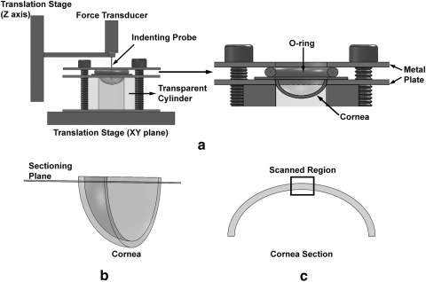
The removed corneas were clamped onto a transparent cylinder between two metal plates with a 12.7-mm-diameter central hole fixed on a three-dimensional Cartesian coordinate positioning system comprising three translation stages (Fig. 1a) driven by manual actuators (SM-25; Newport, Irvine, CA). Two stages were assembled to provide mutually perpendicular x–y axis movement, while a third stage was positioned normal to the x–y plane or along the z-axis. A 250-μm diameter probe with a round tip attached to a force transducer (model F10; Harvard Apparatus, Holliston, MA) was fixed to the translation stage along the z-axis. This assembly was used to displace the cornea, and signals from the force transducer were amplified and filtered (TAM-D, PowerLab 4/30; Harvard Apparatus) and converted to force values with affiliated software (Lab Chart; Harvard Apparatus). Each cornea was displaced through 500 μm in 50-μm steps. Force-displacement plots for each cornea were fitted to a second-order polynomial curve (R2 > 0.98) (Excel; Microsoft, Redmond, WA), and the force/displacement or corneal stiffness was calculated at 500-μm displacement by taking the derivative of the second-order polynomial equation.
Figure 1.
(a) Indentation force measurement apparatus was composed of an x–y translation stage supporting the cornea holder (expanded view), which clamped the cornea between two metal plates with an O-ring. Corneal stiffness was measured with a force transducer with 250-μm diameter indenting probe attached to a z-axis translation stage. (b) After measuring stiffness, the corneas were fixed, bisected through the central cornea, embedded in agarose, and vibratome sectioned to obtain a 300-μm-thick central corneal slice. (c) The central regions of the vibratome sections were scanned with nonlinear optical microscopy.
Assessing Collagen Autofluorescence
After stiffness measurements, the corneas were fixed overnight in 2% paraformaldehyde (Mallinckrodit Baker, Inc., Phillipsburg, NJ) in PBS (pH 7.2) at 4°C. The corneas were then washed in PBS, bisected along the superior–inferior meridian, embedded in 10% low-melting-point agarose (Lonza, Rockland, ME), and sectioned at 300-μm thickness with a vibratome (The Vibratome Company, St. Louis, MO) as shown in Figure 1b. Corneal sections were scanned with a confocal microscope (model LSM 510 Meta; Carl Zeiss, Jena, Germany) with a 40× immersion objective (NA 1.3) and two-photon microscopy (Chameleon femtosecond laser; Coherent Inc., Santa Clara, CA) with 760-nm excitation. Emission spectra were detected (Meta Detector; Carl Zeiss) over the 400- to 450-nm spectrum. To distinguish between collagen autofluorescence and riboflavin fluorescence, we evaluated the spectral emission profiles of riboflavin-soaked, CXL-treated corneas with the lambda mode function of the CLSM (model LSM 510 Meta; Carl Zeiss) over the spectral range from 382 to 682 nm using 760-nm femtosecond laser excitation.
To assess differences in collagen autofluorescence, we tile scanned the center of the corneal section with a 10 × 4 grid pattern to generate a single, 5120 × 2048-pixel image of the cornea at 0.44-μm/pixel resolution covering an area of 2.25 × 0.9 mm (Fig. 1c). Three regions of the image were then analyzed for collagen autofluorescence intensity along an 88-μm-wide region of interest extending from the anterior to the posterior stroma. Using image processing software (Meta Morph; Molecular Devices, Sunnyvale, CA), we calculated the average fluorescence intensity along each line and plotted it as a function of corneal depth. Differences between samples were analyzed by integrating the area under the pixel intensity–depth curve.
Statistics
The significance of differences between groups was calculated with one-way ANOVA on groups with the Holm-Sidak all pair-wise analysis (SigmaStat ver. 3.11; Systat Software Inc., Point Richmond, CA). Linear regression analysis was used to determine the relationship between corneal stiffness and collagen autofluorescence.
Results
Effect of CXL on Corneal Stiffness
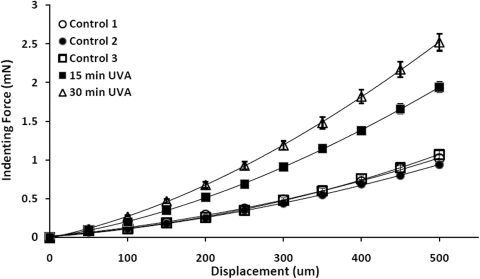
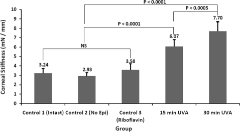
The average force required to displace the cornea over 500 μm in the different treatment groups is shown in Figure 2. For the control groups, displacement of the cornea required a gradual and uniform increase in the indenting force that appeared similar for corneas with intact epithelium (control 1), epithelium removed (control 2), or epithelium removed and soaked with riboflavin (control 3). By comparison, a greater indenting force was necessary to displace the riboflavin-soaked, UVA-exposed corneas, with a 30-minute UVA exposure requiring the greatest force to displace the cornea over 500 μm. To compare differences in corneal stiffness between groups the mean corneal stiffness at 500 μm of displacement was statistically evaluated (Fig. 3). No significant differences were detected between the control groups 1, 2, and 3, averaging 3.23 ± 0.49, 2.93 ± 0.35, and 3.58 ± 0.65 mN/mm, respectively. UVA exposure for 15 minutes significantly increased the corneal stiffness to 6.07 ± 0.73 mN/mm (P < 0.0001). By comparison, UVA exposure for 30 minutes increased corneal stiffness to 7.70 ± 1.00 mN/mm, which was significantly greater than the control (P < 0.0001) and 15-minute UVA exposure (P < 0.0005). Overall, these findings indicate that variations in UVA exposure times result in significant differences in corneal stiffening due to differences in the extent of corneal CXL.
Figure 2.
Force-displacement plots for the five treatment groups (n = 10/group) showing average ± SE for displacements through 500 μm in 50-μm steps.
Figure 3.
Average corneal stiffness (+SD) for each group showing no significant difference (NS) between the control groups, and significantly increased stiffness between the UVA-exposed and control groups.
Effects of CXL on Collagen Autofluorescence
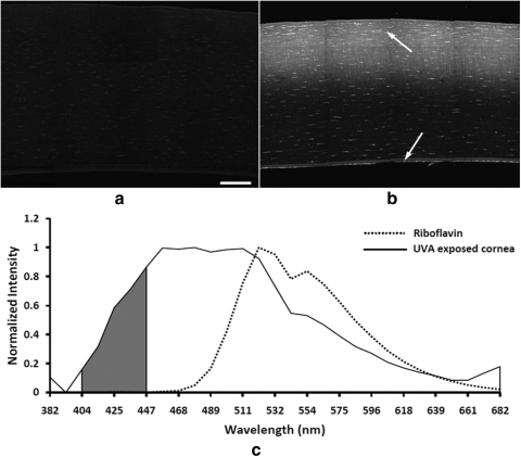
Laser scanning of corneal vibratome sections from control 3 corneas, previously treated with riboflavin and then fixed and washed, did not detect any noticeable autofluorescence signal with two-photon excitation of 760 nm (Fig. 4a). By contrast, vibratome sections from riboflavin-soaked, UVA-exposed corneas showed a strong autofluorescence signal (Fig. 4b), having a spectral emission profile ranging from 390 to 682 nm with an emission shoulder at 425 nm, indicating a peak, with higher intensity peaks at 457 and 511 nm (Fig. 4c). It should be noted that increased cellular autofluorescence of keratocytes and endothelial cells was also detected in UVA-exposed corneas (Fig. 4b, arrows). By contrast, the spectral emission profile of riboflavin alone, when excited by 760 nm two-photon excitation ranged from 460 to 682 nm with peaks at wavelengths longer than 511 nm. These findings indicated that CXL-induced autofluorescence had an emission profile distinctly different from that of riboflavin fluorescence and that collection of the spectrum between 400 and 450 nm was specific for collagen autofluorescence without overlap of any riboflavin fluorescence (Fig. 4c, shaded zone).
Figure 4.
(a) Collagen autofluorescence image (400–450 nm) of control corneas treated with riboflavin. Corneas were fixed and washed to remove excess riboflavin. (b) Collagen autofluorescence image (400–450 nm) of a riboflavin-soaked and 30-minute UVA-exposed cornea showing increased fluorescence in the anterior corneal stroma after removal of the riboflavin. Note the presence of cellular autofluorescence in the corneal stroma and corneal endothelium (arrows). (c) Emission spectra of nonlinear optical signals generated by 760-nm femtosecond laser light from riboflavin and corneas after UVA-induced CXL. Riboflavin showed peak fluorescence at 521 nm, whereas CXL corneas showed peak fluorescence at 425 nm. Bar, 100 μm.
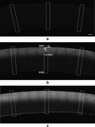
When corneal vibratome sections from control 3–treated corneas were scanned and the fluorescent signal collected over the 400- to 450-nm range, a very weak, uniform signal was detected (Fig. 5a). By contrast, UVA-exposed corneas showed greatly enhanced collagen autofluorescence localized to the anterior cornea that appeared weaker in corneas exposed to 15 minutes of UVA than in those exposed to 30 minutes of UVA (Figs. 5b, 5c). To quantify the amount and localization of autofluorescence, we measured the signal intensity for three different regions (Fig. 5, boxes). Each region of interest was composed of an 88-μm-wide strip of cornea set normal to the anterior corneal surface. As shown in Figure 5b, each pixel inside the 88-μm-wide strip was located with 2D Cartesian coordinates with the x-axis (depth) in the vertical direction and the y-axis (corneal plane) in the horizontal direction. The 0 point along the x-axis was set to the anterior surface, and the average signal intensity along a corneal plane was measured and recorded at each depth, I(x0–n). The resolution of the image was 0.44 μm, and the intensity was averaged over 200 pixels along the 88-μm width. The average intensity Ι(x) along the x-axis was then plotted through the corneal depth for each group. Since the raw collagen autofluorescence depth-intensity plots contained noise associated with increased autofluorescence from stromal cells, noise was reduced by calculating a running average over 15 pixels using the following equation:
 |
where m is the position in the corneal depth (x-axis), Im is the raw signal, I′m is the smoothed signal, and n is the position in the x-axis posterior surface of the endothelium.
Figure 5.
Collagen autofluorescence (400–450 nm) image taken from (a) a control, riboflavin-treated cornea, (b) a 15-minute UVA-exposed cornea, and (c) a 30-minute UVA exposed cornea. Note the presence of autofluorescence in the anterior stroma after UVA exposure (b, c) and the increase intensity after 30-minute UVA exposure (c). To quantify the collagen autofluorescence, the average intensity along each plane (y) was measured and plotted as a function of depth (x). Bar, 100 μm.
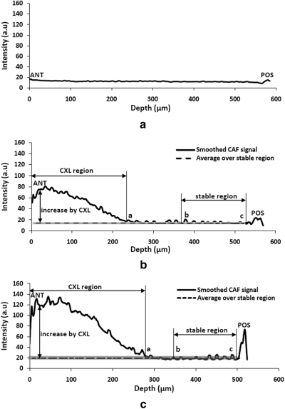
A smoothed plot for each treatment group is shown in Figure 6. While control corneal sections showed a continuously flat profile (Fig. 6a), sections scanned from UVA-treated corneas showed a high collagen autofluorescence intensity region in the anterior stroma (CXL region) and a stable, low-intensity region in the posterior stroma that was presumed to represent the background collagen autofluorescence signal (stable region). A background value for collagen autofluorescence was then determined by measuring the average intensity within the stable region starting at the corneal endothelium (Fig. 6, point c) and extending 150 μm into the posterior stroma (Fig. 6, point b). A range for the background level was then determined by taking the mean background ± SD (Fig. 6, gray line). The CXL region was then defined as the collagen autofluorescence intensity levels that exceeded background range. The point where the collagen autofluorescence intensity curve intersected the background range was defined as the depth of the CXL region, which was not significantly different between the 15- and 30-minute UVA exposures, averaging 259 ± 22 and 271 ± 31 μm or 43.8% and 46.7% of the corneal thickness, respectively (Fig. 6, point a).
Figure 6.
Representative smoothed collagen autofluorescence signals for (a) control, riboflavin-treated, (b) 15-minute UVA-exposed, and (c) 30-minute UVA-exposed corneas starting at the anterior cornea (ANT) and extending to the posterior cornea (POS). Autofluorescence intensity was quantified by first determining the average plus one SD of the background fluorescence level (gray line) within 150 μm of the posterior cornea (stable region, between points b and c). Intensity values above this level were assumed to represent the area of CXL (CXL region), and the area under the curve were integrated to represent the fluorescence increased by CXL (CXL region, between ANT and point a).
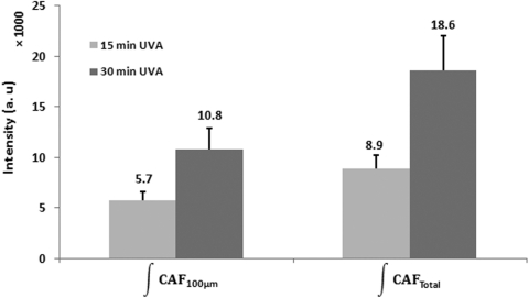
With these parameters, the collagen autofluorescence intensity was integrated within the anterior 100 μm of stroma (CAF100) and over the entire CXL region (CAFtotal) for all three regions, and the average collagen autofluorescence intensity per corneal section was recorded. Overall, there was no significant difference between the regions in the same corneal section, suggesting that collagen autofluorescence is uniform over the surface of the central cornea. In a comparison of the two treatment groups, the intensity of collagen autofluorescence was significantly greater (P < 0.001) in corneas exposed to UVA for 30 versus 15 minutes (Fig. 7). In addition, whereas the values for CAF100 were lower, there was a significant correlation (P < 0.001) between CAF100 and CAFtotal (R2 = 0.86), indicating that measuring collagen autofluorescence in the anterior corneal stroma was highly predictive of the total collagen autofluorescence.
Figure 7.
Plot of collagen autofluorescence within the anterior 100 μm of cornea (CAF100) and the total autofluorescence signal (CAFtotal) for corneas exposed to UVA for 15 or 30 minutes (R2 = 0.86; P < 0.001) between the CAF100 and CAFtotal and that 30 minutes of UVA exposure generated significantly more collagen autofluorescence than did 15 minutes of UVA exposure (P < 0.001).
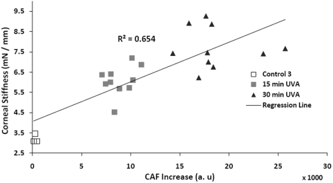
The relationship between the CAFtotal integrated signal intensity and the change in the corneal stiffness with UVA treatment is shown in Figure 8. Regression analysis indicated that there was a significant relationship (P < 0.001; R2 = 0.654). It should be noted that the kinetics of CXL has not been established and that the relationship between collagen autofluorescence and CXL may not be linear.
Figure 8.
Linear regression analysis of collagen autofluorescence and corneal stiffness between control corneas treated with riboflavin alone and corneas treated with UVA for 15 or 30 minutes. Note that there was a significant correlation, with increasing corneal stiffness associated with increasing collagen autofluorescence (P < 0.001).
Discussion
In this study, CXL led to enhanced collagen autofluorescence that was detectable with two-photon excitation by a 760-nm femtosecond laser light. Although normal rabbit corneas showed no detectible collagen autofluorescence, collagen is known to have several autofluorescence emission maximums at 310, 385, 390, and 530 nm.18 More important, oxidation of collagen using singlet oxygen, ozone, or UV light results in the formation of glycosylation-mediated, blue autofluorescence collagen cross-links with an emission maximum of 430 nm.15,19 Since riboflavin is thought to induce CXL through the generation of oxygen free radicals,5,6 our detection of enhanced blue autofluorescence is consistent with the formation of riboflavin-induced collagen cross-links potentially mediated by glycosylation.
Previous studies have used 430-nm collagen autofluorescence to noninvasively assess the accumulation of advanced glycation end products in diabetic patients and collagen content during gestation in rats.20–22 Our findings showing significant differences in the intensity of collagen autofluorescence at different levels of CXL likewise suggest that monitoring collagen autofluorescence using nonlinear optics may be a useful clinical approach to assessing the efficacy of CXL in patients. Currently, corneal stiffening after CXL is measured with destructive biomechanical testing, whereas attempts to measure the biomechanical effects in patients using the ocular response analyzer have remained controversial.23,24 Recently, nonlinear optical microscopy has been used to noninvasively characterize the biomechanical properties of collagen.16,17 These studies show that measuring both collagen content by detecting second harmonic–generated (SHG) signals and CXL by detecting two-photon–excited collagen autofluorescence can be used to predict the bulk elastic modulus of glutaraldehyde cross-linked collagen hydrogels. Since CXL induces changes only in collagen autofluorescence, we propose that monitoring two-photon–induced corneal collagen autofluorescence in patients would be an advanced approach for assessing biomechanical stiffening by CXL in real time. This monitoring could be achieved by using a spectral analyzer (similar to the Meta detector; Carl Zeiss) or more easily by using a narrow bandpass filter covering 400- to 450-nm emission spectra. Although additional study is needed to determine the relationship between collagen autofluorescence and mechanical stiffening in the human cornea, measuring the biomechanical effects of CXL in patients will become increasingly important as new approaches to the application of riboflavin and UVA to the cornea are proposed.
Detection of the collagen autofluorescence signal also showed that CXL was generally limited to the anterior corneal stroma and that 64% and 58% of the total collagen autofluorescence was contained within the first 100 μm for the 15- and 30-minute UVA-exposed corneas, respectively. This finding is consistent with previous studies showing mechanical stiffening and resistance to enzymatic digestion limited to the anterior 200 μm of the stroma.13,14 Cross-linking in this region is also consistent with the measured depth of penetration of riboflavin into the cornea using riboflavin fluorescence.25–27 However, it should be noted that the collagen autofluorescence signal, although greatly reduced, extended deeper into the cornea. This deeper penetration is also consistent with the measured depth of keratocyte cell death after CXL.12 Although keratocyte damage was not assessed in this study, increased keratocyte autofluorescence was also noted, which may be related to cellular damage induced by oxygen free radical formation or UVA exposure. Interestingly, the cellular autofluorescence was also detected in the posterior stroma and corneal endothelium, though at reduce intensity. Corneas treated with UVA alone for 30 minutes without riboflavin also showed a slight increase in cellular autofluorescence, greater than that detected in control corneas, but substantially less than riboflavin-UVA–treated corneas (data not shown). The significance of these findings is not clear, and further study is necessary to determine whether the autofluorescence is linked to potential undetected cellular injury or stress due to UVA and/or riboflavin.
In this study, the material property of the cornea was measured by corneal indentation. Our approach is similar to that used by Ahearne et al.,28 which has been shown to provide a direct and reproducible measurement of the biomechanical stiffness of the human and pig cornea. A more conventional approach is strip extensometry, which is uniaxial and has disadvantages in that measurements only assess those collagen fibrils parallel to the direction of strain,29 are dependent on the direction of the corneal strip,30 and ignore the effect of flattening or stretching a curved surface.31 A more relevant assay is bulge or inflation testing, which is biaxial and axisymmetric. This approach has several technical drawbacks, including potential leakage and difficulty in applying a controlled pressure when considering dissolved and trapped air bubbles in solution. The corneal indentation method used in the present study provides biaxial and axisymmetric stress similar to that obtained by inflation testing, while providing more controlled application of force during testing.28 The corneal indentation method also has disadvantages, in that damage to the endothelial layer occurs during indentation and change of corneal shape. However, the effect on measured corneal stiffness should be negligible, since the cornea is only indented once by 500-μm displacement achieved a small magnitude of indenting force. In addition, the direction of strain is not normal to the corneal surface, as occurs in inflation testing.
It should be noted, however, that the stiffness values recorded using corneal indentation show considerable variation. This variation is comparable to that obtained by other investigators using different approaches6,7 and may in part explain the lower correlation values detected between autofluorescence and stiffness. These findings also suggest that the biomechanical stiffness of the individual corneas may be highly variable and that measurement of collagen autofluorescence would not likely be able to predict the exact change in corneal stiffness. However, measuring collagen autofluorescence in patients before and after CXL would provide important information on the success of treatment (CXL or no CXL) and the relative level of stiffening.
A recent published study also detected increased corneal autofluorescence after CXL in rabbit corneas using an excitation wavelength of 710 nm.32 However, that study evaluated autofluorescence over a broad spectrum (380–530 nm), which includes cellular autofluorescence from NAD(P)H/FAD as well as riboflavin. Although tissues were not collected until 2 weeks after CXL, a contribution by residual riboflavin cannot be excluded. In the present study we specifically collected fluorescence data over the 400- to 450-nm spectrum, effectively excluding riboflavin fluorescence. Furthermore, the present study shows for the first time that there is a proportional increase in collagen autofluorescence associated with longer UVA exposure times and increased corneal stiffening that was not noted in the more recent study.
In conclusion, CXL induces enhanced collagen autofluorescence that can be detected with two-photon–excited fluorescence. Furthermore, the intensity of collagen autofluorescence is related to the time of UVA exposure and the amount of mechanical stiffening of the cornea. These findings suggest that measuring two-photon–induced collagen autofluorescence has the potential of assessing CXL in patients, and future studies must to be conducted to test this hypothesis.
Footnotes
Supported by NIH Grants EY07348, EY018665, EY019719, and EY016663; Research to Prevent Blindness, Inc.; The Skirball Program in Molecular Ophthalmology.
Disclosure: D. Chai, None; R.N. Gaster, None; R. Roizenblatt, None; T. Juhasz, None; D.J. Brown, None; J.V. Jester, None
References
- 1. Rabinowitz YS. Keratoconus. Surv Ophthalmol. 1998;42:297–319 [DOI] [PubMed] [Google Scholar]
- 2. Klintworth GK, Damms T. Corneal dystrophies and keratoconus. Curr Opin Ophthalmol. 1995;6:44–56 [DOI] [PubMed] [Google Scholar]
- 3. Kennedy RH, Bourne WM, Dyer JA. A 48-year clinical and epidemiologic study of keratoconus. Am J Ophthalmol. 1986;101:267–273 [DOI] [PubMed] [Google Scholar]
- 4. Tuft SJ, Moodaley LC, Gregory WM, Davison CR, Buckley RJ. Prognostic factors for the progression of keratoconus. Ophthalmology. 1994;101:439–447 [DOI] [PubMed] [Google Scholar]
- 5. Wollensak G, Spoerl E, Seiler T. Riboflavin/ultraviolet-a-induced collagen crosslinking for the treatment of keratoconus. Am J Ophthalmol. 2003;135:620–627 [DOI] [PubMed] [Google Scholar]
- 6. McCall AS, Kraft S, Edelhauser HF, et al. Mechanisms of corneal tissue cross-linking in response to treatment with topical riboflavin and long-wavelength ultraviolet radiation (UVA). Invest Ophthalmol Vis Sci. 2010;51:129–138 [DOI] [PMC free article] [PubMed] [Google Scholar]
- 7. Wollensak G, Spoerl E, Seiler T. Stress-strain measurements of human and porcine corneas after riboflavin-ultraviolet-A-induced cross-linking. J Cataract Refract Surg. 2003;29:1780–1785 [DOI] [PubMed] [Google Scholar]
- 8. Vinciguerra P, Albe E, Trazza S, et al. Refractive, topographic, tomographic, and aberrometric analysis of keratoconic eyes undergoing corneal cross-linking. Ophthalmology. 2009;116:369–378 [DOI] [PubMed] [Google Scholar]
- 9. Vinciguerra P, Albe E, Trazza S, Seiler T, Epstein D. Intraoperative and postoperative effects of corneal collagen cross-linking on progressive keratoconus. Arch Ophthalmol. 2009;127:1258–1265 [DOI] [PubMed] [Google Scholar]
- 10. Wollensak G. Crosslinking treatment of progressive keratoconus: new hope. Curr Opin Ophthalmol. 2006;17:356–360 [DOI] [PubMed] [Google Scholar]
- 11. Raiskup-Wolf F, Hoyer A, Spoerl E, Pillunat LE. Collagen crosslinking with riboflavin and ultraviolet-A light in keratoconus: long-term results. J Cataract Refract Surg. 2008;34:796–801 [DOI] [PubMed] [Google Scholar]
- 12. Spoerl E, Mrochen M, Sliney D, Trokel S, Seiler T. Safety of UVA-riboflavin cross-linking of the cornea. Cornea. 2007;26:385–389 [DOI] [PubMed] [Google Scholar]
- 13. Kohlhaas M, Spoerl E, Schilde T, Unger G, Wittig C, Pillunat LE. Biomechanical evidence of the distribution of cross-links in corneas treated with riboflavin and ultraviolet A light. J Cataract Refract Surg. 2006;32:279–283 [DOI] [PubMed] [Google Scholar]
- 14. Schilde T, Kohlhaas M, Spoerl E, Pillunat LE. Enzymatic evidence of the depth dependence of stiffening on riboflavin/UVA treated corneas (in German). Ophthalmologe. 2008;105:165–169 [DOI] [PubMed] [Google Scholar]
- 15. Fujimori E. Cross-linking and fluorescence changes of collagen by glycation and oxidation. Biochim Biophys Acta. 1989;998:105–110 [DOI] [PubMed] [Google Scholar]
- 16. Raub CB, Putnam AJ, Tromberg BJ, George SC. Predicting bulk mechanical properties of cellularized collagen gels using multiphoton microscopy. Acta Biomater. 2010;6:4657–4665 [DOI] [PMC free article] [PubMed] [Google Scholar]
- 17. Raub CB, Suresh V, Krasieva T, et al. Noninvasive assessment of collagen gel microstructure and mechanics using multiphoton microscopy. Biophys J. 2007;92:2212–2222 [DOI] [PMC free article] [PubMed] [Google Scholar]
- 18. Richards-Kortum R, Sevick-Muraca E. Quantitative optical spectroscopy for tissue diagnosis. Annu Rev Phys Chem. 1996;47:555–606 [DOI] [PubMed] [Google Scholar]
- 19. Fujimori E. Cross-linking of collagen CNBr peptides by ozone or UV light. FEBS Lett. 1988;235:98–102 [DOI] [PubMed] [Google Scholar]
- 20. Glassman W, Byam-Smith M, Garfield RE. Changes in rat cervical collagen during gestation and after antiprogesterone treatment as measured in vivo with light-induced autofluorescence. Am J Obstet Gynecol. 1995;173:1550–1556 [DOI] [PubMed] [Google Scholar]
- 21. Meerwaldt R, Graaff R, Oomen PH, et al. Simple non-invasive assessment of advanced glycation endproduct accumulation. Diabetologia. 2004;47:1324–1330 [DOI] [PubMed] [Google Scholar]
- 22. Monnier VM, Kohn RR, Cerami A. Accelerated age-related browning of human collagen in diabetes mellitus. Proc Natl Acad Sci U S A. 1984;81:583–587 [DOI] [PMC free article] [PubMed] [Google Scholar]
- 23. Goldich Y, Barkana Y, Morad Y, Hartstein M, Avni I, Zadok D. Can we measure corneal biomechanical changes after collagen cross-linking in eyes with keratoconus?—a pilot study. Cornea. 2009;28:498–502 [DOI] [PubMed] [Google Scholar]
- 24. Vinciguerra P, Albe E, Mahmoud AM, Trazza S, Hafezi F, Roberts CJ. Intra- and postoperative variation in ocular response analyzer parameters in keratoconic eyes after corneal cross-linking. J Refract Surg. 2010;26:669–676 [DOI] [PubMed] [Google Scholar]
- 25. Sondergaard AP, Hjortdal J, Breitenbach T, Ivarsen A. Corneal distribution of riboflavin prior to collagen cross-linking. Curr Eye Res. 2010;35:116–121 [DOI] [PubMed] [Google Scholar]
- 26. Kampik D, Ralla B, Keller S, Hirschberg M, Friedl P, Geerling G. Influence of corneal collagen crosslinking with riboflavin and ultraviolet-a irradiation on excimer laser surgery. Invest Ophthalmol Vis Sci. 2010;51:3929–3934 [DOI] [PubMed] [Google Scholar]
- 27. Spoerl E, Raiskup F, Kampik D, Geerling G. Correlation between UV absorption and riboflavin concentration in different depths of the cornea in CXL. Curr Eye Res. 2010;35:1040–1041; author reply 1042–1043 [DOI] [PubMed] [Google Scholar]
- 28. Ahearne M, Yang Y, Then KY, Liu KK. An indentation technique to characterize the mechanical and viscoelastic properties of human and porcine corneas. Ann Biomed Eng. 2007;35:1608–1616 [DOI] [PubMed] [Google Scholar]
- 29. Studer H, Larrea X, Riedwyl H, Buchler P. Biomechanical model of human cornea based on stromal microstructure. J Biomech. 2010;54:836–842 [DOI] [PubMed] [Google Scholar]
- 30. Elsheikh A, Brown M, Alhasso D, Rama P, Campanelli M, Garway-Heath D. Experimental assessment of corneal anisotropy. J Ref Surg. 2008;24:178–187 [DOI] [PubMed] [Google Scholar]
- 31. Elsheikh A, Anderson K. Comparative study of strip extensometry and inflation tests. J Roy Soc Interface. 2005;2:177–185 [DOI] [PMC free article] [PubMed] [Google Scholar]
- 32. Steven P, Hovakimyan M, Guthoff RF, Huttmann G, Stachs O. Imaging corneal crosslinking by autofluorescence 2-photon microscopy, second harmonic generation, and fluorescence lifetime measurements. J Cataract Refract Surg. 2010;36:2150–2159 [DOI] [PubMed] [Google Scholar]


