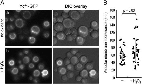
Figure 8 .
(A) Localization of Ycf1–GFP was examined in the YCF1–GFP strain (HPY1955), grown in SC−Trp at 30°, before and after exposure to 2 mM H2O2 for 2 hr. A series of Z sections was captured with the GFP filter and a single, representative Z section is shown. (B) Fluorescence intensity of the vacuolar membrane was plotted and quantified using ImageJ software: pixel intensity in untreated cells, 59.2 ± 18.8 (in a.u.); and in cells treated with H2O2, 71.6 ± 30.1 (in a.u.) (P = 0.03).

