Abstract
Tumors are cellularly and moleculary heterogeneous, with subsets of undifferentiated cancer cells exhibiting stem cell-like features (CSCs). Epithelial to mesenchymal transitions (EMT) are transdifferentiation programs that are required for tissue morphogenesis during embryonic development. The EMT process can be regulated by a diverse array of cytokines and growth factors, such as TGF-β whose activities are dysregulated during malignant tumor progression. Thus, EMT induction in cancer cells results in the acquisition of invasive and metastatic properties. Recent reports indicate that the emergence of CSCs occurs in part as a result of EMT, for example, via cues from tumor stromal components. Recent evidence now indicates that EMT of tumor cells not only causes increased metastasis but also contributes to drug resistance. In this review, we will provide potential mechanistic explanations for the association between EMT induction and the emergence of CSCs. We will also highlight recent studies implicating the function of TGF-β regulated non-coding RNAs in driving EMT and promoting CSC self-renewal. Finally we will discuss how EMT and CSCs may contribute to drug resistance as well as therapeutic strategies to overcome this clinically.
Introduction
Cellular heterogeneity is a histological hallmark of many cancers (Pardal et al., 2003). Despite their clonal origin, tumors are comprised of cells with varied morphological and molecular features. Rare tumor-initiating cells with stem cell-like properties in both hematopoietic and solid malignancies have been identified and classified using distinct cell surface markers. These cancer stem cells (CSCs) can self-renew and differentiate to generate the cellular heterogeneity of the originating tumor (Al-Hajj et al., 2003; Hermann et al., 2007; Lapidot et al., 1994; O’Brien et al., 2007; Ricci-Vitiani et al., 2007; Singh et al., 2004). However, whether CSCs are truly the only cells with de facto tumorigenic potential remains controversial (Gupta et al., 2009a; Quintana et al., 2008).
Regardless, tumors are clearly histologically heterogenous, with subsets of cancer cells exhibiting distinct molecular profiles (Gupta et al., 2009a). Furthermore, cells with different molecular characteristics within the same tumor respond differently to anti-cancer therapeutics, leading to drug resistance (Zhou et al., 2009). Cancer cells may also undergo adaptive changes following therapy, exacerbating drug resistance. In epithelial cancers, these adaptive changes may involve, at least in part, epithelial to mesenchymal transitions (EMT) and the reverse process (MET). These are developmental programs that can be usurped by oncogenically transformed cells during tumor progression (Thiery et al., 2009). Intriguingly, EMT can trigger reversion to a CSC-like phenotype (Mani et al., 2008; Polyak and Weinberg, 2009), providing an association between EMT, CSCs and drug resistance.
Parallels exist between tumorigenesis and the process of wound healing during tissue injury (Dvorak, 1986), both involving the recruitment of mesenchymal stem cells (MSCs), which can differentiate along various epithelial lineages, via MET, and may serve as cell types of origin for a number of cancers. This review addresses the role of epithelial plasticity in cancer. We also relate EMT to CSC biology, an association reinforced by the notion that cancer is a disease of abnormal wound healing and tissue repair, which are accompanied by pathophysiological EMT in adult tissues. We will overview a growing body of evidence suggesting a relationship between EMT, the emergence of cancer stem cells, and drug resistance. This ‘axis of evil’ in the war on cancer represents a significant challenge for the development of improved cancer therapies but provides new opportunities for drug discovery efforts.
Epithelial to mesenchymal transition – an overview
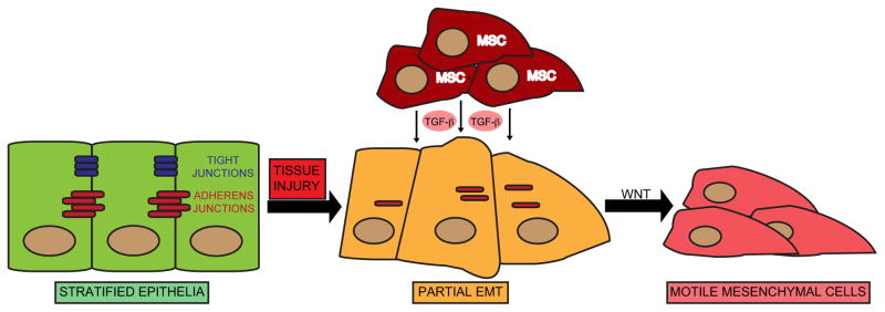
EMT is an evolutionarily conserved developmental process (Thiery, 2003). Conventionally, epithelial cells are defined as surface barrier cells with secretory functions that display distinct apical versus basolateral polarity established by adherens and tight junctions (Figure 1A). Mesenchymal cells serve scaffolding or anchoring functions and have multifunctional roles in tissue repair and wound healing. During embryonic development, certain differentiated polarized epithelial cells, upon extracellular cues, undergo profound morphogenetic changes, referred to collectively as EMT. This is typified by the dissolution of cell-cell junctions and loss of apico-basolateral polarity, resulting in the formation of migratory mesenchymal cells with invasive properties. These mesenchymal cells are recruited to specific sites in the developing embryo where they can differentiate via MET to form epithelial tissues at distal locations.
Figure 1.
(Panel A) EMT in the context of pathophysiological conditions, such as during the repair of injured tissue. Tissue injury promotes the recruitment of mesenchymal stem cells (MSCs), which can secrete EMT-inducing cytokines such as TGF-β, resulting in dissolution of cell-cell contacts and accompanying morphogenetic changes. In cooperation with additional EMT inducers such as Wnt, epithelial cells transform to a fully mesenchymal phenotype, associated with increased motility and invasiveness through basement membranes.
EMT inducers, such as transforming growth factor beta (TGF-β) or receptor tyrosine kinase (RTK) ligands trigger changes in gene expression via complex signaling networks. A paramount consequence of these signaling events is the upregulation of transcriptional repressors, such as the C2H2-type zinc-finger proteins Snail, Slug and Zeb1, Zeb2/SIP1, as well as the bHLH factors Twist and E47 (Peinado et al., 2007). These proteins bind to E-box elements in the promoter of the gene encoding the adherens junction protein E-cadherin, where they recruit histone deacetylases (HDACs) and other co-repressors to facilitate chromatin condensation and subsequent transcriptional repression of E-cadherin expression.
Reduced E-cadherin expression causes adherens junction breakdown, and along with other signaling events such as modulation of Rho GTPase function, loss of cell polarity ensues. This is accompanied by increased stability and cytoplasmic accumulation of the adherens junction component β-catenin. In cooperation with Wnt-mediated signaling, β-catenin forms a nuclear-localized transcriptional complex with TCF/LEF that promotes robust gene expression changes. The canonical EMT program is characterized by complex gene expression changes. Thus, epithelial and mesenchymal cells can be distinguished by expression of a number of classical markers that are used to define either epithelial or mesenchymal character (LaGamba et al., 2005). For instance, epithelial markers include adherens and tight junction proteins such as E-cadherin and ZO-1, whereas mesenchymal markers include the extracellular matrix (ECM) component fibronectin and the intermediate filament protein vimentin (Zeisberg and Neilson, 2009). The loss of polarity and gain of motile characteristics of mesenchymal cells during embryonic development has prompted comparisons to metastatic cancer cells during malignant progression (Yang and Weinberg, 2008). Notably, these properties have also been ascribed to normal stem cells and CSCs.
Operational definitions of cancer stem cells
Cancer stem cells with tumor-initiating potential are often compared to normal stem cells. These are multi-or pluri-potent progenitor cells that can self-renew and divide by asymmetric cell division to give rise to differentiated or committed progenitors (Pardal et al., 2003). Such properties are characteristic of oncogenically transformed cells. Dick and colleagues performed seminal studies identifying and isolating leukemia-initiating cells (LICs) in acute myeloid leukemia (AML), which exhibited a distinct cell surface antigen expression profile (Bonnet and Dick, 1997; Lapidot et al., 1994). These rare CD34+, CD38− cells can form large colonies in immuno-compromised SCID mice and exhibit the self-renewal capacities and multipotency ascribed to normal stem cells, suggesting a hierarchical model for CSC differentiation.
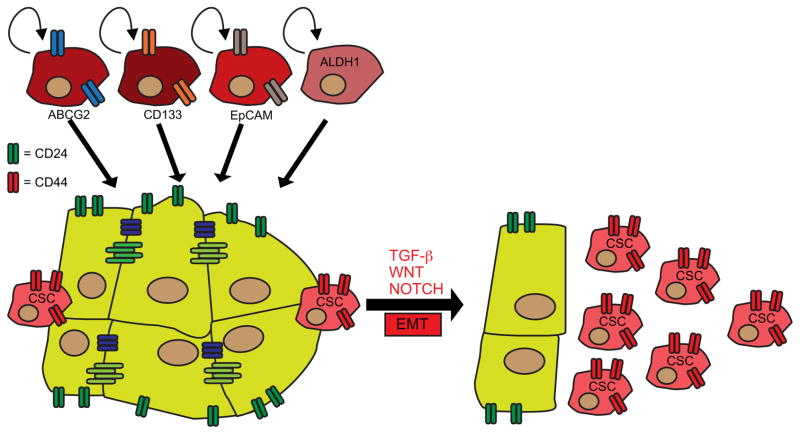
More recent studies have isolated tumor-initiating cells with CSC-like properties in a number of solid malignancies, beginning with the isolation of CD44+,CD24−/low breast cancer initiating cells (Al-Hajj et al., 2003). These cells are thought to exhibit stem cell-like properties because they are capable of reconstituting the heterogeneity of the originating tumor (Figure 2A). These observations were later extended to brain tumors, which exhibit a subpopulation of CD133+ cells with tumor-initiating capabilities (Singh et al., 2004). CD133+ tumor-initiating cells were subsequently isolated from colon cancers (O’Brien et al., 2007; Ricci-Vitiani et al., 2007) and pancreatic cancers (Hermann et al., 2007). Additionally, hepatocellular carcinomas (HCCs) exhibit a tumor-initiating population that can be identified by expression of another cell surface marker, epithelial cell adhesion molecule (EpCAM), which is expressed in normal epithelial progenitor cells (Yamashita et al., 2009). Other markers include aldehyde dehygrogenase 1 (ALDH1), identified as a specific marker for breast cancer-initiating cells (Ginestier et al., 2007). In summary, CSCs can be isolated or identified by distinct marker expression profiles, which may provide clinical utility. Furthermore, since putative CSCs have been implicated in the etiology of a number of diverse cancer types, the CSC model appears to display a universality that extends beyond leukemias.
Figure 2.
(Panel A) Cancer stem cells (CSCs) with tumor-initiating capability can be identified by the expression of a distinct set of marker proteins, such as the ABC family transporter ABCG2, CD133, EpCAM or ALDH1. These CSCs can self-renew and differentiate into a number of cell types to generate the heterogeneity of the originating tumor. (Panel B) EMT can induce a CSC-like phenotype. Inducers of EMT such as TGF-β, Wnt or Notch cause cells to acquire a CD44high CD24low phenotype, reminiscent of breast CSCs.
Origin of cancer stem cells
Do CSCs arise from existing stem/progenitor cells or from acquisition of oncogenic lesions in terminally differentiated cells resulting in dedifferentiation to a primitive stem cell-like state (Figure 2)? Both of these scenarios are likely to exist depending on the context. Early studies of CD34+, CD38− LICs suggested that CSCs arise from normal stem cells that are capable of repopulating the hematopoietic system of SCID mice, since these cells are also CD34+, CD38- (Bonnet and Dick, 1997). Since tumor-initiating cells from various tissue types display properties ascribed to normal stem cells it was hypothesized that tumors originate from these cells. However, differentiated cells can be artificially induced to acquire the pluripotency of embryonic stem cells (so-called iPS cells) (Takahashi and Yamanaka, 2006; Yu et al., 2007b). This has challenged the long-held dogma that terminal differentiation is irreversible. Notably, iPS cells are tumorigenic, forming teratomas that comprise rudimentary tissues from all three germ layers in SCID mice. In addition, the SOX2 transcription factor, which is required for efficient iPS cell reprogramming is amplified in lung and esophageal squamous cancers, underscoring a role for stem cell factors as oncogenes (Bass et al., 2009; McCaughan et al.; Yuan et al.). Recent studies have demonstrated that the p16/INK4a tumor suppressor provides a block to efficient iPS cell reprogramming by inducing a senescence response, which may explain its frequent deletion in human cancers, if the CSC model is valid (Banito et al., 2009; Li et al., 2009a; Utikal et al., 2009). Thus, gain of oncogenic activity and loss of tumor suppressor function cause dedifferentiation, suggesting that CSCs may arise from the acquisition of genetic lesions. In support of this, it has been reported that leukemic stem cells can be generated from committed hematopoietic progenitors through expression of the MLL-AF9 fusion protein (Krivtsov et al., 2006). Thus, oncogenic transformation of partially differentiated cells can lead to the emergence of CSCs.
On the other hand, a recent report that suggests that mesenchymal-like tumors originate from a mesenchymal progenitor cell (Lim et al., 2009). Breast cancers can be subdivided into basal versus luminal subtypes based on hierarchical clustering of gene expression profiles (Sorlie et al., 2001). The basal subtype exhibits a mesenchymal signature whereas the luminal subtype appears to be epithelial (Neve et al., 2006). Lim and colleagues provide evidence that basal subtype breast cancers harboring BRCA1 mutations display a gene expression signature that most closely resembles that of normal luminal progenitor cells in the mammary epithelium. This observation suggests that mesenchymal-like cancers need not develop via dedifferentiation of epithelial-like cells, but instead result from the expansion of pre-neoplastic BRCA1-mutant mesenchymal progenitor cells.
EMT-induced formation of CSCs
Pathophysiological conditions such as tissue injury or tumorigenesis can trigger differentiated cells to acquire a multipotent stem cell like phenotype through EMT induction (Figure 2B). This may mirror developmentally regulated EMT signaling pathways, such as Wnt, Notch and Hedgehog, which drive both normal and CSC renewal and maintenance (Huber et al., 2005; Malanchi et al., 2008; Peacock and Watkins, 2008). Studies also indicate that metastatic cancer cells, which have presumably undergone EMT, may exhibit a CSC phenotype. For instance, disseminated breast cancer cells found in pleural effusions are enriched for a CD44high, CD24low CSC-like population (Al-Hajj et al., 2003). Interestingly, CD44 is a β-catenin/TCF-4 target gene, supporting a role for the EMT-associated Wnt pathway in CSC maintenance (Wielenga et al., 1999). In pancreatic tumors, CD133+ mesenchymal like cells that also express the CXCR4 chemokine receptor, are found predominantly at the invasive front of tumors where they may be primed for metastatic spread (Hermann et al., 2007).
Empirical evidence connecting EMT to the emergence of stem cells has recently been reported by Weinberg and colleagues. Thus, differentiated mammary epithelial cells that have undergone EMT either upon TGFβ treatment or by forced expression of E-cadherin transcriptional repressors, such as Snail, give rise to CD44high, CD24low cells, akin to breast CSCs (Mani et al., 2008). Additionally, stem cells isolated from normal breast tissue or breast cancers express a number of canonical EMT markers. Of clinical significance, studies have shown that EMT-associated emergence of CSCs occurs after an immune response, since CD8+ T-cells can induce dedifferentiation of breast cancer cells, resulting in the formation of CD44high, CD24low stem cell-like cells (Santisteban et al., 2009). Finally, poorly differentiated and aggressive cancers display a gene expression signature related to that of embryonic stem cells (Ben-Porath et al., 2008). In summary, compelling evidence exists relating EMT to the emergence of a CSC-like phenotype, which may be prerequisites for cancer cell metastasis. A major driving force for these processes is the TGF-β signaling pathway, discussed below.
TGF-β modulation of EMT and “stemness”
TGF-β family cytokines are mediators of embryonic development and tissue homeostasis in the adult (Heldin et al., 2009). TGF-β itself is a regulator of many types of physiological and pathophysiological EMT (Derynck et al., 2001; Thiery et al., 2009). Type I and Type II TGF-β receptors (TβRI and RII) are dual specificity kinases, exhibiting both serine/threonine (S/T) and tyrosine kinase activities. Upon ligation of a heterodimeric complex of receptors RI and RII, TGF-β triggers a cascade of signaling events initiated by serine phosphosphorylation of TβRI by RII. This triggers autophosphorylation and activation of TβRI, which subsequently phosphorylates and activates a number of downstream effector proteins, most notably the Smad family of transcription factors.
TβRI activates effector Smads 2 and 3, which subsequently bind to a co-smad, Smad4. The Smad2/3/Smad4 complexes can associate with accessory transcription factors to activate expression of target genes that cause changes in cellular differentiation. Notably, Smads associate with Zeb proteins (Zeb1 and Zeb2/SIP1) to repress expression of E-cadherin during the initiation of EMT (Comijn et al., 2001; Postigo et al., 2003; Verschueren et al., 1999). Interestingly, SIP1 is essential for human embryonic stem cell (hESC) maintenance by Activin, providing further evidence for an association between TGF-β signaling, EMT and “stemness” (Chng et al., 2010).
Genetic evidence implicates TGF-β signaling as a tumor suppressor pathway, with frequent loss of function mutations, LOH and homozygous deletions affecting a number of pathway components, most notably Smad4 in pancreatic cancers. TGF-β has strong growth inhibitory and apoptotic effects resulting in tumor suppression during early stages of tumor progression. However, TGF-β can play a pro-metastatic role during later stages of tumorigenesis. Indeed, metastatic breast cancer stem cells that are CD44high, CD24low strongly express a TGF-β pathway signature and become more epithelial-like following pathway inhibition, suggesting a role for TGF-β in driving breast cancer EMT and promoting CSC maintenance (Shipitsin et al., 2007).
But how do cancer cells with intact Smad signaling components metastasize upon TGF-β exposure without undergoing cell death? There is precedence for this during wound healing, as TGF-β promotes EMT to facilitate the recruitment of inflammatory mediators, which aid in the tissue repair process (Wendt et al., 2009). In order to prevent widespread cytotoxicity, cooperating anti-apoptotic signaling pathways are triggered while TGF-β induces EMT and tissue fibrosis. These pathways, notably those involving PI3K/AKT and NF-κB are frequently activated in human cancers, providing the context for the metastasis-inducing properties of TGF-β without the accompanying cell death.
TGF-β-induced PI3K signaling can block apoptosis potently in mammary epithelial cells treated chronically with TGF-β, which leads to a metastable mesenchymal phenotype and circumvention of TGF-β-induced cell death (Gal et al., 2008). This effect is dependent on the downstream PI3K effector Akt, which can sequester Smad3 in the cytoplasm following insulin treatment of epithelial cells (Conery et al., 2004; Remy et al., 2004). Interestingly, this is independent of Akt’s function as a kinase, which may have clinical implications for the use of Akt kinase inhibitors in certain contexts. PI3K signaling can also circumvent TGF- β-induced growth arrest, specifically in neuroepithelial and glioblastoma cells, by blocking the function of a FoxO/Smad2/3 ternary complex that regulates p21/CIP1 expression (Seoane et al., 2004). Thus, the PI3K/AKT axis appears to be a critical antagonist of TGF-β-induced apoptosis and cell cycle arrest.
TGF-β, microRNAs and cancer cell plasticity
The functional role of non-coding microRNAs in cancer progression has received tremendous attention since the discovery of the let-7 microRNA, which negatively regulates Ras expression (Reinhart et al., 2000). MicroRNAs regulate gene expression by forming duplexes with the 3’ UTRs of target mRNAs, facilitating their degradation by the RISC complex of proteins. They are transcribed as precursors, which are subsequently processed by the DROSHA complex (Baulcombe, 2006).
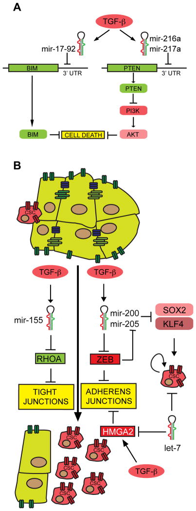
A role for microRNAs in TGF-β signaling has recently been appreciated. Firstly, Smad proteins can directly regulate microRNA processing by binding to the DROSHA complex, which has been shown for mir-21 (Davis et al., 2008). MicroRNAs can regulate TGF-β-induced apoptotic and growth suppressive functionality. For instance, TGF-β can activate Akt in glomerular mesangial cells by inducing expression of mir-216a and mir-217, which target the PI3K negative regulator PTEN (Figure 3A) (Kato et al., 2009). The E2F1-regulated microRNA mir-17-92, which has established oncogenic properties (He et al., 2005), and mir-106b-25 can block TGF-β-mediated growth arrest in gastric cancers by targeting p21/CIP1 and the pro-apoptotic protein BIM (Figure 3A) (Petrocca et al., 2008). Interestingly, transgenic expression of mir-17-92 in the embryonic lung of mice results in the expansion of lung progenitor cells by blocking differentiation (Lu et al., 2007). This occurs by targeting of an Rb-like gene, Rbl2. Along similar lines, loss of RB1 expression in fibroblasts results in reprogramming to an embryonic stem cell-like state via upregulation of Sox2, Oct-4, Klf4 and Nanog (Liu et al., 2009). These reprogrammed cells form spheres and are tumorigenic, exhibiting elevated Zeb1 expression and a CD44high, CD24low CSC-like phenotype. Thus, loss of the Rb tumor suppressor and increased E2F1 activity may stimulate the emergence of CSCs in cooperation with TGF-β-mediated changes in microRNA function.
Figure 3.
(Panel A) TGF-β induced cell death can be blocked by concomitant upregulation of microRNAs. Mir-17–92 can block expression of the pro-apoptotic protein Bim. Pro-survival PI3K signaling can be activated ablation of PTEN expression via mir-216a and mir-217a. (Panel B) Regulation of EMT and CSC “stemness” by microRNAs. TGF-β-induced EMT can be potentiated by expression of mir-155, which targets RhoA, resulting in breakdown of tight junctions. TGF-β-mediated repression of mir-200 family members and mir-205 results in increased Zeb protein expression, leading to E-cadherin repression and breakdown of adherens junctions. Zeb expression also represses mir-200/mir-205 expression, which is associated with CSC renewal via upregulation of the mir-200 targets Sox2 and Klf4. Finally, TGF-β can upregulate the activity of HMGA2, which can aid in E-cadherin repression. HMAG2 is negatively regulated by the let-7 microRNA, a microRNA that can block the self-renewal of breast CSCs. Thus, let-7 mediated depletion of HMGA2 provides a link between TGF-β signaling, EMT and the emergence of CSCs.
Several reports indicate that TGF-β induced EMT in cancer cells is dependent on microRNA function (Figure 3B). Thus, mir-155 regulates epithelial plasticity by targeting RhoA and promoting TGF-β induced EMT as a result of the dissolution of tight junctions (Kong et al., 2008). Most significantly, EMT is regulated by the expression of the mir-200 family and mir-205 (Burk et al., 2008; Gregory et al., 2008; Korpal et al., 2008; Park et al., 2008). These microRNAs target Zeb proteins and are downregulated by TGF-β, resulting in EMT. Interestingly, cancer cells established from a mouse model of lung adenocarcinoma, driven by oncogenic K-Ras and loss of function p53 mutations, display epithelial plasticity (Gibbons et al., 2009). These cells undergo EMT following TGF-β exposure, which is dependent on downregulation of mir-200 with concomitant stabilization of Zeb1 expression.
A role for Zeb1 in modulating CSC biology has now been established (Wellner et al., 2009). Zeb1 represses expression of mir-200c, mir-203 and mir-183, which drives the tumor-initiating properties of pancreatic and colorectal cancer cells by upregulating expression of stem-cell factors Sox2 and Klf4. Another report showed that TGF-β regulates EMT through HMGA2, which forms a complex with Smad proteins to recruit repressors such as SNAIL and SLUG (Thuault et al., 2006). HMGA2 is targeted by the prototypical microRNA let-7, which has also been implicated in the self-renewal and tumorigenecity of breast cancer stem cells (Yu et al., 2007a). In summary, microRNAs play a significant role in TGF-β induced EMT and subsequent dedifferentiation to a stem cell-like state.
EMT-associated resistance to cancer therapeutics
Cancer therapy is often associated with two major forms of drug resistance-- de novo or acquired. Patients who are initially refractory to therapy display intrinsic or “de novo” drug resistance. Patients that initially respond to therapy typically relapse as a consequence of “acquired” drug resistance. The use of so-called “targeted therapeutics”, such as Erlotinib/Tarceva for EGFR-mutation positive non-small cell lung cancer (NSCLC), has lead to the elucidation of various drug resistance mechanisms. Approximately 50–70% of cases of acquired drug resistance in NSCLC patients treated with EGFR inhibitors involve genetic changes including T790M ‘gatekeeper’ mutations in EGFR or amplification of the MET oncogene (Engelman et al., 2007; Sharma et al., 2007). How can we explain drug resistance in the remaining cases? A burgeoning body of literature now suggests that reversible epigenetic changes emerging during acquired drug resistance reflect changes in the differentiation state of the tumor, which is likely to reflect EMT and the emergence of chemorefractory cells with stem cell-like features (Sharma et al.; Voulgari and Pintzas, 2009) (Figure 3).
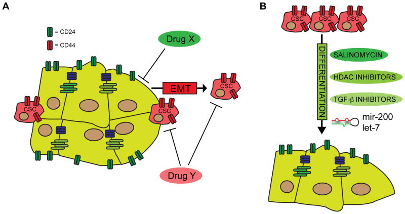
The differentiation state of a tumor may also contribute to de novo resistance. For instance, elevated E-cadherin expression is associated with sensitivity to EGFR kinase inhibitors, with drug-resistant cells being more mesenchymal-like (Witta et al., 2006). Urothelial cancers are subdivided into two groups, superficial and muscle-invasive. The latter group, associated with extremely poor prognoses and high mortality rates, exhibits mesenchymal-like properties, underscored by the observation that these tumors express Zeb2/SIP1, which protects cells from DNA damage-induced apoptosis (Sayan et al., 2009). Basal subtype mesenchymal-like breast cancers are also associated with higher mortality rates. Even in epithelial-like luminal breast cancers with HER2/neu amplification, cells with CSC features (CD44high, CD24low) are resistant to neoadjuvant chemotherapy and HER2 pharmacological inhibition (Li et al., 2008). In fact, chemotherapy leads to an increase in the number of CD44high, CD24low CSCs, which represents a potentially important mechanism of acquired drug resistance in this setting (Figure 4A). However, some reports also suggest that basal, mesenchymal-like cancers are initially more sensitive to neoadjuvant chemotherapy than epithelial luminal tumors (Carey et al., 2007; Liedtke et al., 2008; Paik et al., 2006; Yauch et al., 2005). Yet, despite the initial responses, patients with basal cancers are given the worst prognoses, suggesting that these mesenchymal-like cancers are more prone to developing drug resistance. It is possible that epithelial-like cancers are initially more sensitive to targeted therapies, such as EGFR and HER2 antagonists, whereas mesenchymal cancers are more sensitive to DNA damaging agents such as doxorubicin.
Figure 4.
(Panel A) Effective cancer therapy may require combination treatments. A putative “Drug X” may selectively kill bulk tumor cells that are well-differentiated. This could be combined with “Drug Y” to kill cells with a CSC phenotype. Cancer cells treated with Drug X may undergo EMT to acquire CSC-like properties, in which case co-administration of Drug Y would kill both pre-existing CSCs and those that emerge via treatment with Drug X. (Panel B) A novel strategy to eliminate CSCs could be to induce their differentiation, which could lead to intrinsic apoptotic cell death or could sensitize the resulting differentiated cells to existing therapies. This differentiation switch could be facilitated with compounds such as salinomycin, HDAC inhibitors or TGF-β pathway inhibitors. Finally, differentiation could also be promoted by administration of therapeutic microRNAs, such as mir-200 family members.
Strategies to modulate EMT and eliminate CSCs
Targeting CSCs therapeutically is likely to be challenging, since both bulk tumor cells and CSCs must be eliminated, potentially demanding combination drug therapies (Figure 4B). Since CSCs are molecularly distinct from bulk tumor cells, one can target their activity by exploiting these molecular differences. For instance, cell surface marker expression could be used for antibody-directed therapy to target proteins such as CD133, CD44 or EpCAM (Deonarain et al., 2009). ABC transporters, which are ATP-dependent drug efflux pumps, could be targeted by ATP-competitive agents (Kuhnle et al., 2009; Robey et al., 2007). Another strategy to selectively target CSCs has emerged from high-throughput compound screening efforts. In one study, the antibiotic salinomycin was identified, which preferentially killed breast CSCs (Gupta et al., 2009b). Interestingly, salinomycin induced the differentiation of mesenchymal-like cancers in vivo, as assessed by increased E-cadherin and reduced vimentin expression. Thus, a proof of principle has been established for the notion that poorly differentiated cancers with a high CSC content can be treated by facilitating differentiation to a more epithelial-like state.
This can be reinforced by the results with HDAC inhibitors, such as n-butyrate, which can induce the differentiation of erthythroleukemic cells (Leder et al., 1975; Riggs et al., 1977). HDAC-dependent histone deacetylation, which is elevated in some cancers, is required to repress expression of epithelial genes such as E-cadherin during EMT. Furthermore, HDACs can affect the activity of non-histone substrates, such as HIF-1 (Jeong et al., 2002; Kong et al., 2006) and NF-κB (Ashburner et al., 2001), which have been implicated in driving EMT and drug resistance. Thus, HDAC inhibition could be a useful strategy to induce the differentiation of mesenchymal-like cancer cells and CSCs, which could trigger apoptotic responses or chemosensitize these cells to other therapies. Several HDAC inhibitors, such as Trichostatin A (TSA) and Vorinostat, have now entered clinical trials for various cancers either as single agents or in combination with targeted and conventional therapeutics (Lane and Chabner, 2009). Finally, activation of the AMP-activated kinase (AMPK) with the diabetes drug metformin results in selective killing of cancer stem cells in combination with chemotherapy (Hirsch et al., 2009). This suggests that targeting mTOR activity, which is negatively regulated by AMPK, may be a strategy to block the renewal of CSCs.
The feasibility of manipulating epithelial plasticity is reinforced by studies showing that depletion of Zeb1, either chemically or by RNAi, in mesenchymal-like cells results in a partial epithelial metaplasia and either gain of K-Ras dependency or drug sensitivity (Li et al., 2009b; Singh et al., 2009). Thus, inhibitors of TGF-β pathway components that promote Zeb1 expression could prove effective in sensitizing chemoresistant mesenchymal-like tumors to anti-cancer therapeutics. Epithelial trans-differentiation could also be achieved by re-expression of mir-200 family members, which may limit the expansion of CSCs by virtue of Zeb1 blockade. Effective delivery of therapeutic microRNAs has been achieved in mice with liver cancer, so this may eventually prove to be feasible in humans (Kota et al., 2009).
The PI3K/PTEN/mTOR signaling pathway plays a prominent role in mediating drug resistance. Loss of the PTEN tumor suppressor in many malignancies is associated with extremely poor prognoses and de novo chemoresistance (Keniry and Parsons, 2008). A direct relationship has been demonstrated in breast cancers that exhibit PTEN loss, since these tumors are resistant to the HER2/Erb2-targeting antibody trastuzumab (Herceptin) (Nagata et al., 2004). In fact, PTEN activity is directly responsible for the growth inhibitory effects of trastuzumab. PI3K-mediated chemoresistance in cancer cells may be a function of TGF-β pathway activation, and resultant CSC maintenance. Indeed, HER2 amplified breast cancer cells, upon TGF-β exposure, lose sensitivity to HER2 inhibition as a result of PI3K activation by the ErbB3 protein (Wang et al., 2008).
Recent reports have linked PI3K activity directly to CSC expansion and maintenance. For example, the mTOR and stat3 pathways cooperate to promote the proliferation of breast CSCs (Zhou et al., 2007). PTEN loss in hematopoietic progenitor cells leads to myeloproliferative disorder followed by leukemia as a result of preferential expansion of leukemia-initiating stem cells (LICs) with the accompanying loss of normal hematopoietic stem cells (HSCs) (Yilmaz et al., 2006). Therapeutically, rapamycin mediated inhibition of mTOR, which is hyperactivated as a consequence of PTEN loss, results in loss of LICs and gain of normal HSCs. PI3K/AKT signaling is also required for the maintenance of CD44+ and CD133+ prostate cancer CSCs (Dubrovska et al., 2009). PTEN-deficient CSCs are likely to be highly refractory to therapeutics. In medulloblastomas, where radiation therapy results in the eradication of bulk tumor cells, therapy-resistant PTEN-deficient CSCs, displaying elevated Akt activity, remain viable (Hambardzumyan et al., 2008). In summary, inhibition of PI3K/Akt/mTOR signaling components may prevent CSC self-renewal and circumvent CSC-mediated resistance to cancer therapeutics.
Concluding remarks
Since cancers are heterogenous, future drug discovery efforts aimed at increasing patient survival will undoubtedly need to consider the plasticity of cancer cells. At least one source of such plasticity is EMT and the emergence of dedifferentiated cells with CSC-like properties. Several key signaling pathways contribute to this process, namely TGF-βand Wnt, known inducers of EMT and promoters of stem cell maintenance. Recent studies have implicated microRNA functionality in these processes, and the dysregulation of microRNA expression is likely to be a major contributing factor in the etiology of some cancers.
Targeting EMT pathways and CSC maintenance is a promising therapeutic strategy. This appears to be feasible, since several studies have successfully shown that pharmacological agents can modulate the differentiation state of a tumor. Moreover, CSCs can be eliminated or functionally antagonized by inducing their differentiation. Thus, “differentiation-inducing” agents such as salinomycin or HDAC inhibitors may have therapeutic value. Targeting the TGF-β and Wnt pathways provides additional means to eliminating CSCs. These pathways can strongly activate anti-apoptotic signaling such as those mediated by PI3K and NF-κB. Thus, PI3K or Akt inhibition to block EMT and the emergence of CSCs may prove useful. Finally, since a role for microRNAs in mediating EMT and CSC maintenance is now becoming fully appreciated, therapeutic delivery of microRNAs may represent yet an additional strategy to potentially disrupt this axis of evil in the war on cancer.
References
- Al-Hajj M, Wicha MS, Benito-Hernandez A, Morrison SJ, Clarke MF. Prospective identification of tumorigenic breast cancer cells. Proc Natl Acad Sci U S A. 2003;100:3983–8. doi: 10.1073/pnas.0530291100. [DOI] [PMC free article] [PubMed] [Google Scholar]
- Ashburner BP, Westerheide SD, Baldwin AS., Jr The p65 (RelA) subunit of NF-kappaB interacts with the histone deacetylase (HDAC) corepressors HDAC1 and HDAC2 to negatively regulate gene expression. Mol Cell Biol. 2001;21:7065–77. doi: 10.1128/MCB.21.20.7065-7077.2001. [DOI] [PMC free article] [PubMed] [Google Scholar]
- Banito A, Rashid ST, Acosta JC, Li S, Pereira CF, Geti I, et al. Senescence impairs successful reprogramming to pluripotent stem cells. Genes Dev. 2009;23:2134–9. doi: 10.1101/gad.1811609. [DOI] [PMC free article] [PubMed] [Google Scholar]
- Bass AJ, Watanabe H, Mermel CH, Yu S, Perner S, Verhaak RG, et al. SOX2 is an amplified lineage-survival oncogene in lung and esophageal squamous cell carcinomas. Nat Genet. 2009;41:1238–42. doi: 10.1038/ng.465. [DOI] [PMC free article] [PubMed] [Google Scholar]
- Baulcombe DC. Short silencing RNA: the dark matter of genetics? Cold Spring Harb Symp Quant Biol. 2006;71:13–20. doi: 10.1101/sqb.2006.71.052. [DOI] [PubMed] [Google Scholar]
- Ben-Porath I, Thomson MW, Carey VJ, Ge R, Bell GW, Regev A, et al. An embryonic stem cell-like gene expression signature in poorly differentiated aggressive human tumors. Nat Genet. 2008;40:499–507. doi: 10.1038/ng.127. [DOI] [PMC free article] [PubMed] [Google Scholar]
- Bonnet D, Dick JE. Human acute myeloid leukemia is organized as a hierarchy that originates from a primitive hematopoietic cell. Nat Med. 1997;3:730–7. doi: 10.1038/nm0797-730. [DOI] [PubMed] [Google Scholar]
- Burk U, Schubert J, Wellner U, Schmalhofer O, Vincan E, Spaderna S, et al. A reciprocal repression between ZEB1 and members of the miR-200 family promotes EMT and invasion in cancer cells. EMBO Rep. 2008;9:582–9. doi: 10.1038/embor.2008.74. [DOI] [PMC free article] [PubMed] [Google Scholar]
- Carey LA, Dees EC, Sawyer L, Gatti L, Moore DT, Collichio F, et al. The triple negative paradox: primary tumor chemosensitivity of breast cancer subtypes. Clin Cancer Res. 2007;13:2329–34. doi: 10.1158/1078-0432.CCR-06-1109. [DOI] [PubMed] [Google Scholar]
- Chng Z, Teo A, Pedersen RA, Vallier L. SIP1 Mediates Cell-Fate Decisions between Neuroectoderm and Mesendoderm in Human Pluripotent Stem Cells. Cell Stem Cell. 2010;6:59–70. doi: 10.1016/j.stem.2009.11.015. [DOI] [PubMed] [Google Scholar]
- Comijn J, Berx G, Vermassen P, Verschueren K, van Grunsven L, Bruyneel E, et al. The two-handed E box binding zinc finger protein SIP1 downregulates E-cadherin and induces invasion. Mol Cell. 2001;7:1267–78. doi: 10.1016/s1097-2765(01)00260-x. [DOI] [PubMed] [Google Scholar]
- Conery AR, Cao Y, Thompson EA, Townsend CM, Jr, Ko TC, Luo K. Akt interacts directly with Smad3 to regulate the sensitivity to TGF-beta induced apoptosis. Nat Cell Biol. 2004;6:366–72. doi: 10.1038/ncb1117. [DOI] [PubMed] [Google Scholar]
- Davis BN, Hilyard AC, Lagna G, Hata A. SMAD proteins control DROSHA-mediated microRNA maturation. Nature. 2008;454:56–61. doi: 10.1038/nature07086. [DOI] [PMC free article] [PubMed] [Google Scholar]
- Deonarain MP, Kousparou CA, Epenetos AA. Antibodies targeting cancer stem cells: a new paradigm in immunotherapy? MAbs. 2009;1:12–25. doi: 10.4161/mabs.1.1.7347. [DOI] [PMC free article] [PubMed] [Google Scholar]
- Derynck R, Akhurst RJ, Balmain A. TGF-beta signaling in tumor suppression and cancer progression. Nat Genet. 2001;29:117–29. doi: 10.1038/ng1001-117. [DOI] [PubMed] [Google Scholar]
- Dubrovska A, Kim S, Salamone RJ, Walker JR, Maira SM, Garcia-Echeverria C, et al. The role of PTEN/Akt/PI3K signaling in the maintenance and viability of prostate cancer stem-like cell populations. Proc Natl Acad Sci U S A. 2009;106:268–73. doi: 10.1073/pnas.0810956106. [DOI] [PMC free article] [PubMed] [Google Scholar]
- Dvorak HF. Tumors: wounds that do not heal. Similarities between tumor stroma generation and wound healing. N Engl J Med. 1986;315:1650–9. doi: 10.1056/NEJM198612253152606. [DOI] [PubMed] [Google Scholar]
- Engelman JA, Zejnullahu K, Mitsudomi T, Song Y, Hyland C, Park JO, et al. MET amplification leads to gefitinib resistance in lung cancer by activating ERBB3 signaling. Science. 2007;316:1039–43. doi: 10.1126/science.1141478. [DOI] [PubMed] [Google Scholar]
- Gal A, Sjoblom T, Fedorova L, Imreh S, Beug H, Moustakas A. Sustained TGF beta exposure suppresses Smad and non-Smad signalling in mammary epithelial cells, leading to EMT and inhibition of growth arrest and apoptosis. Oncogene. 2008;27:1218–30. doi: 10.1038/sj.onc.1210741. [DOI] [PubMed] [Google Scholar]
- Gibbons DL, Lin W, Creighton CJ, Rizvi ZH, Gregory PA, Goodall GJ, et al. Contextual extracellular cues promote tumor cell EMT and metastasis by regulating miR-200 family expression. Genes Dev. 2009;23:2140–51. doi: 10.1101/gad.1820209. [DOI] [PMC free article] [PubMed] [Google Scholar]
- Ginestier C, Hur MH, Charafe-Jauffret E, Monville F, Dutcher J, Brown M, et al. ALDH1 is a marker of normal and malignant human mammary stem cells and a predictor of poor clinical outcome. Cell Stem Cell. 2007;1:555–67. doi: 10.1016/j.stem.2007.08.014. [DOI] [PMC free article] [PubMed] [Google Scholar]
- Gregory PA, Bert AG, Paterson EL, Barry SC, Tsykin A, Farshid G, et al. The miR-200 family and miR-205 regulate epithelial to mesenchymal transition by targeting ZEB1 and SIP1. Nat Cell Biol. 2008;10:593–601. doi: 10.1038/ncb1722. [DOI] [PubMed] [Google Scholar]
- Gupta PB, Chaffer CL, Weinberg RA. Cancer stem cells: mirage or reality? Nat Med. 2009a;15:1010–2. doi: 10.1038/nm0909-1010. [DOI] [PubMed] [Google Scholar]
- Gupta PB, Onder TT, Jiang G, Tao K, Kuperwasser C, Weinberg RA, et al. Identification of selective inhibitors of cancer stem cells by high-throughput screening. Cell. 2009b;138:645–59. doi: 10.1016/j.cell.2009.06.034. [DOI] [PMC free article] [PubMed] [Google Scholar]
- Hambardzumyan D, Becher OJ, Rosenblum MK, Pandolfi PP, Manova-Todorova K, Holland EC. PI3K pathway regulates survival of cancer stem cells residing in the perivascular niche following radiation in medulloblastoma in vivo. Genes Dev. 2008;22:436–48. doi: 10.1101/gad.1627008. [DOI] [PMC free article] [PubMed] [Google Scholar]
- He L, Thomson JM, Hemann MT, Hernando-Monge E, Mu D, Goodson S, et al. A microRNA polycistron as a potential human oncogene. Nature. 2005;435:828–33. doi: 10.1038/nature03552. [DOI] [PMC free article] [PubMed] [Google Scholar]
- Heldin CH, Landstrom M, Moustakas A. Mechanism of TGF-beta signaling to growth arrest, apoptosis, and epithelial-mesenchymal transition. Curr Opin Cell Biol. 2009;21:166–76. doi: 10.1016/j.ceb.2009.01.021. [DOI] [PubMed] [Google Scholar]
- Hermann PC, Huber SL, Herrler T, Aicher A, Ellwart JW, Guba M, et al. Distinct populations of cancer stem cells determine tumor growth and metastatic activity in human pancreatic cancer. Cell Stem Cell. 2007;1:313–23. doi: 10.1016/j.stem.2007.06.002. [DOI] [PubMed] [Google Scholar]
- Hirsch HA, Iliopoulos D, Tsichlis PN, Struhl K. Metformin selectively targets cancer stem cells, and acts together with chemotherapy to block tumor growth and prolong remission. Cancer Res. 2009;69:7507–11. doi: 10.1158/0008-5472.CAN-09-2994. [DOI] [PMC free article] [PubMed] [Google Scholar]
- Huber MA, Kraut N, Beug H. Molecular requirements for epithelial-mesenchymal transition during tumor progression. Curr Opin Cell Biol. 2005;17:548–58. doi: 10.1016/j.ceb.2005.08.001. [DOI] [PubMed] [Google Scholar]
- Jeong JW, Bae MK, Ahn MY, Kim SH, Sohn TK, Bae MH, et al. Regulation and destabilization of HIF-1alpha by ARD1-mediated acetylation. Cell. 2002;111:709–20. doi: 10.1016/s0092-8674(02)01085-1. [DOI] [PubMed] [Google Scholar]
- Kato M, Putta S, Wang M, Yuan H, Lanting L, Nair I, et al. TGF-beta activates Akt kinase through a microRNA-dependent amplifying circuit targeting PTEN. Nat Cell Biol. 2009;11:881–9. doi: 10.1038/ncb1897. [DOI] [PMC free article] [PubMed] [Google Scholar]
- Keniry M, Parsons R. The role of PTEN signaling perturbations in cancer and in targeted therapy. Oncogene. 2008;27:5477–85. doi: 10.1038/onc.2008.248. [DOI] [PubMed] [Google Scholar]
- Kong W, Yang H, He L, Zhao JJ, Coppola D, Dalton WS, et al. MicroRNA-155 is regulated by the transforming growth factor beta/Smad pathway and contributes to epithelial cell plasticity by targeting RhoA. Mol Cell Biol. 2008;28:6773–84. doi: 10.1128/MCB.00941-08. [DOI] [PMC free article] [PubMed] [Google Scholar]
- Kong X, Lin Z, Liang D, Fath D, Sang N, Caro J. Histone deacetylase inhibitors induce VHL and ubiquitin-independent proteasomal degradation of hypoxia-inducible factor 1alpha. Mol Cell Biol. 2006;26:2019–28. doi: 10.1128/MCB.26.6.2019-2028.2006. [DOI] [PMC free article] [PubMed] [Google Scholar]
- Korpal M, Lee ES, Hu G, Kang Y. The miR-200 family inhibits epithelial-mesenchymal transition and cancer cell migration by direct targeting of E-cadherin transcriptional repressors ZEB1 and ZEB2. J Biol Chem. 2008;283:14910–4. doi: 10.1074/jbc.C800074200. [DOI] [PMC free article] [PubMed] [Google Scholar]
- Kota J, Chivukula RR, O'Donnell KA, Wentzel EA, Montgomery CL, Hwang HW, et al. Therapeutic microRNA delivery suppresses tumorigenesis in a murine liver cancer model. Cell. 2009;137:1005–17. doi: 10.1016/j.cell.2009.04.021. [DOI] [PMC free article] [PubMed] [Google Scholar]
- Krivtsov AV, Twomey D, Feng Z, Stubbs MC, Wang Y, Faber J, et al. Transformation from committed progenitor to leukaemia stem cell initiated by MLL-AF9. Nature. 2006;442:818–22. doi: 10.1038/nature04980. [DOI] [PubMed] [Google Scholar]
- Kuhnle M, Egger M, Muller C, Mahringer A, Bernhardt G, Fricker G, et al. Potent and selective inhibitors of breast cancer resistance protein (ABCG2) derived from the p-glycoprotein (ABCB1) modulator tariquidar. J Med Chem. 2009;52:1190–7. doi: 10.1021/jm8013822. [DOI] [PubMed] [Google Scholar]
- LaGamba D, Nawshad A, Hay ED. Microarray analysis of gene expression during epithelial-mesenchymal transformation. Dev Dyn. 2005;234:132–42. doi: 10.1002/dvdy.20489. [DOI] [PubMed] [Google Scholar]
- Lane AA, Chabner BA. Histone deacetylase inhibitors in cancer therapy. J Clin Oncol. 2009;27:5459–68. doi: 10.1200/JCO.2009.22.1291. [DOI] [PubMed] [Google Scholar]
- Lapidot T, Sirard C, Vormoor J, Murdoch B, Hoang T, Caceres-Cortes J, et al. A cell initiating human acute myeloid leukaemia after transplantation into SCID mice. Nature. 1994;367:645–8. doi: 10.1038/367645a0. [DOI] [PubMed] [Google Scholar]
- Leder A, Orkin S, Leder P. Differentiation of erythroleukemic cells in the presence of inhibitors of DNA synthesis. Science. 1975;190:893–4. doi: 10.1126/science.1059262. [DOI] [PubMed] [Google Scholar]
- Li H, Collado M, Villasante A, Strati K, Ortega S, Canamero M, et al. The Ink4/Arf locus is a barrier for iPS cell reprogramming. Nature. 2009a;460:1136–9. doi: 10.1038/nature08290. [DOI] [PMC free article] [PubMed] [Google Scholar]
- Li X, Lewis MT, Huang J, Gutierrez C, Osborne CK, Wu MF, et al. Intrinsic resistance of tumorigenic breast cancer cells to chemotherapy. J Natl Cancer Inst. 2008;100:672–9. doi: 10.1093/jnci/djn123. [DOI] [PubMed] [Google Scholar]
- Li Y, VandenBoom TG, 2nd, Kong D, Wang Z, Ali S, Philip PA, et al. Up-regulation of miR-200 and let-7 by natural agents leads to the reversal of epithelial-to-mesenchymal transition in gemcitabine-resistant pancreatic cancer cells. Cancer Res. 2009b;69:670–12. doi: 10.1158/0008-5472.CAN-09-1298. [DOI] [PMC free article] [PubMed] [Google Scholar]
- Liedtke C, Mazouni C, Hess KR, Andre F, Tordai A, Mejia JA, et al. Response to neoadjuvant therapy and long-term survival in patients with triple-negative breast cancer. J Clin Oncol. 2008;26:1275–81. doi: 10.1200/JCO.2007.14.4147. [DOI] [PubMed] [Google Scholar]
- Lim E, Vaillant F, Wu D, Forrest NC, Pal B, Hart AH, et al. Aberrant luminal progenitors as the candidate target population for basal tumor development in BRCA1 mutation carriers. Nat Med. 2009;15:907–13. doi: 10.1038/nm.2000. [DOI] [PubMed] [Google Scholar]
- Liu Y, Clem B, Zuba-Surma EK, El-Naggar S, Telang S, Jenson AB, et al. Mouse fibroblasts lacking RB1 function form spheres and undergo reprogramming to a cancer stem cell phenotype. Cell Stem Cell. 2009;4:336–47. doi: 10.1016/j.stem.2009.02.015. [DOI] [PMC free article] [PubMed] [Google Scholar]
- Lu Y, Thomson JM, Wong HY, Hammond SM, Hogan BL. Transgenic over-expression of the microRNA miR-17-92 cluster promotes proliferation and inhibits differentiation of lung epithelial progenitor cells. Dev Biol. 2007;310:442–53. doi: 10.1016/j.ydbio.2007.08.007. [DOI] [PMC free article] [PubMed] [Google Scholar]
- Malanchi I, Peinado H, Kassen D, Hussenet T, Metzger D, Chambon P, et al. Cutaneous cancer stem cell maintenance is dependent on beta-catenin signalling. Nature. 2008;452:650–3. doi: 10.1038/nature06835. [DOI] [PubMed] [Google Scholar]
- Mani SA, Guo W, Liao MJ, Eaton EN, Ayyanan A, Zhou AY, et al. The epithelial-mesenchymal transition generates cells with properties of stem cells. Cell. 2008;133:704–15. doi: 10.1016/j.cell.2008.03.027. [DOI] [PMC free article] [PubMed] [Google Scholar]
- McCaughan F, Pole JC, Bankier AT, Konfortov BA, Carroll B, Falzon M, et al. Progressive 3q Amplification Consistently Targets SOX2 in Preinvasive Squamous Lung Cancer. Am J Respir Crit Care Med. doi: 10.1164/rccm.201001-0005OC. [DOI] [PMC free article] [PubMed] [Google Scholar]
- Nagata Y, Lan KH, Zhou X, Tan M, Esteva FJ, Sahin AA, et al. PTEN activation contributes to tumor inhibition by trastuzumab, and loss of PTEN predicts trastuzumab resistance in patients. Cancer Cell. 2004;6:117–27. doi: 10.1016/j.ccr.2004.06.022. [DOI] [PubMed] [Google Scholar]
- Neve RM, Chin K, Fridlyand J, Yeh J, Baehner FL, Fevr T, et al. A collection of breast cancer cell lines for the study of functionally distinct cancer subtypes. Cancer Cell. 2006;10:515–27. doi: 10.1016/j.ccr.2006.10.008. [DOI] [PMC free article] [PubMed] [Google Scholar]
- O’Brien CA, Pollett A, Gallinger S, Dick JE. A human colon cancer cell capable of initiating tumour growth in immunodeficient mice. Nature. 2007;445:106–10. doi: 10.1038/nature05372. [DOI] [PubMed] [Google Scholar]
- Paik S, Tang G, Shak S, Kim C, Baker J, Kim W, et al. Gene expression and benefit of chemotherapy in women with node-negative, estrogen receptor-positive breast cancer. J Clin Oncol. 2006;24:3726–34. doi: 10.1200/JCO.2005.04.7985. [DOI] [PubMed] [Google Scholar]
- Pardal R, Clarke MF, Morrison SJ. Applying the principles of stem-cell biology to cancer. Nat Rev Cancer. 2003;3:895–902. doi: 10.1038/nrc1232. [DOI] [PubMed] [Google Scholar]
- Park SM, Gaur AB, Lengyel E, Peter ME. The miR-200 family determines the epithelial phenotype of cancer cells by targeting the E-cadherin repressors ZEB1 and ZEB2. Genes Dev. 2008;22:894–907. doi: 10.1101/gad.1640608. [DOI] [PMC free article] [PubMed] [Google Scholar]
- Peacock CD, Watkins DN. Cancer stem cells and the ontogeny of lung cancer. J Clin Oncol. 2008;26:2883–9. doi: 10.1200/JCO.2007.15.2702. [DOI] [PMC free article] [PubMed] [Google Scholar]
- Peinado H, Olmeda D, Cano A. Snail, Zeb and bHLH factors in tumour progression: an alliance against the epithelial phenotype? Nat Rev Cancer. 2007;7:415–28. doi: 10.1038/nrc2131. [DOI] [PubMed] [Google Scholar]
- Petrocca F, Visone R, Onelli MR, Shah MH, Nicoloso MS, de Martino I, et al. E2F1-regulated microRNAs impair TGFbeta-dependent cell-cycle arrest and apoptosis in gastric cancer. Cancer Cell. 2008;13:272–86. doi: 10.1016/j.ccr.2008.02.013. [DOI] [PubMed] [Google Scholar]
- Polyak K, Weinberg RA. Transitions between epithelial and mesenchymal states: acquisition of malignant and stem cell traits. Nat Rev Cancer. 2009;9:265–73. doi: 10.1038/nrc2620. [DOI] [PubMed] [Google Scholar]
- Postigo AA, Depp JL, Taylor JJ, Kroll KL. Regulation of Smad signaling through a differential recruitment of coactivators and corepressors by ZEB proteins. EMBO J. 2003;22:2453–62. doi: 10.1093/emboj/cdg226. [DOI] [PMC free article] [PubMed] [Google Scholar]
- Quintana E, Shackleton M, Sabel MS, Fullen DR, Johnson TM, Morrison SJ. Efficient tumour formation by single human melanoma cells. Nature. 2008;456:593–8. doi: 10.1038/nature07567. [DOI] [PMC free article] [PubMed] [Google Scholar]
- Reinhart BJ, Slack FJ, Basson M, Pasquinelli AE, Bettinger JC, Rougvie AE, et al. The 21-nucleotide let-7 RNA regulates developmental timing in Caenorhabditis elegans. Nature. 2000;403:901–6. doi: 10.1038/35002607. [DOI] [PubMed] [Google Scholar]
- Remy I, Montmarquette A, Michnick SW. PKB/Akt modulates TGF-beta signalling through a direct interaction with Smad3. Nat Cell Biol. 2004;6:358–65. doi: 10.1038/ncb1113. [DOI] [PubMed] [Google Scholar]
- Ricci-Vitiani L, Lombardi DG, Pilozzi E, Biffoni M, Todaro M, Peschle C, et al. Identification and expansion of human colon-cancer-initiating cells. Nature. 2007;445:111–5. doi: 10.1038/nature05384. [DOI] [PubMed] [Google Scholar]
- Riggs MG, Whittaker RG, Neumann JR, Ingram VM. n-Butyrate causes histone modification in HeLa and Friend erythroleukaemia cells. Nature. 1977;268:462–4. doi: 10.1038/268462a0. [DOI] [PubMed] [Google Scholar]
- Robey RW, Shukla S, Steadman K, Obrzut T, Finley EM, Ambudkar SV, et al. Inhibition of ABCG2-mediated transport by protein kinase inhibitors with a bisindolylmaleimide or indolocarbazole structure. Mol Cancer Ther. 2007;6:1877–85. doi: 10.1158/1535-7163.MCT-06-0811. [DOI] [PubMed] [Google Scholar]
- Santisteban M, Reiman JM, Asiedu MK, Behrens MD, Nassar A, Kalli KR, et al. Immune-induced epithelial to mesenchymal transition in vivo generates breast cancer stem cells. Cancer Res. 2009;69:2887–95. doi: 10.1158/0008-5472.CAN-08-3343. [DOI] [PMC free article] [PubMed] [Google Scholar]
- Sayan AE, Griffiths TR, Pal R, Browne GJ, Ruddick A, Yagci T, et al. SIP1 protein protects cells from DNA damage-induced apoptosis and has independent prognostic value in bladder cancer. Proc Natl Acad Sci U S A. 2009;106:14884–9. doi: 10.1073/pnas.0902042106. [DOI] [PMC free article] [PubMed] [Google Scholar]
- Seoane J, Le HV, Shen L, Anderson SA, Massague J. Integration of Smad and forkhead pathways in the control of neuroepithelial and glioblastoma cell proliferation. Cell. 2004;117:211–23. doi: 10.1016/s0092-8674(04)00298-3. [DOI] [PubMed] [Google Scholar]
- Sharma SV, Bell DW, Settleman J, Haber DA. Epidermal growth factor receptor mutations in lung cancer. Nat Rev Cancer. 2007;7:169–81. doi: 10.1038/nrc2088. [DOI] [PubMed] [Google Scholar]
- Sharma SV, Lee DY, Li B, Quinlan MP, Takahashi F, Maheswaran S, et al. A chromatin-mediated reversible drug-tolerant state in cancer cell subpopulations. Cell. 141:69–80. doi: 10.1016/j.cell.2010.02.027. [DOI] [PMC free article] [PubMed] [Google Scholar]
- Shipitsin M, Campbell LL, Argani P, Weremowicz S, Bloushtain-Qimron N, Yao J, et al. Molecular definition of breast tumor heterogeneity. Cancer Cell. 2007;11:259–73. doi: 10.1016/j.ccr.2007.01.013. [DOI] [PubMed] [Google Scholar]
- Singh A, Greninger P, Rhodes D, Koopman L, Violette S, Bardeesy N, et al. A gene expression signature associated with "K-Ras addiction" reveals regulators of EMT and tumor cell survival. Cancer Cell. 2009;15:489–500. doi: 10.1016/j.ccr.2009.03.022. [DOI] [PMC free article] [PubMed] [Google Scholar]
- Singh SK, Hawkins C, Clarke ID, Squire JA, Bayani J, Hide T, et al. Identification of human brain tumour initiating cells. Nature. 2004;432:396–401. doi: 10.1038/nature03128. [DOI] [PubMed] [Google Scholar]
- Sorlie T, Perou CM, Tibshirani R, Aas T, Geisler S, Johnsen H, et al. Gene expression patterns of breast carcinomas distinguish tumor subclasses with clinical implications. Proc Natl Acad Sci U S A. 2001;98:10869–74. doi: 10.1073/pnas.191367098. [DOI] [PMC free article] [PubMed] [Google Scholar]
- Takahashi K, Yamanaka S. Induction of pluripotent stem cells from mouse embryonic and adult fibroblast cultures by defined factors. Cell. 2006;126:663–76. doi: 10.1016/j.cell.2006.07.024. [DOI] [PubMed] [Google Scholar]
- Thiery JP. Epithelial-mesenchymal transitions in development and pathologies. Curr Opin Cell Biol. 2003;15:740–6. doi: 10.1016/j.ceb.2003.10.006. [DOI] [PubMed] [Google Scholar]
- Thiery JP, Acloque H, Huang RY, Nieto MA. Epithelial-mesenchymal transitions in development and disease. Cell. 2009;139:871–90. doi: 10.1016/j.cell.2009.11.007. [DOI] [PubMed] [Google Scholar]
- Thuault S, Valcourt U, Petersen M, Manfioletti G, Heldin CH, Moustakas A. Transforming growth factor-beta employs HMGA2 to elicit epithelial-mesenchymal transition. J Cell Biol. 2006;174:175–83. doi: 10.1083/jcb.200512110. [DOI] [PMC free article] [PubMed] [Google Scholar]
- Utikal J, Polo JM, Stadtfeld M, Maherali N, Kulalert W, Walsh RM, et al. Immortalization eliminates a roadblock during cellular reprogramming into iPS cells. Nature. 2009;460:1145–8. doi: 10.1038/nature08285. [DOI] [PMC free article] [PubMed] [Google Scholar]
- Verschueren K, Remacle JE, Collart C, Kraft H, Baker BS, Tylzanowski P, et al. SIP1, a novel zinc finger/homeodomain repressor, interacts with Smad proteins and binds to 5'-CACCT sequences in candidate target genes. J Biol Chem. 1999;274:20489–98. doi: 10.1074/jbc.274.29.20489. [DOI] [PubMed] [Google Scholar]
- Voulgari A, Pintzas A. Epithelial-mesenchymal transition in cancer metastasis: mechanisms, markers and strategies to overcome drug resistance in the clinic. Biochim Biophys Acta. 2009;1796:75–90. doi: 10.1016/j.bbcan.2009.03.002. [DOI] [PubMed] [Google Scholar]
- Wang SE, Xiang B, Guix M, Olivares MG, Parker J, Chung CH, et al. Transforming growth factor beta engages TACE and ErbB3 to activate phosphatidylinositol-3 kinase/Akt in ErbB2-overexpressing breast cancer and desensitizes cells to trastuzumab. Mol Cell Biol. 2008;28:5605–20. doi: 10.1128/MCB.00787-08. [DOI] [PMC free article] [PubMed] [Google Scholar]
- Wellner U, Schubert J, Burk UC, Schmalhofer O, Zhu F, Sonntag A, et al. The EMT-activator ZEB1 promotes tumorigenicity by repressing stemness-inhibiting microRNAs. Nat Cell Biol. 2009;11:1487–95. doi: 10.1038/ncb1998. [DOI] [PubMed] [Google Scholar]
- Wendt MK, Allington TM, Schiemann WP. Mechanisms of the epithelial-mesenchymal transition by TGF-beta. Future Oncol. 2009;5:1145–68. doi: 10.2217/fon.09.90. [DOI] [PMC free article] [PubMed] [Google Scholar]
- Wielenga VJ, Smits R, Korinek V, Smit L, Kielman M, Fodde R, et al. Expression of CD44 in Apc and Tcf mutant mice implies regulation by the WNT pathway. Am J Pathol. 1999;154:515–23. doi: 10.1016/S0002-9440(10)65297-2. [DOI] [PMC free article] [PubMed] [Google Scholar]
- Witta SE, Gemmill RM, Hirsch FR, Coldren CD, Hedman K, Ravdel L, et al. Restoring E-cadherin expression increases sensitivity to epidermal growth factor receptor inhibitors in lung cancer cell lines. Cancer Res. 2006;66:944–50. doi: 10.1158/0008-5472.CAN-05-1988. [DOI] [PubMed] [Google Scholar]
- Yamashita T, Ji J, Budhu A, Forgues M, Yang W, Wang HY, et al. EpCAM-positive hepatocellular carcinoma cells are tumor-initiating cells with stem/progenitor cell features. Gastroenterology. 2009;136:1012–24. doi: 10.1053/j.gastro.2008.12.004. [DOI] [PMC free article] [PubMed] [Google Scholar]
- Yang J, Weinberg RA. Epithelial-mesenchymal transition: at the crossroads of development and tumor metastasis. Dev Cell. 2008;14:818–29. doi: 10.1016/j.devcel.2008.05.009. [DOI] [PubMed] [Google Scholar]
- Yauch RL, Januario T, Eberhard DA, Cavet G, Zhu W, Fu L, et al. Epithelial versus mesenchymal phenotype determines in vitro sensitivity and predicts clinical activity of erlotinib in lung cancer patients. Clin Cancer Res. 2005;11:8686–98. doi: 10.1158/1078-0432.CCR-05-1492. [DOI] [PubMed] [Google Scholar]
- Yilmaz OH, Valdez R, Theisen BK, Guo W, Ferguson DO, Wu H, et al. Pten dependence distinguishes haematopoietic stem cells from leukaemia-initiating cells. Nature. 2006;441:475–82. doi: 10.1038/nature04703. [DOI] [PubMed] [Google Scholar]
- Yu F, Yao H, Zhu P, Zhang X, Pan Q, Gong C, et al. let-7 regulates self renewal and tumorigenicity of breast cancer cells. Cell. 2007a;131:1109–23. doi: 10.1016/j.cell.2007.10.054. [DOI] [PubMed] [Google Scholar]
- Yu J, Vodyanik MA, Smuga-Otto K, Antosiewicz-Bourget J, Frane JL, Tian S, et al. Induced pluripotent stem cell lines derived from human somatic cells. Science. 2007b;318:1917–20. doi: 10.1126/science.1151526. [DOI] [PubMed] [Google Scholar]
- Yuan P, Kadara H, Behrens C, Tang X, Woods D, Solis LM, et al. Sex determining region Y-Box 2 (SOX2) is a potential cell-lineage gene highly expressed in the pathogenesis of squamous cell carcinomas of the lung. PLoS One. 5:e9112. doi: 10.1371/journal.pone.0009112. [DOI] [PMC free article] [PubMed] [Google Scholar]
- Zeisberg M, Neilson EG. Biomarkers for epithelial-mesenchymal transitions. J Clin Invest. 2009;119:1429–37. doi: 10.1172/JCI36183. [DOI] [PMC free article] [PubMed] [Google Scholar]
- Zhou BB, Zhang H, Damelin M, Geles KG, Grindley JC, Dirks PB. Tumour-initiating cells: challenges and opportunities for anticancer drug discovery. Nat Rev Drug Discov. 2009;8:806–23. doi: 10.1038/nrd2137. [DOI] [PubMed] [Google Scholar]
- Zhou J, Wulfkuhle J, Zhang H, Gu P, Yang Y, Deng J, et al. Activation of the PTEN/mTOR/STAT3 pathway in breast cancer stem-like cells is required for viability and maintenance. Proc Natl Acad Sci U S A. 2007;104:16158–63. doi: 10.1073/pnas.0702596104. [DOI] [PMC free article] [PubMed] [Google Scholar]


