Abstract
Background:
Knowledge about root canal morphology and its frequent variations can exert considerable influence on the success of endodontic treatment. The aim of this study was to survey the root canal morphology of mandibular first premolar teeth in a Gujarati population by decalcification and clearing technique.
Methods:
One hundred thirty eight extracted mandibular first premolar teeth were collected from a Gujarati population. After decalcifying and clearing, the teeth were examined for tooth length, number of cusps and roots, number and shape of canal orifices and canal types.
Results:
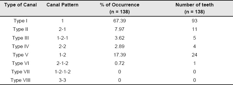
The average length of mandibular first premolar teeth was 21.2 mm. All the teeth had 2 cusps. One hundred thirty four teeth (97.1%) had one root, and just 4 teeth (2.89%) had two roots. Mesial invagination of root was found in 21 teeth (15.21%). One canal orifice was found in 122 teeth (88.4%) and two canal orifices in 16 teeth (11.59%). Shape of orifices was found to be round in 46 teeth (33.33%), oval in 72 teeth (52.17%) and flattened ribbion in 20 teeth (14.49%). According to Vertucci's classification, Type I canal system was found in 93 teeth (67.39%), Types II,III,IV,V,and VI in 11 teeth (7.97%), 5 teeth (3.62%), 4 teeth (2.89%), 24 teeth (17.39%), and 1 tooth (0.72%) respectively.
Conclusion:
Mandibular first premolar teeth were mostly found to have one root and Type I canal system.
Keywords: Canal orifice, Decalcification, Length of tooth, Mandibular first premolar, Root canal system
Introduction
Over the years it has been established that successful endodontic treatment can be achievable with accurate diagnosis & treatment planning, along with having the knowledge of root canal morphology and its frequent variations. Peter et al.1 reported that the original geometry of canal, before shaping and cleaning procedures, had more influence on the changes, that occurred during preparation, than the instrumentation technique itself. Thus, they emphasized on the importance of root canal anatomy.
The wide range of studies conducted on root canal anatomy, from the early work of Hess and Zurcher,2 to more recent, demonstrating anatomic complexities of the root canal systems, have all emphasized on the fact that a root with tapering canal and a single foramen is an exception rather than a rule.
The root canal system is complex and canal may branch, divide and rejoin taking various pathways to the apex. Weine3 categorized the root canal system into four basic types. Vertucci4 found numerous complex canal systems and identified eight pulp canal configurations.
All races and ethnic groups have some degree of dental anatomic variations. Asian populations present one of the widest variations in coronal shape, external root form and internal canal space morphology.
Amongst all the teeth, mandibular first premolar is quite difficult to treat, and has high flare up and failure rates, the major contributory factor is attributed to the Variations in root canal anatomy.5,6 Though a few studies on these teeth have been carried out in India, no study on the variations in root canal anatomy of Mandibular first premolar has been carried out in the central region of Gujarat, India, where the failure rate of endodontic treatment of these teeth is quite high.
Materials and Methods
For this in-vitro study 138 permanent extracted mandibular first premolar teeth were collected from the Out Patient Department of K M Shah Dental College and Hospital, Sumandeep Vidyapeeth, Vadodara, India, of the local population. Teeth were collected irrespective of age and sex of the patients.
The inclusion criteria were intact teeth extracted for (i) Orthodontic treatment, (ii) Periodontal diseases, (iii) Periapical diseases, (iv) Extreme mobility.
The exclusion criteria were (i) Grossly decayed or carious teeth, (ii) Fractured teeth (iii), Teeth with crazy shapes, (iv) Root canal treated teeth, (v) Teeth with full coverage restoration.
Teeth were cleaned from all debris, attached tissue and calculus using an Ultrasonic Scaler and were preserved in 10% of formalin solution. Teeth were measured for length, using Electronic Vernier Caliper (MEKA Electronic Vernier Caliper 150 mm, Model no. 06912, Hangzhou Meka Tools Co. Ltd., Zhejiang, China), from the tip of the crown to the apex of the root. For curved roots, tangents were drawn to the curved portion of the tooth and end length was measured by connecting the points of tangency.
Access cavities were prepared using No. 2 Round diamond point (Mani, Japan) in a High Speed Air Rotor Handpiece (NSK Standard, NSK Company, Japan) with air-water spray. Oval shaped access cavity was prepared which extended bucally up to the tip of buccal cusp, lingually up to lingual cusp inclination, walls were diverged occlusally with No. 2 Endo Access bur (Maillefer, Dentsply, Switzerland) for better visualization of the orifices. Shape and number of the canal orifices were observed under a Dental Operating Microscope (Seiler i Q 100-180, Seiler Precision Microscopes, St. Louis, U.S.A.) at 12X magnification. Then teeth were placed in 5.2% of sodium hypochlorite solution (Merck Limited, Mumbai, India) for 24 hours. Teeth were then decalcified and were made transparent by technique reported by Robertson et al.7
Following the placement in Sodium hypochlorite solution, teeth were washed with running water for 2 hours, then placed in 5% of Nitric Acid solution (SDFCL, Mumbai, India) for 72 hours. Nitric Acid solution was renewed every 24 hours.
Teeth were then washed with running water and placed in ascending grades of Isopropyl alcohol (SDFCL, Mumbai, India) i.e. 70%, 80%, 90% and 100% successively for 12 hours each, for a total duration of 48 hours for dehydration.
Teeth were rendered transparent by placing in Methyl Salicylate (SDFCL, Mumbai, Maharashtra, India). Methylene Blue Dye (SDFCL, Mumbai, India) was injected into these decalcified and transparent teeth through the access opening till the dye exited through the apical foramen; thus the entire pulp space were colored. These teeth were then observed under Dental Operating Microscope at 12X magnification and root canal systems were identified according to Vertucci's Classification.4
Results
Tooth length: In this study the average length of the mandibular first premolars was found to be 21.2 mm with the longest tooth having a length of 26 mm and shortest 17 mm.
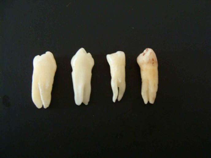
Number of Cusps & Roots: Two cusps were found in all the 138 teeth. One hundre thirty four teeth (97.10%) had one root and two roots were found in 4 teeth had two roots [2.89%]. Mesial invagination of root was found in 21 teeth (15.21%) (Table 1 and Figure 1).
Table 1.
Number of roots in mandibular first premolar

Figure 1.

Two rooted mandibular first premolar teeth.
Canal Orifice: In the mandibular first premolar teeth one canal orifice was found in 122 teeth (88.40%) and two canal orifices were found in 16 teeth (11.59%)
The shape of the canal orifice was found to be round in 46 teeth (33.33%), oval in 72 teeth (52.17%) and flattened ribbion in 20 teeth (14.40%) (Table 2).
Table 2.
Number and shape of orifice in mandibular first premolar

Canal Type: According to Vertucci's classification of canal types, Type I canal system was found in 93 teeth (67.39%), Type II in 11 teeth (7.97%,), Type III in 5 teeth (3.62%) and Type IV in 4 teeth (2.89%), Type V in 24 teeth (17.39%), and Type VI in 1 tooth (0.76%) (Table 3 and Figure 2).
Table 3.
Canal system types according to Vertucci's classification

Figure 2.
Canal type and canal pattern according to vertucci,s classification (A) TypeI, (B) TypeII, (C) Type III, (D) Type IV, (E) Type V, (F) Type VI.
Discussion
One of the predominant causes of the failure of root canal treatment in mandibular first premolar is due to the variations in root canal anatomy. In the Central Gujarat Region of India the failure rate of mandibular first premolar has been quite high with the variations in root canal anatomy being one of the attributable causes. Since no previous study on the root canal morphology of these teeth had been conducted, this study was taken up.
Although many studies on root canal anatomy have been carried out using macroscopic section,8 radiography,9 direct observation with microscope,10 decalcification and clearing,11,12 3D reconstruction,13 computed tomography,14,15 decalcification and clearing technique has provided the most detailed information along with being simple and inexpensive. In this study the average length of mandibular first premolar teeth was found to be 21.2 mm whereas Cohen & Hargreaves6 and Velumurgan & Sandhya16 reported the average length of the teeth as 21.6 mm.
All the mandibular first premolar teeth examined in this study were found to have 2 cusps which is in keeping with the established external morphology of this tooth.17,18 Evagination manifested as an extra cusp or enamel pearl, although commonly reported in mandibular premolars in people of Asian descent was not found.
In this study 97.10% of the mandibular first premolar teeth were found to have one root (134 teeth) whereas 2.89% teeth were found to have two roots (4 teeth) which is similar to the finding of Zillich & Dawson,19 Vertucci,4 Rahimi et al.20 and Velumurgan & Sandhya.16
Mesial invagination of the root that arises as a result of invagination of Hertwig's epithelial root sheath and manifested as accentuation of the normal longitudinal root groove was found in 21teeth (15.21%) which was consistent with the finding of Robinson et al.14, who found invagination in 15% mandibular first premolars using spiral computed tomography and that of Velumurgan & Sandhya16 who reported 14% of mandibular first premolars with mesial invagination of the root.
According to Vertucci's classification,4 Root canal system of mandibular first premolars was found to be predominantly (67.39%) Type I (Single canal extends from pulp chamber to apex). Vertucci's reported Type I canal system in 70%, Velmurugan & Sandhya16 in 72%, while Zillich & Dawnson19 reported an incidence ranging from 67.2% to 86.3%. Type II canal system (Two separate canals leave pulp chamber and join short of apex to form one canal) was found in 7.97% teeth whereas Vertucci4 reported 0%, Velmurugan & Sandhya16 6% and Rahimi et al.20 5.6 %. Type III Canal system (One canal leaves pulp chamber and divides into two canals in the root, and finally merge into one and exit) was found in 3.62% teeth which was similar to 4% in the findings of Vertucci4 and 3% of Velmurugan & Sandhya.16 Type IV canal system (Two separate canals extend from pulp chamber to apex) was found in 2.89% mandibular first premolars, Vertucci4 reported this system in 1.5% teeth, Velmurugan & Sandhya16 in 10% and Rahimi et al.20 in 22 %. Type V Canal system (One canal leaves pulp chamber, divides short of apex into two) was found in 17.39% teeth, Vertucci4 found this system in 24% teeth while Velumurgan & Sandhya16 reported an incidence of 8%. Type VI canal system (Two canals leave pulp chamber merge in the root and devide again short of apex to exit as two distinct canals) was found in 0.72% teeth. Types VII (One canal leaves pulp chamber, divides and then rejoin in root and finallydevides into two canals short of the apex) and VIII (Three separate canals extend from pulp chamber to the apex) were not identified in any of the teeth.
The difference in the incidence of I, II, III, IV, V, canal system in the present study and those reported by Vertucci4, Velmurugan & Sandhya16 and Rahimi et al.20 could be attributed to racial difference since this study was carried out on a Gujarati Indian population whereas Vertucci4 carried out study on Caucasian population, Rahimi et al.20 in Iranian population and Velmurugan & Sandhya16 on South Indian population.
Conclusion
It can be concluded from the results of this study that mandibular first premolar teeth present a wide variety of radicular features, with one root and one canal being found in majority of cases. Presence of two roots, two canals, mesial invagination of root is also found in a number of cases, which complicates the endodontic treatmen.
References
- 1.Peters OA, Schönenberger K, Laib A. Effects of four Ni-Ti preparation techniques on root canal geometry assessed by micro computed tomography. Int Endod J. 2001;34(3):221–30. doi: 10.1046/j.1365-2591.2001.00373.x. [DOI] [PubMed] [Google Scholar]
- 2.Hess W, Zurcher E. New York: William Wood & Co; 1925. The anatomy of root canals of the teeth of the Permanent & Deciduous Dentition. [Google Scholar]
- 3.Weine FS. 5th edn. St. Louis: Mosby– Yearbook lnc; 1996. Endodontic Therapy; p. 243. [Google Scholar]
- 4.Vertucci FJ. Root canal anatomy of the human permanent teeth. Oral Surg Oral Med Oral Pathol. 1984;58:589–99. doi: 10.1016/0030-4220(84)90085-9. [DOI] [PubMed] [Google Scholar]
- 5.Ingle JI, Taintor JF. 3rd ed. Philadelphia: Lea & Fibiger; 1985. Endodontic; pp. 27–52. [Google Scholar]
- 6.Cohen S, Hargreaves KM. 9th ed. St. Louis: Mosby Elservier; 2006. Pathways of the Pulp; p. 216. [Google Scholar]
- 7.Robertson D, Leeb J, Mckee M, Brewer E. A clearing technique for the study of root canal systems. J Endod. 1980;6(1):421–4. doi: 10.1016/S0099-2399(80)80218-4. [DOI] [PubMed] [Google Scholar]
- 8.Baisden MK, Kulild JC, Weller RN. Root canal configuration of the mandibular first premolar. J Endod. 1992;18(10):505–8. doi: 10.1016/S0099-2399(06)81352-X. [DOI] [PubMed] [Google Scholar]
- 9.Pineda F, Kuttler Y. Mesiodistal and Buccolingual roentgenographic investigation of 7,275 root canals. Oral Surg Oral Med Oral Pathol. 1972;33(1):101–10. doi: 10.1016/0030-4220(72)90214-9. [DOI] [PubMed] [Google Scholar]
- 10.Sempira HN, Hartwell GR. Frequency of second mesiobuccal canals in maxillary molars as determined by use of an operating microscope: a clinical study. J Endod. 2000;26(11):673–4. doi: 10.1097/00004770-200011000-00010. [DOI] [PubMed] [Google Scholar]
- 11.Caliskan MK, Pehlivan Y, Sepetcioglu F, Turkun M, Tuncer SS. Root canal morphology of human permanent teeth in a Turkish population. J Endod. 1995;21(4):200–4. doi: 10.1016/S0099-2399(06)80566-2. [DOI] [PubMed] [Google Scholar]
- 12.Rwenyonyi CM, Kutesa AM, Muwazi LM, Buwembo W. Root and canal morphology of maxillary first and second permanent molar teeth in a Ugandan population. Int Endod J. 2007;40(9):679–83. doi: 10.1111/j.1365-2591.2007.01265.x. [DOI] [PubMed] [Google Scholar]
- 13.Mikrogeorgis G, Lyroudia KL, Nikopoulos N, Pitas I, Molyvdas I, Lambrianidis TH. 3D computer-aided reconstruction of six teeth with morphological abnormalities. Int Endod J. 1999;32(2):88–93. doi: 10.1046/j.1365-2591.1999.00189.x. [DOI] [PubMed] [Google Scholar]
- 14.Robinson S, Czerny C, Gahleitner A, Bernhart T, Kainberger FM. Dental CT evaluation of mandibular first premolar root configurations and canal variations. Oral Surg Oral Med Oral Pathol Oral Radio Endo. 2002;93(3):328–32. doi: 10.1067/moe.2002.120055. [DOI] [PubMed] [Google Scholar]
- 15.Reuben J, Velmurugan N, Kandaswamy D. The evaluation of root canal morphology of the mandibular first molar in an Indian population using spiral computed tomography scan: an in vitro study. J Endod. 2008;34(2):212–5. doi: 10.1016/j.joen.2007.11.018. [DOI] [PubMed] [Google Scholar]
- 16.Velmurugan N, Sandhya R. Root canal morphology of mandibular first premolar in an Indian population: a laboratory study. Int Endo J. 2009;42(1):54–58. doi: 10.1111/j.1365-2591.2008.01494.x. [DOI] [PubMed] [Google Scholar]
- 17.Ash M, Nelson S. 8th Ed. Philadelphia: Saunders; 2003. Wheeler's Dental Anatomy, Physiology and Occlusion. [Google Scholar]
- 18.Fuller J, Denely G. 2nd ed. Chicago: Year Book Medical Publishers Inc; 1984. Concise Dental Anatomy & Morphology. [Google Scholar]
- 19.Zillich R, Dowson J. Root canal morphology of mandibular first & second premolars. Oral Surg Oral Med Oral Pathol. 1973;36(5):738–44. doi: 10.1016/0030-4220(73)90147-3. [DOI] [PubMed] [Google Scholar]
- 20.Rahimi S, Shahi S, Yavari H, Manafi H, Eskandarzadeh N. Root canal configuration of mandibular first & second premolars in an Iranian population. Journal of Dental Research, Dental Clinics, Dental Prospects. 2007;1(2):59–64. doi: 10.5681/joddd.2007.010. [DOI] [PMC free article] [PubMed] [Google Scholar]


